. 2023 Sep;29(9):1339-1354.
doi: 10.1261/rna.079582.123. Epub 2023 May 23.

Structural basis for RNA-duplex unwinding by the DEAD-box helicase DbpA

Affiliations

Structural basis for RNA-duplex unwinding by the DEAD-box helicase DbpA

Jan Philip Wurm. RNA. 2023 Sep.

Abstract

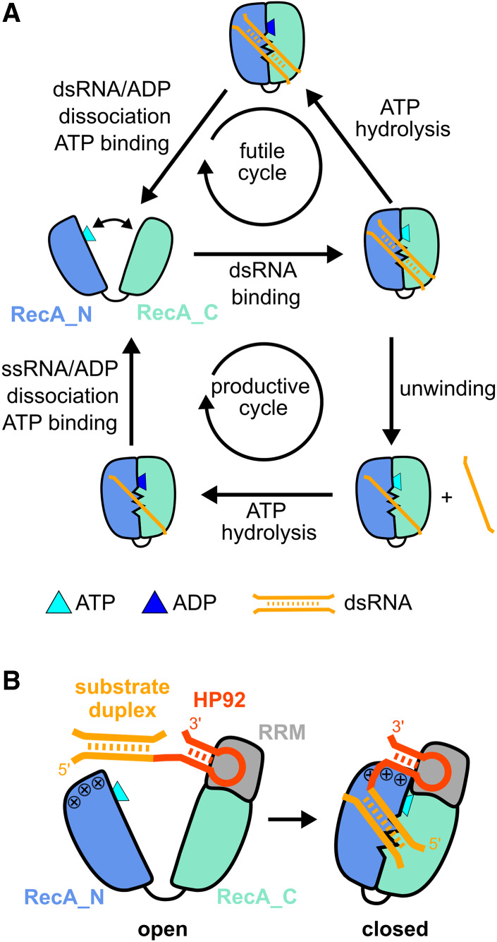
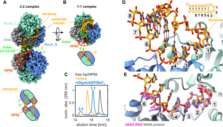
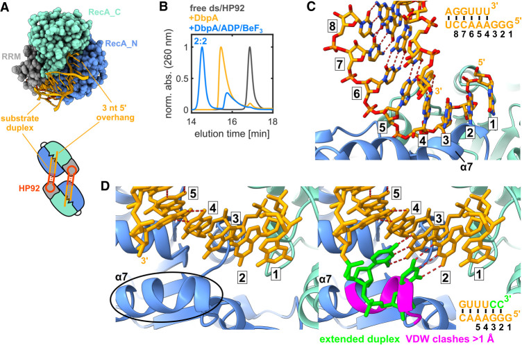
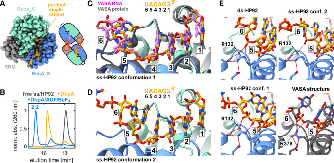
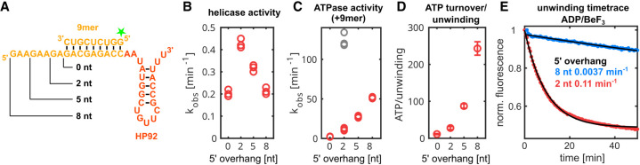
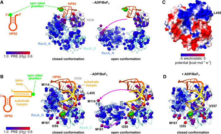
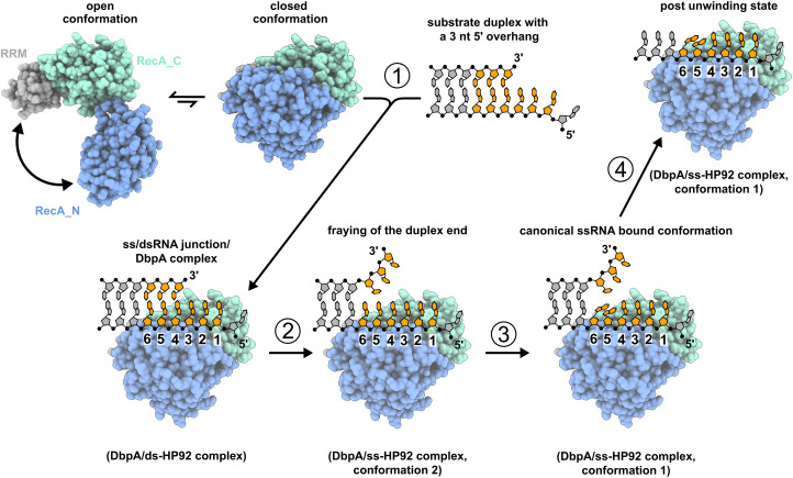
DEAD-box RNA helicases are implicated in most aspects of RNA biology, where these enzymes unwind short RNA duplexes in an ATP-dependent manner. During the central step of the unwinding cycle, the two domains of the helicase core form a distinct closed conformation that destabilizes the RNA duplex, which ultimately leads to duplex melting. Despite the importance of this step for the unwinding process no high-resolution structures of this state are available. Here, I used nuclear magnetic resonance spectroscopy and X-ray crystallography to determine structures of the DEAD-box helicase DbpA in the closed conformation, complexed with substrate duplexes and single-stranded unwinding product. These structures reveal that DbpA initiates duplex unwinding by interacting with up to three base-paired nucleotides and a 5' single-stranded RNA duplex overhang. These high-resolution snapshots, together with biochemical assays, rationalize the destabilization of the RNA duplex and are integrated into a conclusive model of the unwinding process.

Keywords: DEAD-box helicase; NMR spectroscopy; RNA; molecular mechanism; ribosome biogenesis.


Figures

FIGURE 1.
FIGURE 1.
Unwinding cycle of DEAD-box helicases and activation of DbpA by HP92 RNA. (A) Schematic diagram of futile cycles (top) and productive unwinding cycles (bottom) of DEAD-box helicases. The helicase core (RecA_N domain blue, RecA_C domain cyan) alternates between an open conformation in the apo or ATP-bound state and a closed conformation in the presence of ATP and RNA. (B) Domain orientation of DbpA in the open (left) and closed state (right). The C-terminal RNA recognition motif (RRM) (gray) orients HP92 (red) such that the stem of HP92 forms favorable interactions with a positively charged patch (indicated by + signs) on the RecA_N domain in the closed state. This stabilizes the closed state and enables the recruitment of the substrate duplex (orange, located 5′ to HP92) to the active site of the helicase core.
FIGURE 2.
FIGURE 2.
The DbpA/hp-HP92/ADPBeF3 complex represents a trapped unwinding intermediate. (A) Fluorescence-based unwinding assays in the presence of different ATP analogs. A 5′ fluorescein (green star) labeled 9mer RNA is hybridized to an RNA containing HP92 (top). Unwinding can be followed by a decrease in fluorescence intensity (bottom). Mono-exponential fits to the fluorescence time traces are shown in black for ATP, ADP/BeF3, and ATPγS. (B) Ile region of methyl TROSY spectra of ILMVA-labeled DbpA in the free state (black) and bound to hp-HP92 RNA (blue). (C) Sequence plots of chemical shift perturbations (CSPs) induced by binding of the hp-HP92 RNA (top) and differences between CSPs (ΔCSPs) induced by binding of the ss-HP92 RNA and the hp-HP92 (bottom). DbpA domains are indicated at the top. (D) Methyl TROSY spectra of DbpA/hp-HP92 complex prior to (blue) and after addition of ADP/BeF3 (orange). (E) Sequence plots of CSPs induced by binding of ADP/BeF3 to the DbpA/hp-HP92 RNA complex (top) and ΔCSPs between binding of ADP/BeF3 to the ss-HP92/DbpA and to the hp-HP92/DbpA complexes (bottom). (F) ΔCSPs from (E) plotted onto a model of the closed state of DbpA bound to ssRNA (Wurm et al. 2021) (red, large ΔCSPs, blue, small ΔCSPs). The ssRNA is shown in orange. (G) Sequence of hp-HP92 RNA. Nucleotides with assigned imino proton signals are numbered and shown in red (HP92) or green (substrate hairpin). (H) 1H-1D imino proton spectra (left) and 1H15N-HMQC spectra (right) of uridine 15N-labeled hp-HP92 RNA prior to (black) and after addition of DbpA (blue). (I) Same spectra as in (H), but for the hp-HP92/DbpA complex prior to (blue) and after addition of ADP/BeF3 (orange).
FIGURE 3.
FIGURE 3.
Crystal structure of the hp-HP92/DbpA complex in the closed state. (A) Overall structure (top) and schematic diagram (bottom) of the complex between two DbpA molecules (chains A/B) and two hp-HP92 RNAs (chains F/G), as observed in the crystal. HP92 (red), the substrate hairpin (orange), and the 3 nt linker that base pairs with the 3′ overhang of HP92 (green) are shown in ribbon representation. The RecA_N (blue), RecA_C (cyan), and RRM (gray) domains of DbpA are shown in sphere representation. (B) Model of the 1:1 complex obtained by connecting HP92 and the substrate hairpin bound to one DbpA molecule by a flexible 3 nt linker (green). (C) SEC chromatograms of free hp-HP92 (black), the hp-HP92/DbpA complex (orange), and the hp-HP92/DbpA/ADP/BeF3 (blue) complex. (D) Close-up of the interaction between substrate hairpin (chain F, orange) and the active site of the helicase core (chain B, RecA_N blue, RecA_C cyan). The nucleotides that interact with the active site are numbered 1–6. The substrate hairpin is shown in the upper-right, with identical numbering. The distorted base pair between the adenosine in position 4 and the opposite uridine is indicated by a dashed line. (E) Comparison between the substrate hairpin bound to DbpA (only nt 1–7 are shown for clarity) and the complex between a 6 nt ssRNA (pink) and the DEAD-box helicase VASA (gray; PDB ID 2db3).
FIGURE 4.
FIGURE 4.
Crystal structure of the ds-HP92/DbpA complex in the closed state. (A) Interaction of the substrate duplex including a 3 nt 5′ overhang with the active site of the helicase core. For clarity, only the substrate duplex (chain D) and one DbpA molecule (chain B) are shown (top). Schematic diagram of the 2:2 complex observed in the crystal (bottom). (B) SEC chromatograms of free ds-HP92 (black), the ds-HP92/DbpA complex (orange), and the ds-HP92/DbpA/ADP/BeF3 (blue) complex. (C) Close-up of the interaction between the substrate duplex (chain D, orange) and the active site of the helicase core (chain B, RecA_N blue, RecA_C cyan). The nucleotides that interact with the active site are numbered 1–6. The substrate duplex is shown in the upper-right, with identical numbering. (D) Close-up of the active site in the vicinity of α-helix α7 (left). Extension of the duplex by the addition of 2 nt at the 3′ end (green) leads to severe clashes with α-helix α7 of the RecA_N domain (right). Residues that show van der Waals clashes >1 Å with the RNA are shown in pink.
FIGURE 5.
FIGURE 5.
Crystal structure of the ss-HP92/DbpA complex in the closed state. (A) Interaction of the ssRNA with the active site of the helicase core. For clarity, only the ssRNA (chain L) and one DbpA molecule (chain I) are shown (left). Schematic diagram of the 2:2 complex observed in the crystal (right). (B) SEC chromatograms of free ss-HP92 (black), the ss-HP92/DbpA complex (orange), and the ss-HP92/DbpA/ADP/BeF3 complex (blue). (C) Close-up of the interaction between the ssRNA region (chain L, orange) in conformation 1 and the active site of the helicase core (chain I, RecA_N blue, RecA_C cyan). For comparison, the complex between a 6 nt RNA (pink) and the DEAD-box helicase VASA (gray; PDB ID 2db3) is shown. The nucleotides that interact with the active site are numbered 1–6. The ssRNA sequence is shown on top with identical numbering. (D) Close-up of the interaction between the ssRNA region (chain C, orange) in conformation 2 and the active site of the helicase core (chain A, RecA_N blue, RecA_C cyan). (E) Comparison of the interactions between nucleotides in positions 5 and 6 for DbpA and VASA. The top row and the bottom-left panel show complexes between DbpA and ds-HP92 and ss-HP92 in conformation 1 and 2. The lower-right panel shows the VASA/ssRNA complex (PDB ID 2db3). Hydrogen bonds formed by R132 (DbpA) or the corresponding R378 (VASA) are indicated by dashed-red lines.
FIGURE 6.
FIGURE 6.
The length of the ssRNA 5′ overhang influences helicase and ATPase activity of DbpA. (A) RNA constructs used for activity assays. The substrate duplex (orange) is located 5′ to HP92 (red). The varying length of the 5′ overhangs is indicated. For helicase assays, the 9mer RNA contained a fluorescein label at the 5′ end (indicated by a green star). (B) Unwinding rates observed in single-turnover experiments are plotted versus the 5′ overhang length. Results from three measurements are shown. (C) ATPase turnover rates are plotted versus the 5′ overhang length. Rates were determined in the absence (gray) and presence of the 9mer RNA (red). Results from three measurements are shown. (D) The number of hydrolyzed ATP molecules for each unwinding event are plotted versus the 5′ overhang length. The values represent mean and standard deviation calculated from the experiments shown in panels B and C. (E) Single-turnover unwinding of RNA constructs with a 5′ overhang of 2 nt (red) or 8 nt (blue) in the presence of ADP/BeF3 is followed by fluorescence intensity measurements. Exponential fits to fluorescence time traces are shown in black and the unwinding rates obtained from the fits are given.
FIGURE 7.
FIGURE 7.
DbpA transiently samples the closed state in the absence of ATP. (A) PRE experiments were performed with spin labeled HP92 RNA and ILMVA-labeled DbpA in the absence of ADP/BeF3. The RNA construct is shown on the left. The position of the 4-thiouridine residue that carries the nitroxide spin label is indicated by a green circle. The methyl groups of DbpA are colored according to the decrease in signal intensity, due to the spatial proximity of the spin label from blue (no effect) to red (strong reduction). The closed conformation (left structure) and an arbitrary open conformation (right structure) are depicted. The pink arrow indicates the reorientation of the RecA_N domain between the two structures. The C1′ atom of the spin labeled 4-thiouridine is shown as a green sphere. (B) PRE experiments with an hp-HP92 RNA containing a spin label in the loop of the substrate hairpin (green circle; left). Methyl groups are colored as in (A) for the closed conformation (left structure) and for an arbitrary open conformation (right structure). The pink arrows indicate the reorientation of the RecA_N domain. The methyl groups of M114 and M161 that exhibit the largest PREs in the RecA_N domain are labeled. The RNA is shown in the conformation observed in the DbpA/hp-HP92 complex, where the substrate interacts with the active site of the helicase core. (C) DbpA in the closed conformation colored according to its electrostatic surface potential (blue = positive, red = negative). (D) PRE experiments with identical RNA as in (B), but in the presence of ADP/BeF3. Methyl groups are colored as in (A) and DbpA is shown in the closed conformation.
FIGURE 8.
FIGURE 8.
Model of the unwinding mechanism for duplexes with 5′ ssRNA overhangs. HP92 and ATP are omitted for clarity. The nucleotides that interact with the active site of DbpA are colored orange. DbpA transiently adopts the closed conformation in the absence of substrate RNA (upper left). (1) The closed conformation binds to the ss/dsRNA junction, whereby nt 1–3 are single-stranded and nt 4–6 are still part of the duplex (as depicted in the structure of the DbpA/ds-HP92 complex). (2) Duplex breathing leads to a ssRNA-bound state with continuous base stacking (DbpA/ss-HP92 complex in conformation 2). (3) The nucleotides in positions 5/6 rearrange to a conformation that is incompatible with duplex formation (DbpA/ss-HP92 complex in conformation 1). (4) The loss of these three base pairs leads to the dissociation of the upper RNA strand.

Similar articles

Cited by

References

    1. Afonine PV, Grosse-Kunstleve RW, Echols N, Headd JJ, Moriarty NW, Mustyakimov M, Terwilliger TC, Urzhumtsev A, Zwart PH, Adams PD. 2012. Towards automated crystallographic structure refinement with phenix.refine. Acta Crystallogr D Biol Crystallogr 68: 352–367. 10.1107/S0907444912001308 - DOI - PMC - PubMed
    1. Andreou AZ, Harms U, Klostermeier D. 2019. Single-stranded regions modulate conformational dynamics and ATPase activity of eIF4A to optimize 5′-UTR unwinding. Nucleic Acids Res 47: 5260–5275. 10.1093/nar/gkz254 - DOI - PMC - PubMed
    1. Ballut L, Marchadier B, Baguet A, Tomasetto C, Séraphin B, Le Hir H. 2005. The exon junction core complex is locked onto RNA by inhibition of eIF4AIII ATPase activity. Nat Struct Mol Biol 12: 861–869. 10.1038/nsmb990 - DOI - PubMed
    1. Bowers HA, Maroney PA, Fairman ME, Kastner B, Lührmann R, Nilsen TW, Jankowsky E. 2006. Discriminatory RNP remodeling by the DEAD-box protein DED1. RNA 12: 903–912. 10.1261/rna.2323406 - DOI - PMC - PubMed
    1. Chen Z, Li Z, Hu X, Xie F, Kuang S, Zhan B, Gao W, Chen X, Gao S, Li Y, et al. 2020. Structural basis of human helicase DDX21 in RNA binding, unwinding, and antiviral signal activation. Adv Sci 7: 2000532. 10.1002/advs.202000532 - DOI - PMC - PubMed

Publication types

LinkOut - more resources