. 2023 May 23;6(8):e202302133.
doi: 10.26508/lsa.202302133. Print 2023 Aug.

The CHARGE syndrome-associated protein FAM172A controls AGO2 nuclear import

Affiliations

The CHARGE syndrome-associated protein FAM172A controls AGO2 nuclear import

Sephora Sallis et al. Life Sci Alliance. .

Abstract

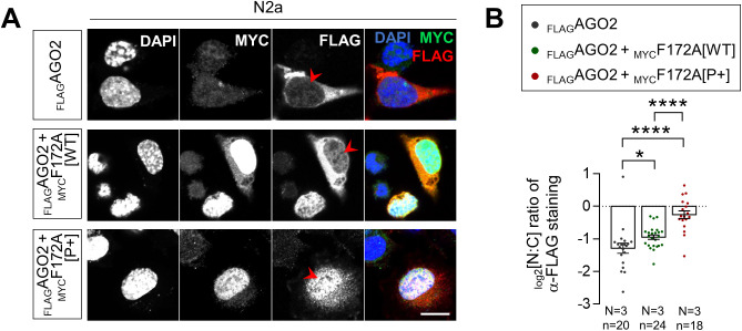
CHARGE syndrome is a neural crest-related disorder mainly caused by mutation of the chromatin remodeler-coding gene CHD7 Alternative causes include mutation of other chromatin and/or splicing factors. One of these additional players is the poorly characterized FAM172A, which we previously found in a complex with CHD7 and the small RNA-binding protein AGO2 at the chromatin-spliceosome interface. Focusing on the FAM172A-AGO2 interplay, we now report that FAM172A is a direct binding partner of AGO2 and, as such, one of the long sought-after regulators of AGO2 nuclear import. We show that this FAM172A function mainly relies on its classical bipartite nuclear localization signal and associated canonical importin-α/β pathway, being enhanced by CK2-induced phosphorylation and abrogated by a CHARGE syndrome-associated missense mutation. Overall, this study thus strengthens the notion that noncanonical nuclear functions of AGO2 and associated regulatory mechanisms might be clinically relevant.


Conflict of interest statement

The authors declare that they have no conflict of interest.

Figures

Figure 1.
Figure 1.. FAM172A influences AGO2 nuclear localization.
(A) Immunofluorescence analysis of AGO2 distribution in WT and Fam172aTp/Tp e10.5 MEFs at moderate density (40,000 cells/cm2), with nuclei stained using DAPI. (A, B) Quantification of relative fluorescence intensity of anti-AGO2 staining in the nucleus and cytoplasm (N:C ratio, expressed in log 2 scale) using images such as those displayed in (A). Yellow and white dashed lines in zoomed-in views in (A) delineate measured areas for the nucleus and cytoplasm, respectively. (C) Western blot analysis of AGO2 protein levels in cytoplasmic and nuclear fractions of WT and Fam172aTp/Tp e10.5 MEFs grown at high density (200,000 cells/cm2) and exposed to leptomycin B showing a specific decrease in the nucleus of Fam172aTp/Tp cells (N = 3). GAPDH and Lamin A/C are used as loading control for cytoplasmic and nuclear fractions, respectively. (C, D) Quantitative analysis of relative anti-AGO2 signals in mutant versus WT cells (with average in WT set at 1) after normalization with relevant loading control (GAPDH for total and cytoplasmic fractions; Lamin A/C for nuclear fraction), as determined via densitometry (ImageJ) using images such as those displayed in (C). (E) The diagram of mouse FAM172A isoforms based on Ensembl/Uniprot databases. (F) Western blot analysis of FAM172A isoforms in trunk (T) or head (H) extracts from embryos at the indicated developmental stages, using GAPDH as loading control (N = 3). (G) Immunofluorescence staining of endogenous FAM172A and transfected MYC-tagged version of the 371aa-long isoform (MYCF172A[371]) in e10.5 MEFs, with nuclei stained using DAPI (N = 3). The arrow in the left panel points the endoplasmic reticulum. (H) Immunofluorescence analysis of AGO2 distribution in Fam172aTp/Tp e10.5 MEFs transfected or not with MYCF172A[371]. Red arrowheads compare fluorescence intensity in nuclei of transfected and non-transfected cells. (H, I) Quantification of relative fluorescence intensity of anti-AGO2 staining in the nucleus and cytoplasm (N:C ratio, expressed in log 2 scale) using images such as those displayed in (H). Scale bar, 20 μm. N = number of biological replicates, n = number of cells. *P ≤ 0.05 and ****P ≤ 0.0001; t-test. (Further data can be found in Fig S1).
Figure S1.
Figure S1.. FAM172A influences the nuclear localization of AGO2, but not AGO1.
(A) Immunofluorescence analysis of AGO2 distribution in WT and Fam172Tp/Tp e10.5 MEFs at high density (200,000 cells/cm2), with the nuclei stained using DAPI. (A, B) Quantification of relative fluorescence intensity for anti-AGO2 staining in the nucleus and cytoplasm (N:C ratio, expressed in log 2 scale) using images such as those displayed in (A). Yellow and white dashed lines in zoomed-in views in A delineate areas of measurement for the nucleus and cytoplasm, respectively. (C) Western blot analysis of AGO2 protein levels in nuclear and cytoplasmic fractions of WT and Fam172aTp/Tp e10.5 MEFs grown at high density (200,000 cells/cm2) showing a specific decrease in the nucleus of Fam172aTp/Tp cells (N = 3). Lamin A/C and GAPDH are used as loading control for nuclear and cytoplasmic fractions, respectively. (C, D) Quantitative analysis of relative anti-AGO2 signals in mutant versus WT cells after normalization with relevant loading control (GAPDH for total and cytoplasmic fractions; Lamin A/C for nuclear fraction), as determined via densitometry (ImageJ) using images such as those displayed in (C). (E) Immunofluorescence analysis of AGO1 distribution in WT and Fam172Tp/Tp e10.5 MEFs at high density (200,000 cells/cm2), with the nuclei stained using DAPI. (E, F) Quantification of relative fluorescence intensity for anti-AGO1 staining in the nucleus and cytoplasm (N:C ratio, expressed in log 2 scale) using images such as those displayed in (E). (G) Immunofluorescence analysis of FAM172A distribution in WT e10.5 MEFs transfected or not with FLAGAGO2 (N = 3). Red arrowheads compare fluorescence intensity in nuclei of transfected and non-transfected cells. (H) Western blot analysis of FAM172A isoforms in NIH 3T3 fibroblasts, Neuro2a (N2a) neuroblasts, and R1 embryonic stem cells (N = 3). Scale bar, 20 μm. N = number of biological replicates, n = number of cells. **P ≤ 0.01, ****P ≤ 0.0001; t-test.
Figure 2.
Figure 2.. FAM172A directly interacts with AGO2.
(A) In vitro co-IP of recombinant MBPFAM172A (WT versus E229Q versions) and HisAGO2 using MBP as bait (N = 3). MBP alone was used as negative control. The red arrowhead points to reduced amount of co-immunoprecipitated HisAGO2 when using the E229Q version of FAM172A. (B, C) Co-IP of transfected MYCFAM172A and FLAGAGO2 (full-length versus indicated N-term and C-term truncations; see panel (B)) in N2a cells using FLAG as bait (N = 3). The red arrowhead in panel C points to reduced amount of co-immunoprecipitated MYCFAM172A when using the C-term half of AGO2. (D) Immunofluorescence analysis of AGO2 distribution in Fam172aTp/Tp e10.5 MEFs transfected or not with E229Q-mutated MYCFAM172A, with nuclei stained using DAPI. Red arrowheads compare relative fluorescence intensity in nuclei of transfected and non-transfected cells. (D, E) Quantification of relative fluorescence intensity of anti-AGO2 staining in the nucleus and cytoplasm (N:C ratio, expressed in the log 2 scale) using images such as those displayed in (D). Data for Fam172aTp/Tp and Fam172aTp/Tp + MYCF172A[WT] conditions are the same as initially displayed in Fig 1I (both assays were performed at the same time), being duplicated here for comparison purposes only. (F) Immunofluorescence analysis of AGO2 distribution in human LCLs derived from an individual with FAM172A[E228Q]-associated CHARGE syndrome and parental control, with the nuclei stained using DAPI (N = 3 technical replicates). (F, G) Quantification of relative fluorescence intensity of anti-AGO2 staining in nucleus and cytoplasm (N:C ratio, expressed in the log 2 scale) using images such as those displayed in (F). Scale bar, 20 μm. N = number of biological replicates, n = number of cells. ***P ≤ 0.001 and ****P ≤ 0.0001; t-test. (Further data can be found in Fig S2).
Figure S2.
Figure S2.. Subcellular distribution of FLAGAGO2 deletion mutants and E229Q-mutated MYCFAM172A.
(A) Immunofluorescence analysis of the subcellular distribution of full-length and indicated deletion mutants of FLAGAGO2 proteins in N2a cells co-transfected with MYCFAM172A, with the nuclei stained using DAPI. Red arrowheads compare relative fluorescence intensity in nuclei. (A, B) Quantification of relative fluorescence intensity for anti-FLAG staining in the nucleus and cytoplasm (N:C ratio, expressed in log 2 scale) using images such as those displayed in (A). (C) Immunofluorescence analysis of the subcellular distribution of WT- and E229Q-mutated MYCFAM172A proteins in transfected N2a cells, with nuclei stained using DAPI. Red arrowheads compare relative fluorescence intensity in cytoplasm. (C, D) Quantification of relative fluorescence intensity for anti-MYC staining in the nucleus and cytoplasm (N:C ratio, expressed in log 2 scale) using images such as those displayed in (C). Scale bar, 20 μm. N = number of biological replicates, n = number of cells.*P ≤ 0.05, ****P ≤ 0.0001; t-test.
Figure 3.
Figure 3.. The NLS of FAM172A is required for AGO2 nuclear import.
(A) Diagram of the BiFC assay based on structural complementation between two non-fluorescent N-terminal and C-terminal halves of a yellow fluorescent protein (Venus) that are respectively fused to AGO2 (N-VenusAGO2) and FAM172A (C-VenusF172A). (B) Comparison of BiFC fluorescence intensity between N-VenusAGO2–C-VenusF172A[WT] and N-VenusAGO2–C-VenusF172A[mNLS]. Mean fluorescence intensity was determined using flow cytometry 24 h after transfection in N2a cells. (C) Immunofluorescence analysis of MYC-tagged WT and NLS-mutated FAM172A proteins in transfected N2a cells, with nuclei stained using DAPI. Red arrowheads compare relative fluorescence intensity in the nucleus for WT- and NLS-mutated FAM172A. (C, D) Quantification of relative fluorescence intensity of anti-MYC staining in the nucleus and cytoplasm (N:C ratio, expressed in the log 2 scale) using images such as those displayed in (C). (E) 5-h-long time-lapse recordings of BiFC signal generated using N-VenusAGO2 and C-VenusF172A (comparing WT and mNLS versions) in N2a cells treated with leptomycin B or vehicle (ethanol) only. Red arrowheads compare relative fluorescence intensity in the nucleus. (E, F) Quantification of relative BiFC signal intensity in the nucleus and cytoplasm (N:C ratio, expressed in log 2 scale) using images such as those displayed in (E). (G) Five-hour-long time-lapse recordings of BiFC signal generated using N-VenusAGO2 and C-VenusF172A in N2a cells treated with leptomycin B with or without ivermectin or vehicle (ethanol) only. Red arrowheads compare relative fluorescence intensity in the nucleus. (G, H) Quantification of relative BiFC signal intensity in the nucleus and cytoplasm (N:C ratio, expressed in log 2 scale) using images such as those displayed in (G). (I) Immunofluorescence analysis of AGO2 distribution in Fam172aTp/Tp e10.5 MEFs transfected or not with MYCF172A[mNLS], with the nuclei stained using DAPI. Red arrowheads compare relative fluorescence intensity in nuclei of transfected and non-transfected cells. (I, J) Quantification of relative fluorescence intensity of anti-AGO2 staining in the nucleus and cytoplasm (N:C ratio, expressed in log 2 scale) using images such as those displayed in (I). (E, G) Data for Fam172aTp/Tp and Fam172aTp/Tp + MYCF172A[WT] conditions are the same as initially displayed in Fig 1I (both assays were performed at the same time), being duplicated here for comparison purposes only. Scale bar, 20 μm (E, G). N = number of biological replicates, n = number of cells. *P ≤ 0.05, **P ≤ 0.01, ***P ≤ 0.001, and ****P ≤ 0.0001; t-test. (Further data can be found in Fig S3).
Figure S3.
Figure S3.. FAM172A–AGO2 interaction is direct and specific.
(A) Distribution of the AGO2–FAM172A BiFC signal in living N2a cells 48 h after transfection with indicated combinations of N-Venus-tagged AGO2 and C-Venus-tagged FAM172A constructs, with nuclei stained using Hoechst (N = 3). (B, C) Comparison of overall BiFC fluorescence intensity between N-VenusAGO2–C-VenusF172A[WT] and N-VenusAGO2–C-VenusF172A[E229Q] (B) or after competition with a 5-time excess of FLAGAGO2 (C; also displaying percentage of positive cells in right panel). Mean fluorescence intensity and percentage of positive cells were determined using flow cytometry 24 h after transfection in N2a cells. (D) Immunofluorescence analysis of C-VenusFAM172A and N-VenusAGO2 distribution in transfected N2a using a GFP antibody that recognize both halves of Venus, with nuclei stained using DAPI (N = 3). Red arrowheads point to transfected cells. (E) Distribution of N-VenusAGO2–C-VenusF172A BiFC signal in living NIH3T3 cells 48 h after transfection, with nuclei stained using Hoechst (N = 3). Scale bar, 20 μm. N = number of biological replicates, n = number of cells. *P ≤ 0.05, **P ≤ 0.01 and ***P ≤ 0.001; t-test.
Figure S4.
Figure S4.. Sequence of CK2-NLS loop and predicted 3D structure of FAM172A.
(A) The diagram of all mutated versions of FAM172A used in this study. (B) PyMOL vizualisation of AlphaFold-predicted 3D structure of FAM172A.
Figure 4.
Figure 4.. CK2 phosphorylates FAM172A.
(A, B) Representative CID-MS/MS spectra of most frequently phosphorylated peptides after in vitro co-incubation of recombinant FAM172A with recombinant CK2 and ATP. (C, D) Proportion of overall phosphorylation (C) and relative frequency of phosphorylated residues (D) after in vitro CK2-induced phosphorylation. (E, F) Representative CID-MS/MS spectra of phosphorylated FAM172A peptides after anti-MYC immunoprecipitation from MYCFAM172A-transfected N2a cells.
Figure 5.
Figure 5.. AGO2 nuclear import depends on the status of FAM172A phosphorylation.
(A) 5-h-long time-lapse recordings of BiFC signal generated using N-VenusAGO2 and C-VenusF172A in N2a cells treated with leptomycin B or vehicle (ethanol) only, with or without 4,5,6,7-tetrabromobenzotriazole. Red arrowheads compare relative fluorescence intensity in the nucleus. (A, B) Quantification of relative BiFC signal intensity in the nucleus and cytoplasm (N:C ratio, expressed in log 2 scale) using images such as those displayed in (A). (C) 5-h-long time-lapse recordings of the BiFC signal generated using N-VenusAGO2 and C-VenusF172A (comparing phosphodead [P−] and phosphomimetic [P+] versions) in N2a cells treated with leptomycin B or vehicle (ethanol) only. Red arrowheads compare relative fluorescence intensity in the nucleus. (C, D) Quantification of relative BiFC signal intensity in the nucleus and cytoplasm (N:C ratio, expressed in log 2 scale) using images such as those displayed in (C). Data for C-VenusF172A[WT] are the same as initially displayed in Fig 3F (both assays were performed at the same time), being duplicated here for comparison purposes only. (E) Comparison of overall BiFC fluorescence intensity between N-VenusAGO2–C-VenusF172A[WT], N-VenusAGO2–C-VenusF172A[P-], and N-VenusAGO2–C-VenusF172A[P+]. Mean fluorescence intensity was determined using flow cytometry 24 h after transfection in N2a cells. (F) Immunofluorescence analysis of the distribution of MYC-tagged WT, phosphodead (P−), and phosphomimetic (P+) versions of murine FAM172A protein in transfected N2a cells, with nuclei stained using DAPI. (F, G) Quantification of relative fluorescence intensity of anti-MYC staining in the nucleus and cytoplasm (N:C ratio, expressed in the log 2 scale) using images such as those displayed in (F). Data for the MYCF172A[WT] condition are the same as initially displayed in Fig 3D (both assays were performed at the same time), being duplicated here for comparison purposes only. (A, C, F) Scale bar, 20 μm (A, C) and 10 μm (F). N = number of biological replicates, n = number of cells. *P ≤ 0.05, **P ≤ 0.01, ***P ≤ 0.001, and ****P ≤ 0.0001; t-test. (Further data can be found in Fig S5).
Figure S5.
Figure S5.. Phosphomimetic MYCFAM172A stimulates the nuclear entry of FLAGAGO2.
(A) Immunofluorescence analysis of full-length FLAGAGO2 distribution as a function of the presence of WT and phosphomimetic versions of MYCFAM172A in transfected N2a, with nuclei stained using DAPI. Red arrowheads compare relative fluorescence intensity in the nuclei (N = 3). (A, B) Quantification of relative fluorescence intensity for anti-FLAG staining in the nucleus and cytoplasm (N:C ratio, expressed in log 2 scale) using images such as those displayed in (A). Scale bar, 20 μm. N = number of biological replicates, n = number of cells. *P ≤ 0.05 and ****P ≤ 0.0001; t-test.
Figure S6.
Figure S6.. AGO2 overexpression can functionally compensate for the loss of FAM172A ex vivo.
(A, B) Quantitative analysis of cell proliferation (A) and alternative splicing of Cd44 (B) in e10.5 Fam172aTp/Tp MEFs, after co-transfection with a GFP expression vector and either MYCFAM172A (WT, E229Q-mutant or NLS-mutant) or FLAGAGO2 (WT, NLS-containing, and deletion mutants) expression vectors. Cell proliferation is expressed in % of GFP-positive cells based on double-immunofluorescence staining of GFP and the proliferation marker Ki67. Alternative splicing of Cd44 was analyzed by RT–qPCR after FACS-mediated recovery of GFP-positive cells. Expression levels of variable exons 8/9 (vE8/9) were normalized against levels of a constant exons 4/5 (cE4/5), and splicing fold change was then determined by comparison to the reference value (dashed line) obtained with WT control embryos (N = 5 per condition). (C) Immunofluorescence analysis of the distribution of NLS-containing FLAGAGO2 in transfected N2a cells, with nuclei stained using DAPI. Scale bar, 20 μm. *P ≤ 0.05, **P ≤ 0.01, ***P ≤ 0.001 and ****P ≤ 0.0001; t-test.
Figure S7.
Figure S7.. MYCFAM172A interacts with various endogenous importins.
(A, B, C, D) Co-IP of transfected MYCFAM172A with endogenous importin-5 (A, B) or endogenous importin-β1 and importin-α1 (C, D) in N2a cells, using MYC as bait (N = 3). (B, D) Binding was increased by short-term formaldehyde-induced crosslink, and an excess of MYC peptide was used to control specificity (B, D). (E) Impact of the NLS mutation on importin-α1 recruitment could be evidenced once, in the absence of crosslink.
Figure S8.
Figure S8.. FAM172A can homodimerize.
(A) Co-IP of transfected MYCFAM172A with HAFAM172A in N2a cells using MYC as bait (N = 3). Binding was increased by short-term formaldehyde-induced crosslink, and an excess of the MYC peptide was used to control specificity. (B) Five-hour-long time-lapse recordings of the BiFC signal generated using N-VenusF172A and C-VenusF172A in N2a cells treated with ivermectin or vehicle (ethanol) only. Red arrowheads compare relative fluorescence intensity in the cytoplasm. (B, C) Quantification of relative BiFC signal intensity in the nucleus and cytoplasm (N:C ratio, expressed in log 2 scale) using images such as those displayed in (B). Scale bar, 20 μm. N = number of biological replicates, n = number of cells. **P ≤ 0.01, ***P ≤ 0.001; t-test.
Figure S9.
Figure S9.. FAM172A also interacts with AGO1 and Nucleolin (NCL).
(A) In vitro co-IP of recombinant MBPFAM172A and HisAGO1, using MBP as bait (N = 3). MBP alone was used as negative control. (B) Co-IP of transfected MYCFAM172A and either FLAGAGO1 or FLAGAGO2 in N2a cells, using FLAG as bait (N = 3). Numbers between immunoblots refer to the amount of co-immunoprecipitated MYCFAM172A after normalization for the amount of immunoprecipitated FLAGAGO proteins, as determined via densitometry. (C) Co-IP of transfected MYCFAM172A and FLAGAGO2 with endogenous NCL in N2a cells, using NCL as bait (N = 3).

References

    1. Pauli S, Bajpai R, Borchers A (2017) Charged with neural crest defects. Am J Med Genet C Semin Med Genet 175: 478–486. 10.1002/ajmg.c.31584 - DOI - PubMed
    1. Pilon N (2021) Treatment and prevention of neurocristopathies. Trends Mol Med 27: 451–468. 10.1016/j.molmed.2021.01.009 - DOI - PubMed
    1. Vissers LE, van Ravenswaaij CM, Admiraal R, Hurst JA, de Vries BB, Janssen IM, van der Vliet WA, Huys EH, de Jong PJ, Hamel BC, et al. (2004) Mutations in a new member of the chromodomain gene family cause charge syndrome. Nat Genet 36: 955–957. 10.1038/ng1407 - DOI - PubMed
    1. Belanger C, Berube-Simard FA, Leduc E, Bernas G, Campeau PM, Lalani SR, Martin DM, Bielas S, Moccia A, Srivastava A, et al. (2018) Dysregulation of cotranscriptional alternative splicing underlies charge syndrome. Proc Natl Acad Sci U S A 115: E620–E629. 10.1073/pnas.1715378115 - DOI - PMC - PubMed
    1. Moccia A, Srivastava A, Skidmore JM, Bernat JA, Wheeler M, Chong JX, Nickerson D, Bamshad M, Hefner MA, Martin DM, et al. (2018) Genetic analysis of charge syndrome identifies overlapping molecular biology. Genet Med 20: 1022–1029. 10.1038/gim.2017.233 - DOI - PMC - PubMed

Publication types