. 2023 May 23;14(1):2962.
doi: 10.1038/s41467-023-38751-8.

An mRNA-based T-cell-inducing antigen strengthens COVID-19 vaccine against SARS-CoV-2 variants

Affiliations

An mRNA-based T-cell-inducing antigen strengthens COVID-19 vaccine against SARS-CoV-2 variants

Wanbo Tai et al. Nat Commun. .

Abstract

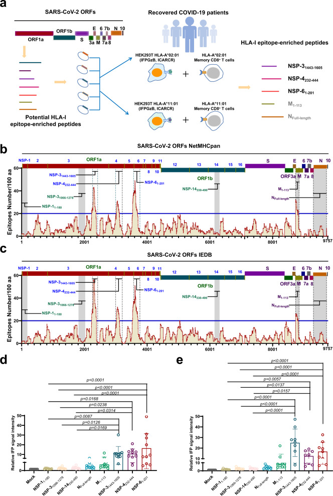
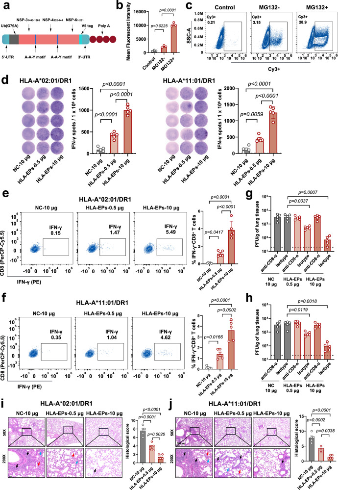
Herd immunity achieved through mass vaccination is an effective approach to prevent contagious diseases. Nonetheless, emerging SARS-CoV-2 variants with frequent mutations largely evaded humoral immunity induced by Spike-based COVID-19 vaccines. Herein, we develop a lipid nanoparticle (LNP)-formulated mRNA-based T-cell-inducing antigen, which targeted three SARS-CoV-2 proteome regions that enriched human HLA-I epitopes (HLA-EPs). Immunization of HLA-EPs induces potent cellular responses to prevent SARS-CoV-2 infection in humanized HLA-A*02:01/DR1 and HLA-A*11:01/DR1 transgenic mice. Of note, the sequences of HLA-EPs are highly conserved among SARS-CoV-2 variants of concern. In humanized HLA-transgenic mice and female rhesus macaques, dual immunization with the LNP-formulated mRNAs encoding HLA-EPs and the receptor-binding domain of the SARS-CoV-2 B.1.351 variant (RBDbeta) is more efficacious in preventing infection of SARS-CoV-2 Beta and Omicron BA.1 variants than single immunization of LNP-RBDbeta. This study demonstrates the necessity to strengthen the vaccine effectiveness by comprehensively stimulating both humoral and cellular responses, thereby offering insight for optimizing the design of COVID-19 vaccines.


Conflict of interest statement

The authors declare no competing interests.

Figures

Fig. 1
Fig. 1. Identification of HLA-I epitope-enriched peptides that activate CD8+ T lymphocytes from convalescent COVID-19 patients.
a Schematic diagram of experimental design. The epitopes for 78 HLA-I alleles were predicted by both the NetMHCpan and IEDB bioinformatic tools. The peptides enriching HLA-I epitopes were ectopically expressed to assess the capability to activate CD8+ T lymphocytes from convalescent COVID-19 patients through a reporter cell-based epitope expression system. b, c Identification of the fragments enriching human cytotoxic T-lymphocyte epitopes in the SARS-CoV-2 proteome. The distribution of potential cytotoxic HLA-I epitopes was predicted by in silico analyses of NetMHCpan (b) and IEDB (c). Four regions, NSP-31443–1605, NSP-4232–444, NSP-61–201, and M1–113, with more than 20 effective epitopes (affinity concentration >10 nM) per 100 amino acids were selected as HLA-I epitope-enriched peptides for further investigation, and the three regions, NSP-11–180, NSP-31066-1278, and NSP-14330–490 with fewest predicted effective epitopes were set as controls. The N protein was included in further investigation. The horizontal lines denote the threshold for selecting the HLA-I epitope enrichment regions. d, e Activation of CD8+ T lymphocytes from convalescent COVID-19 patients. HEK293T cells were transfected to stably express the pHAGE_EF1a_ICADCR, pHAGE_EF1a_IFPGZB, and pHAGE_EF1aHLA vectors. Either HLA-A*02:01 (d) or HLA-A*11:01 (e) overexpressing HEK293T cells were transfected with the genes encoding the NSP-11–180, NSP-31066–1278, NSP-14330–490, NFull-length, M1–113, NSP-31443–1605, NSP-4232–444, or NSP-61–201, respectively. At 36 h post-transfection, 1 × 106 HEK293T cells were co-cultured with 2 × 105 HLA genotype-matched memory CD8+ T cells from convalescent COVID-19 patients (n = 10 for HLA-A*02:01, and n = 9 for HLA-A*11:01) for 12 h, and activation of CD8+ T cells was characterized by the relative IFP signal intensity in the co-cultred HEK293T cells using the flow cytometric measurement. All data are presented as the mean ± S.E.M from the indicated number of biological repeats. Source data are provided as a Source Data file. Experiments were repeated twice independently with similar results. Both independent experiments contain two technical repeats (d, e). Adjusted p values were determined by one-way ANOVA with Tukey’s multiple comparison post-hoc two-sided tests.
Fig. 2
Fig. 2. Immunogenicity of HLA-EPs in both HLA-A*02:01/DR1 and HLA-A*11:01/DR1 transgenic mice.
a Schematic diagram of the HLA-EPs mRNA construct. b, c The expression of HLA-EPs in HEK293T cells. The results are shown by the mean fluorescent intensity (MFI) of HLA-EPs positive cells (b) and the representative flow cytometry plots (c) of 3 biological replicates containing 3 technical replicates. d–f The proliferation of epitope-specific splenocytes producing IFN-γ following stimulation with the peptide pool. The proliferation of epitope-specific, IFN-γ producing splenocytes was measured by ELISpot. Each point represents the mean of 3 technical replicates, with a limit of detection (LOD) = 2, and representative dots are shown (d). The frequency of IFN-γ positive CD8+ T cells was measured by an ICS (e, f). The representative flow cytometry plots are presented, and symbols represent individual mice. gj The CD8+ T-cell-dependent protection of LNP-HLA-EPs against SARS-CoV-2. Both vaccinated HLA-A*02:01/DR1 and HLA-A*11:01/DR1 transgenic mice with or without CD8+ T-cell depletion, were infected with the SARS-CoV-2 B.1.351 variant (1.4 × 105 PFU/mouse). Viral titers in lung tissues of HLA-A*02:01/DR1 and HLA-A*11:01/DR1 transgenic mice, with and without CD8+ T-cell depletion after 4 days post-inoculation (n = 5), were determined by a plaque assay with two technical replicates (g, h). The LOD was 10 PFU per gram of tissue. Histopathological changes in the lungs of challenged mice were evaluated by an H&E staining (i, j). Images derived from one representative animal in each group with sites of inflammatory cell infiltration (black arrows), blood clots (blue arrows), and alveolar deformation (red arrows) are presented. Scale bar is 50 μm. All data (b, dj) are presented as the mean ± S.E.M. from the indicated number of biological repeats, and statistical significance was calculated via one-way ANOVA with Tukey’s multiple comparison post hoc two-sided tests (b, df, i, j) or two-way ANOVA with Bonferroni’s multiple comparisons (g, h). P values were adjusted for multiple comparisons. Source data are provided as a Source Data file. Data are representative of one (i, j), two (dh), or three (a, b) independent experiments with similar results.
Fig. 3
Fig. 3. Protection of HLA-A*02:01/DR transgenic mice from SARS-CoV-2 infection by dual immunization with SARS-CoV-2 HLA-EPs and RBD antigens.
a Schematic diagram of the immunization, sample collection, and challenge schedule. bd The humoral immune activation and antibody production after immunization. The lymph nodes collected (n = 5) were analyzed for GC B cells and Tfh cells (b). SARS-CoV-2 RBD-specific IgG antibodies in the sera collected 21 days after the booster vaccination were detected with ELISA (c). The neutralizing antibodies against SARS-CoV-2 variants were detected with live viruses-based neutralization assays (d). The dashed lines represent that the LOD was 100 for ELISA, and that the lower limit of quantification was 8 for live virus neutralization. eh The cellular immune response. Splenocytes of immunized HLA-A*02:01/DR1 transgenic mice (n = 5) collected were restimulated ex vivo and subjected to IFN-γ ELISpot (e) (LOD = 5) and IL-4 ELISpot (f) (LOD = 2). The production of IFN-γ and TNF-α by CD8+ T cells (g) and CD4+ T cells was analyzed by an ICS (h). i, j The protective efficacy of the vaccine against the SARS-CoV-2 B.1.351 (Beta) and B.1.1.529 (Omicron BA.1) variants. Vaccinated HLA-A*02:01/DR1 transgenic mice (n = 5) were challenged with the SARS-CoV-2 B.1.351 variant or B.1.1.529 variant, and viral titers at 4 days post-inoculation in the lung (i-upper panel) and trachea (i-bottom panel) tissues were determined by a plaque assay, with LODs being 20 and 100. The histopathological changes of the lungs at 4 days post-inoculation by the B.1.351 (j-upper panel) or B.1.1.529 (j-bottom panel) variant were evaluated after H&E staining. Images derived from one representative animal in each group with sites of inflammatory cell infiltration (black arrows), blood clots (blue arrows), and alveolar deformation (red arrows) are presented. Scale bar is 50 μm. Data (bj) are shown as the mean ± S.E.M from individual mice. Source data are provided as a Source Data file. One (b, j) or two (ci) independent experiments were performed with 2 technical replicates. Adjusted p values for statistical analysis were calculated via one-way ANOVA with Tukey’s multiple comparison post-hoc two-sided tests (b, ej) or two-way ANOVA with Bonferroni’s multiple comparisons (c, d).
Fig. 4
Fig. 4. Immunological features and protection against SARS-CoV-2 infection conferred by dual immunization with HLA-EPs and RBD antigens in nonhuman primates.
a Schematic diagram of the immunization, sample collection, and challenge schedule. bd The humoral immune response against SARS-CoV-2. SARS-CoV-2 RBD-specific IgG antibodies in the sera of macaques were detected by ELISA using the RBD of the SARS-CoV-2 B.1.351 variant (b). The mean ± S.E.M. of RBD-specific IgG titers from 5 biological replicates were plotted against the days post-initial immunization when the sera were collected. The dashed line represents the LOD. The neutralizing antibodies were detected with pseudovirus-based (c) and live virus-based (d) neutralization assays. Pseudoviruses of six representative variants, Wuhan-Hu-1 (wild-type), B.1.1.7 (Alpha), B.1.351 (Beta), P.1 (Gamma), B.1.617.2 (Delta) and B.1.1.529 (Omicron BA.1), and infectious viruses of the six variants were exploited in the assays. The dashed lines represent the lower limit of quantification of 50 and 8, respectively. e, f The Th1-biased cellular immune response. PBMCs collected 14 days post-booster immunization were re-stimulated ex vivo and subjected to IFN-γ ELISpot (LOD = 5) (e) and IL-4 ELISpot (LOD = 2) (f), and representative dots are shown (right panels). g, h The protective efficacy of the vaccine against SARS-CoV-2. Macaques (n = 3) were challenged with 7.0 × 105 PFU of SARS-CoV-2 (GDPCC-nCoV84, Beta variant) 3 weeks post-booster immunization. The copy number of SARS-CoV-2 sgRNA was determined by qRT-PCR in throat swabs (left panel) and anal swabs (right panel) obtained on Days 0, 1, 3, 5, and 7 post-inoculation (g). The LOD was established based on the standard curves at 100 copies per swab. The subgenomic copies in selected lung lobes collected from all macaques 7 days post-inoculation were determined by qRT-PCR (h). Data (ch) are shown as the mean ± S.E.M from individual macaques. Source data are provided as a Source Data file. Experiments were repeated twice independently with similar results. Both independent experiments contain 3 technical repeats (bh). Statistical significance was calculated via one-way ANOVA with Tukey’s multiple comparison post-hoc two-sided tests (e, f, h) or two-way ANOVA with Bonferroni’s multiple comparisons (bd, g). P values were adjusted for multiple comparisons.

References

    1. World Health Organization. Weekly epidemiological update on COVID-19. https://www.who.int/publications/m/item/weekly-epidemiological-update-on... (2023).
    1. Kemp SA, et al. SARS-CoV-2 evolution during treatment of chronic infection. Nature. 2021;592:277–282. doi: 10.1038/s41586-021-03291-y. - DOI - PMC - PubMed
    1. Polack FP, et al. Safety and efficacy of the BNT162b2 mRNA Covid-19 vaccine. N. Engl. J. Med. 2020;383:2603–2615. doi: 10.1056/NEJMoa2034577. - DOI - PMC - PubMed
    1. Chagla Z. The BNT162b2 (BioNTech/Pfizer) vaccine had 95% efficacy against COVID-19 ≥7 days after the 2nd dose. Ann. Intern. Med. 2021;174:Jc15. doi: 10.7326/ACPJ202102160-015. - DOI - PubMed
    1. Yang J, et al. A vaccine targeting the RBD of the S protein of SARS-CoV-2 induces protective immunity. Nature. 2020;586:572–577. doi: 10.1038/s41586-020-2599-8. - DOI - PubMed

Publication types

Supplementary concepts