. 2023 May 23;14(1):2975.
doi: 10.1038/s41467-023-38799-6.

Adolescent stress impairs postpartum social behavior via anterior insula-prelimbic pathway in mice

Affiliations

Adolescent stress impairs postpartum social behavior via anterior insula-prelimbic pathway in mice

Kyohei Kin et al. Nat Commun. .

Abstract

Adolescent stress can be a risk factor for abnormal social behavior in the postpartum period, which critically affects an individual social functioning. Nonetheless, the underlying mechanisms remain unclear. Using a mouse model with optogenetics and in vivo calcium imaging, we found that adolescent psychosocial stress, combined with pregnancy and delivery, caused hypofunction of the glutamatergic pathway from the anterior insula to prelimbic cortex (AI-PrL pathway), which altered PrL neuronal activity, and in turn led to abnormal social behavior. Specifically, the AI-PrL pathway played a crucial role during recognizing the novelty of other mice by modulating "stable neurons" in PrL, which were constantly activated or inhibited by novel mice. We also observed that glucocorticoid receptor signaling in the AI-PrL pathway had a causal role in stress-induced postpartum changes. Our findings provide functional insights into a cortico-cortical pathway underlying adolescent stress-induced postpartum social behavioral deficits.


Conflict of interest statement

The authors declare no competing interests.

Figures
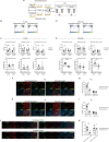
Fig. 1
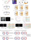
Fig. 1. Social isolation in late adolescence (SILA) induced postpartum behavioral changes in social novelty (SN)-trial and reduced neural activity in the anterior insula (AI) and prelimbic cortex (PrL).
A Experimental design for social interaction test (SIT) experiments. B Representative heatmaps of the mouse track during sociability (S)-trials. C Unstressed and stressed dams showed sociability toward a mouse in the S-trials (two-way mixed ANOVA). N = 8 mice. D Representative heatmaps of the mouse track during the SN-trials. E Stressed dams did not show a novel mouse preference in the SN-trials, compared to unstressed dams (two-way mixed ANOVA). Social preference indexes for S- and SN-trials were shown in Fig. 1C, E (Student’s t-test, Cohen’s D = 4.766 for total interaction time in the SN-trial, and Cohen’s D = 1.478 for interaction time per visit in the SN-trial). N = 8 mice. F Representative images of c-Fos+ (green), Vglut1+ or Vgat+ (red), DAPI (blue), and colocalized cells in PrL of unstressed or stressed dams. Scale bar, 50 µm. G Stressed dams showed a decreased number of Vglut1+c-Fos+ cells, but not Vgat+c-Fos+, in PrL after the SN-trial (two-way mixed ANOVA). N = 8 mice. H Representative images of c-Fos+ (green), Vglut1+ or Vgat+ (red), DAPI (blue), and colocalized cells in AI of unstressed or stressed dams. Scale bar, 50 µm. I Stressed dams showed a decreased number of Vglut1+c-Fos+ cells, but not Vgat+c-Fos+, in AI after the SN-trials (two-way mixed ANOVA). N = 8 mice. J Representative images of EGFP+ (green), c-Fos+ (far red), Vglut1+ or Vgat+ (red), DAPI (blue), and colocalized cells in AI of unstressed or stressed dams injected with AAV-retro-hSyn-EGFP into PrL. Scale bar, 50 µm. K Stressed dams showed a decreased number of EGFP+Vglut1+c-Fos+ cells, but not EGFP+Vgat+c-Fos+, in AI after the SN-trial. Scale bar, 50 µm. (two-way mixed ANOVA). N = 8 mice. All data were represented as mean ± SEM. * = post hoc Bonferroni, p < 0.05. ** = post hoc Bonferroni, p < 0.01. # = ANOVA main effect for the brain region, p < 0.05. See Supplemental Table 3 for details on the statistical analyses.
Fig. 2
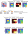
Fig. 2. AI-PrL pathway hypofunction caused SILA-induced reduction of PrL neuronal activity, leading to altered postpartum social novelty recognition.
A Strategy of in vivo microendoscopic calcium recording of PrL through optogenetic manipulation of the AI-PrL pathway. B Representative image showing Chrimson (red) in the AI-PrL pathway and GCaMP6f (green) underneath a GRIN lens. Scale bars, 1 mm and 100 µm. C Experimental timeline of SIT without and with optogenetic manipulation. D Representative heatmaps of the mouse track during the SN-trials in unstressed dams expressing eNPHR. E Optogenetic inhibition of the AI-PrL pathway decreased social novelty preference (two-way mixed ANOVA, Wilcoxon signed-rank test). N = 8 mice. F Representative heatmaps of the mouse track during the SN-trials in stressed dams expressing Chrimson. G Optogenetic activation of the AI-PrL pathway increased social novelty preference (two-way mixed ANOVA, Wilcoxon signed-rank test). N = 8 mice. H Imaging field of view showing raw calcium fluorescence and regions of interest (ROIs). Scale bar, 100 µm. I Receiver operating characteristic (ROC) curves computed from three example neurons that were categorized as novel-excited [area under ROC curve (auROC) = 0.88], novel-unresponsive (auROC = 0.50), or novel-suppressed (auROC = 0.16). J Representative calcium traces from novel-excited, novel-suppressed, and novel-unresponsive neurons. K, L Representative extracted ROIs and calcium traces from PrL. M PrL activity during interaction with a familiar mouse did not show any differences between stressed and unstressed dams expressing control viruses (Chi-squared test). N Fractions of novel-excited and novel-suppressed cells in stressed dams expressing control viruses were significantly decreased and increased, respectively, compared to unstressed dams expressing control viruses (Chi-squared test). O Optogenetic inhibition of the AI-PrL pathway in unstressed dams decreased and increased the fractions of novel-excited and novel-suppressed neurons in PrL, respectively (Chi-squared test). Optogenetic activation in stressed dams increased and decreased the fractions of novel-excited and novel-suppressed neurons, respectively (Chi-squared test). All data are represented as mean ± SEM. For ANOVAs, * = post hoc Bonferroni, p < 0.05. ** = post hoc Bonferroni, p < 0.01. ns non-significant. See Supplemental Table 3 for details on the statistical analyses.
Fig. 3
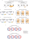
Fig. 3. AI-PrL pathway affected the activity of stable neurons in PrL during the SN-trials.
A Stable and dynamic neurons defined by longitudinal registration of calcium imaging over two consecutive days (SN-trials with and without light stimulation) from animals expressing only control viruses, not opsin. Even without optogenetic manipulation, several PrL neurons showed the same neuronal activity patterns during SN-trials on two consecutive days (defined as “stable neurons”), while others showed different activity patterns (defined as “dynamic neurons”). B Significant differences in the patterns of PrL activity changes between SN-trials on two consecutive days were observed between stressed and unstressed dams expressing control viruses (Chi-squared test). C Scheme of our hypothesis regarding the effect of the AI-PrL pathway on PrL activity during SN-trials. D Significant differences in the patterns of PrL activity changes from SN-trials without light stimulation to SN-trials with light stimulation were observed between unstressed dams expressing control viruses and eNPHR (Chi-squared test). E Significant differences in the patterns of PrL activity changes from SN-trials without light stimulation to SN-trials with light stimulation were observed between stressed dams expressing control viruses and Chrimson (Chi-squared test). **p < 0.01. See Supplemental Table 3 for details on the statistical analyses.
Fig. 4
Fig. 4. AI-PrL pathway was involved in SILA-induced changes in postpartum PrL neuronal activity and social novelty recognition behavior during interactions.
A Experimental timeline for SIT with behavioral closed-loop optogenetic manipulation. B Representative heatmaps of the track during SN-trials in unstressed dams expressing eNPHR. C Optogenetic inhibition during interaction, but not during exploration, in unstressed dams decreased total interaction time and interaction time per visit with novel mice, but not the number of visits (two-way mixed ANOVA, Wilcoxon signed-rank test). N = 8 mice. D Representative heatmaps of the track during SN-trials in stressed dams expressing Chrimson. E Optogenetic activation during interaction, but not during exploration, in stressed dams increased total interaction time and interaction time per visit with novel mice, but not the number of visits (two-way mixed ANOVA). N = 8 mice. F Optogenetic activation during interaction in stressed dams increased and decreased the fractions of novel-excited and novel-suppressed neurons in PrL, respectively. Optogenetic inhibition during interaction in unstressed dams decreased and increased the fractions of novel-excited and novel-suppressed neurons in PrL, respectively. These changes were not observed with optogenetic manipulation during exploration (Chi-squared test). For ANOVAs, * indicates statistical significance for post hoc Bonferroni comparisons. *p < 0.05, **p < 0.01. ns non-significant (p > 0.05). All data are represented as mean ± SEM. See Supplemental Table 3 for details on the statistical analyses.
Fig. 5
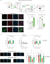
Fig. 5. Activation of GR signaling in the AI-PrL pathway played a causal role in postpartum PrL dysfunction and subsequent behavioral changes in social novelty recognition.
A Scheme of AI-PrL pathway-specific GR-KO using the CRE-DOG method. B Representative images of EGFP+ (green), GR+ (red), DAPI (blue), and colocalized cells in AI of GRfl/fl mice with CRE-DOG. Scale bar, 100 µm. C GR deletion progressed over time along with expression levels of EGFP (one-way ANOVA, Welch’s ANOVA). N = 4 mice. D AI-PrL pathway-specific GR-KO ameliorated SILA-induced behavioral changes in SN-trials (two-way ANOVA, Mann–Whitney U-test). N = 6 mice. E Representative images of c-Fos + (red or green), DAPI (blue), and colocalized cells in AI and PrL of stressed dams with or without AI-PrL pathway-specific GR-KO. Scale bar, 50 μm. F AI-PrL pathway-specific GR-KO normalized the decreased neural activity of AI and PrL in stressed dams (two-way ANOVA). N = 6 mice. * indicates statistical significance for post hoc Bonferroni comparisons. *p < 0.05, **p < 0.01. All data were represented as mean ± SEM. See Supplemental Table 3 for details on the statistical analyses.

Update of

Similar articles

Cited by

References

    1. Brunton PJ, Russell JA. The expectant brain: adapting for motherhood. Nat. Rev. Neurosci. 2008;9:11–25. doi: 10.1038/nrn2280. - DOI - PubMed
    1. Meltzer-Brody S, et al. Postpartum psychiatric disorders. Nat. Rev. Dis. Prim. 2018;4:18022. doi: 10.1038/nrdp.2018.22. - DOI - PubMed
    1. Guintivano J, et al. Adverse life events, psychiatric history, and biological predictors of postpartum depression in an ethnically diverse sample of postpartum women. Psychol. Med. 2018;48:1190–1200. doi: 10.1017/S0033291717002641. - DOI - PMC - PubMed
    1. Viguera AC, et al. Episodes of mood disorders in 2,252 pregnancies and postpartum periods. Am. J. Psychiatry. 2011;168:1179–1185. doi: 10.1176/appi.ajp.2011.11010148. - DOI - PubMed
    1. O’Hara MW, McCabe JE. Postpartum depression: current status and future directions. Annu. Rev. Clin. Psychol. 2013;9:379–407. doi: 10.1146/annurev-clinpsy-050212-185612. - DOI - PubMed

Publication types