

Echo2Pheno: a deep-learning application to uncover echocardiographic phenotypes in conscious mice
- PMID: 37221250
- PMCID: PMC10290584
- DOI: 10.1007/s00335-023-09996-x
Echo2Pheno: a deep-learning application to uncover echocardiographic phenotypes in conscious mice
Abstract
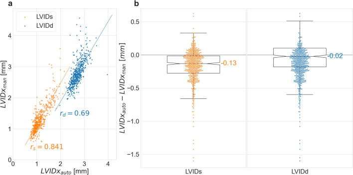
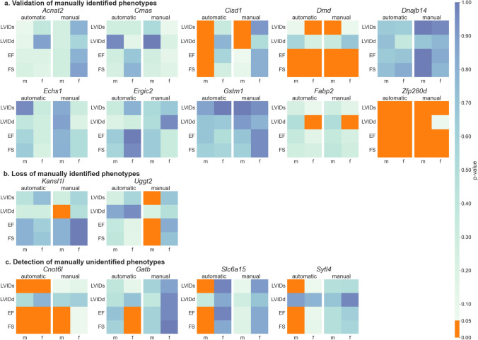
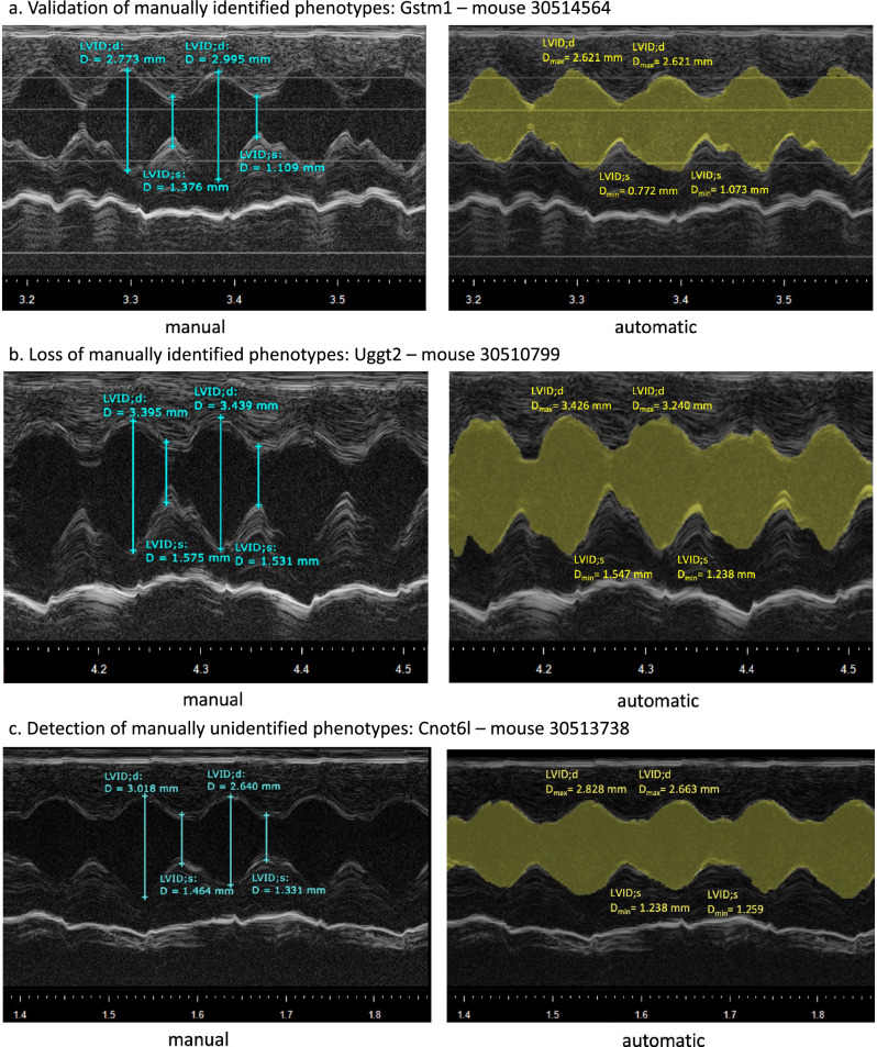
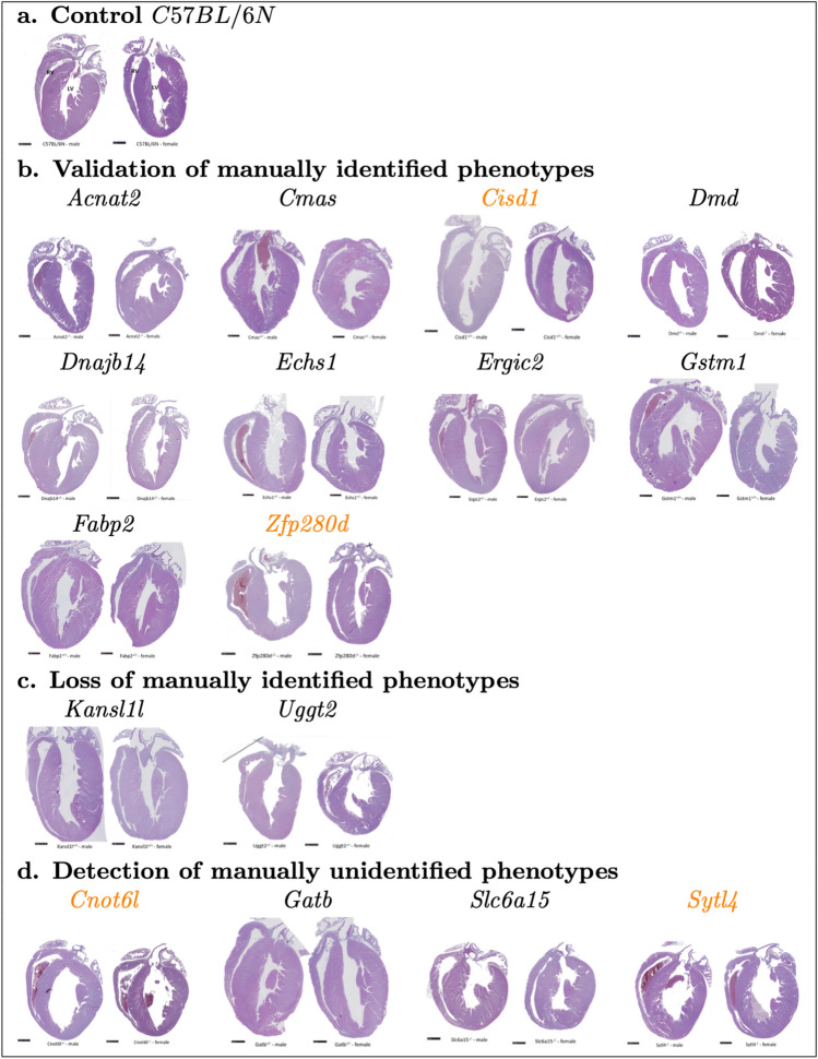
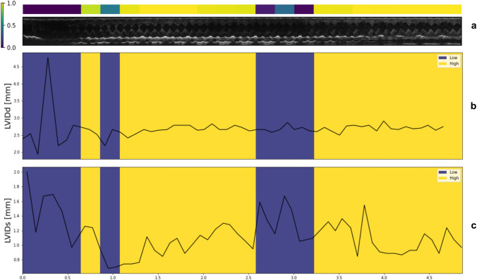
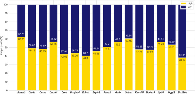
Echocardiography, a rapid and cost-effective imaging technique, assesses cardiac function and structure. Despite its popularity in cardiovascular medicine and clinical research, image-derived phenotypic measurements are manually performed, requiring expert knowledge and training. Notwithstanding great progress in deep-learning applications in small animal echocardiography, the focus has so far only been on images of anesthetized rodents. We present here a new algorithm specifically designed for echocardiograms acquired in conscious mice called Echo2Pheno, an automatic statistical learning workflow for analyzing and interpreting high-throughput non-anesthetized transthoracic murine echocardiographic images in the presence of genetic knockouts. Echo2Pheno comprises a neural network module for echocardiographic image analysis and phenotypic measurements, including a statistical hypothesis-testing framework for assessing phenotypic differences between populations. Using 2159 images of 16 different knockout mouse strains of the German Mouse Clinic, Echo2Pheno accurately confirms known cardiovascular genotype-phenotype relationships (e.g., Dystrophin) and discovers novel genes (e.g., CCR4-NOT transcription complex subunit 6-like, Cnot6l, and synaptotagmin-like protein 4, Sytl4), which cause altered cardiovascular phenotypes, as verified by H&E-stained histological images. Echo2Pheno provides an important step toward automatic end-to-end learning for linking echocardiographic readouts to cardiovascular phenotypes of interest in conscious mice.
© 2023. The Author(s).
Conflict of interest statement
The authors declare no competing interests.
Figures
Similar articles
-
Deep Learning for Segmentation Using an Open Large-Scale Dataset in 2D Echocardiography.IEEE Trans Med Imaging. 2019 Sep;38(9):2198-2210. doi: 10.1109/TMI.2019.2900516. Epub 2019 Feb 22. IEEE Trans Med Imaging. 2019. PMID: 30802851
-
Automated interpretation of systolic and diastolic function on the echocardiogram: a multicohort study.Lancet Digit Health. 2022 Jan;4(1):e46-e54. doi: 10.1016/S2589-7500(21)00235-1. Epub 2021 Dec 1. Lancet Digit Health. 2022. PMID: 34863649
-
Clinically Feasible and Accurate View Classification of Echocardiographic Images Using Deep Learning.Biomolecules. 2020 Apr 25;10(5):665. doi: 10.3390/biom10050665. Biomolecules. 2020. PMID: 32344829 Free PMC article.
-
Italian Society of Cardiovascular Echography (SIEC) Consensus Conference on the state of the art of contrast echocardiography.Ital Heart J. 2004 Apr;5(4):309-34. Ital Heart J. 2004. PMID: 15185894 Review.
-
Deep learning-based automatic segmentation of images in cardiac radiography: A promising challenge.Comput Methods Programs Biomed. 2022 Jun;220:106821. doi: 10.1016/j.cmpb.2022.106821. Epub 2022 Apr 19. Comput Methods Programs Biomed. 2022. PMID: 35487181 Review.
Cited by
-
High expression of CNOT6L contributes to the negative development of type 2 diabetes.Sci Rep. 2024 Oct 21;14(1):24723. doi: 10.1038/s41598-024-76095-5. Sci Rep. 2024. PMID: 39433858 Free PMC article.
-
Realistic Aspects of Cardiac Ultrasound in Rats: Practical Tips for Improved Examination.J Imaging. 2024 Sep 6;10(9):219. doi: 10.3390/jimaging10090219. J Imaging. 2024. PMID: 39330439 Free PMC article. Review.
References
-
- Anaya-Isaza A, Mera-Jiménez L, Zequera-Diaz M. An overview of deep learning in medical imaging. Inform Med Unlocked. 2021;26:100723. doi: 10.1016/j.imu.2021.100723. - DOI
-
- Arora G, et al. Differences in left ventricular ejection fraction using teichholz formula and volumetric methods by cmr: implications for patient stratification and selection of therapy. J Cardiovasc Magn Reson. 2010;12(1):1–2. - PubMed
Publication types
MeSH terms
Substances
LinkOut - more resources
Full Text Sources
