

Tolerability and tropism of recombinant adeno-associated virus vectors in the African green monkey (Chlorocebus sabaeus) anterior chamber
- PMID: 37221271
- PMCID: PMC11287778
- DOI: 10.1038/s41434-023-00407-z
Tolerability and tropism of recombinant adeno-associated virus vectors in the African green monkey (Chlorocebus sabaeus) anterior chamber
Abstract
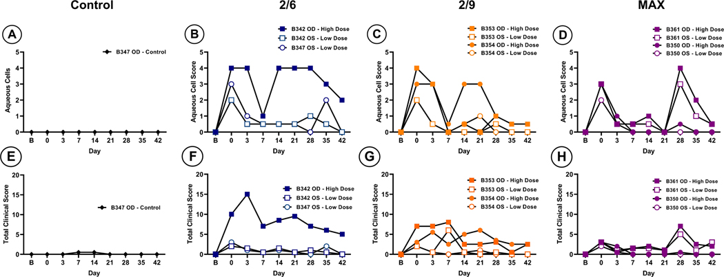
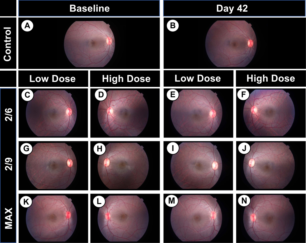
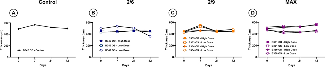
While many studies have investigated the use of recombinant adeno-associated vectors (rAAV) in the posterior chamber for treatment of inherited retinal diseases, fewer studies have looked at rAAV's ability to transduce cells within the anterior chamber. This study focuses on evaluating the tropism and tolerability of three rAAV serotypes-rAAV2/6, rAAV2/9, and rAAV2/2[MAX]-expressing a green fluorescent protein (GFP) reporter following intracameral injection in the non-human primate (NHP) African green monkey (Chlorocebus sabaeus) model. Injection of high dose (1 × 10¹² vg/eye) rAAV vector resulted in transient inflammation characterized by aqueous flare and cellular infiltrate that resolved without intervention in all serotypes. Post-mortem histology revealed widespread expression of GFP in cells of the trabecular meshwork and iris in high dose rAAV2/6, rAAV2/9, and particularly rAAV2/2[MAX] eyes, indicating that rAAV vectors of these serotypes have broad tropism for cells of the anterior chamber and may facilitate the treatment of blinding disorders, such as glaucoma.
© 2023. The Author(s), under exclusive licence to Springer Nature Limited.
Figures


Similar articles
-
Differential targeting of feline photoreceptors by recombinant adeno-associated viral vectors: implications for preclinical gene therapy trials.Gene Ther. 2014 Oct;21(10):913-20. doi: 10.1038/gt.2014.65. Epub 2014 Jul 24. Gene Ther. 2014. PMID: 25056608 Free PMC article.
-
Cellular tropism and transduction properties of seven adeno-associated viral vector serotypes in adult retina after intravitreal injection.Gene Ther. 2009 Apr;16(4):521-32. doi: 10.1038/gt.2008.178. Epub 2008 Dec 18. Gene Ther. 2009. PMID: 19092858
-
Improvement of Photoreceptor Targeting via Intravitreal Delivery in Mouse and Human Retina Using Combinatory rAAV2 Capsid Mutant Vectors.Invest Ophthalmol Vis Sci. 2017 Dec 1;58(14):6429-6439. doi: 10.1167/iovs.17-22281. Invest Ophthalmol Vis Sci. 2017. PMID: 29260200 Free PMC article.
-
Custom adeno-associated virus capsids: the next generation of recombinant vectors with novel tropism.Hum Gene Ther. 2005 Apr;16(4):408-16. doi: 10.1089/hum.2005.16.408. Hum Gene Ther. 2005. PMID: 15871672 Review.
-
Recombinant adeno-associated viral vectors in the nervous system.Hum Gene Ther. 2005 Jul;16(7):781-91. doi: 10.1089/hum.2005.16.781. Hum Gene Ther. 2005. PMID: 16000060 Review.
References
-
- Pascolini D, Mariotti SP. Global estimates of visual impairment: 2010. British Journal of Ophthalmology. 2012. May;96(5):614–8. - PubMed
-
- Liu J, Saghizadeh M, Tuli SS, Kramerov AA, Lewin AS, Bloom DC, et al. Different tropism of adenoviruses and adeno-associated viruses to corneal cells: implications for corneal gene therapy. 2008. [cited 2022 Dec 12]; Available from: http://www.molvis.org/molvis/v14/a245 - PMC - PubMed
Publication types
MeSH terms
Grants and funding
LinkOut - more resources
Full Text Sources
