

Longitudinal Assessment of the Choroidal Vascularity Index in Eyes with Branch Retinal Vein Occlusion-Associated Cystoid Macular Edema
- PMID: 37221425
- PMCID: PMC10287880
- DOI: 10.1007/s40123-023-00731-y
Longitudinal Assessment of the Choroidal Vascularity Index in Eyes with Branch Retinal Vein Occlusion-Associated Cystoid Macular Edema
Abstract
Introduction: Cystoid macular edema (CME) is the most common cause of central vision loss in eyes with branch retinal vein occlusion (BRVO eyes). In recent literature, choroidal vascularity index (CVI) has been proposed to be an enhanced depth imaging optical coherence tomography (EDI-OCT) metric that may help characterize choroidal vascular changes in the setting of retinal ischemia, and potentially prognose visual outcomes and treatment patterns for patients with BRVO-related CME. This study sought to further characterize choroidal vascular changes in BRVO by comparing the CVI, subfoveal choroidal thickness (SFCT), and central subfield thickness (CST) in BRVO eyes with CME compared to unaffected fellow eyes.
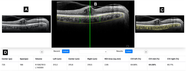
Methods: This was a retrospective cohort study. Subjects included treatment-naïve BRVO eyes with CME diagnosed within 3 months of onset of symptoms and unaffected fellow eyes. EDI-OCT images were collected at baseline and at the 12-month follow-up visit. CVI, SFCT, and CST were measured. Demographics, treatment patterns, and best-corrected visual acuity (VA) were abstracted. Median CVI, SFCT, CST, and VA were compared between the two cohorts. Longitudinal relationships between these variables were analyzed.
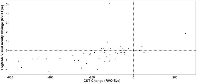
Results: A total of 52 treatment-naïve eyes with BRVO and CME and 48 unaffected fellow eyes were identified. Baseline CVI was lower in eyes with BRVO than in fellow eyes (64.7% vs. 66.4%, P = 0.003). At 12 months, there was no difference in CVI between BRVO eyes and fellow eyes (65.7% vs 65.8%, P = 0.536). In BRVO eyes, there was a strong correlation between reduced CST and improved VA over the 12-month study period (r = 0.671, P < 0.001).
Conclusion: There are differences in CVI in treatment-naïve BRVO eyes with CME at presentation compared to fellow eyes, but these differences resolve over time. Anatomic changes in macular thickness in BRVO eyes with CME may be correlated with VA outcomes.
Keywords: Branch retinal vein occlusion; Choroidal vascularity index; Cystoid macular edema; Optical coherence tomography.
Plain language summary
Our study evaluated a novel ocular optical coherence tomography imaging metric, the choroidal vascularity index, in eyes that developed cystoid macular edema, a condition which can significantly impair acuity of central vision, after being diagnosed with branch retinal vein occlusion. In each patient, we compared the choroidal vascularity index in eyes that developed treatment-naïve, newly diagnosed branch retinal vein occlusion with cystoid macular edema to the non-diseased fellow eye. We made comparisons at the time of diagnosis (baseline) and at the 12-month follow up, and analyzed changes over time. We found that at the baseline visit, branch retinal vein occlusion eyes with cystoid macular edema had a significantly lower choroidal vascularity index than their unaffected fellow eyes, but that the differences between eyes resolved by the 12-month follow-up visit. Our findings suggest that choroidal vascularity may be compromised in the acute phase of branch retinal vein occlusion, but that this phenomenon resolves over time. Future research should further evaluate whether imaging characteristics of choroidal vascularity may be associated with changes in anatomic and visual outcomes in retinal diseases.
© 2023. The Author(s).
Conflict of interest statement
No conflicting relationship or competing interest exists for any author.
Figures
References
-
- Campochiaro PA. Anti-vascular endothelial growth factor treatment for retinal vein occlusions. Ophthalmologica. 2012;227(Suppl 1):30–5. - PubMed
-
- Noma H, Funatsu H, Yamasaki M, et al. Pathogenesis of macular edema with branch retinal vein occlusion and intraocular levels of vascular endothelial growth factor and interleukin-6. Am J Ophthalmol. 2005;140(2):256.e1-.e7. - PubMed
Grants and funding
LinkOut - more resources
Full Text Sources
