

Imaging the brain and vascular reactions to headache treatments: a systematic review
- PMID: 37221469
- PMCID: PMC10207747
- DOI: 10.1186/s10194-023-01590-5
Imaging the brain and vascular reactions to headache treatments: a systematic review
Abstract
Background: Neuroimaging studies have made an important contribution to our understanding of headache pathophysiology. This systematic review aims to provide a comprehensive overview and critical appraisal of mechanisms of actions of headache treatments and potential biomarkers of treatment response disclosed by imaging studies.
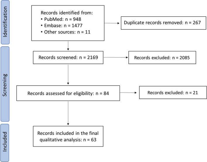
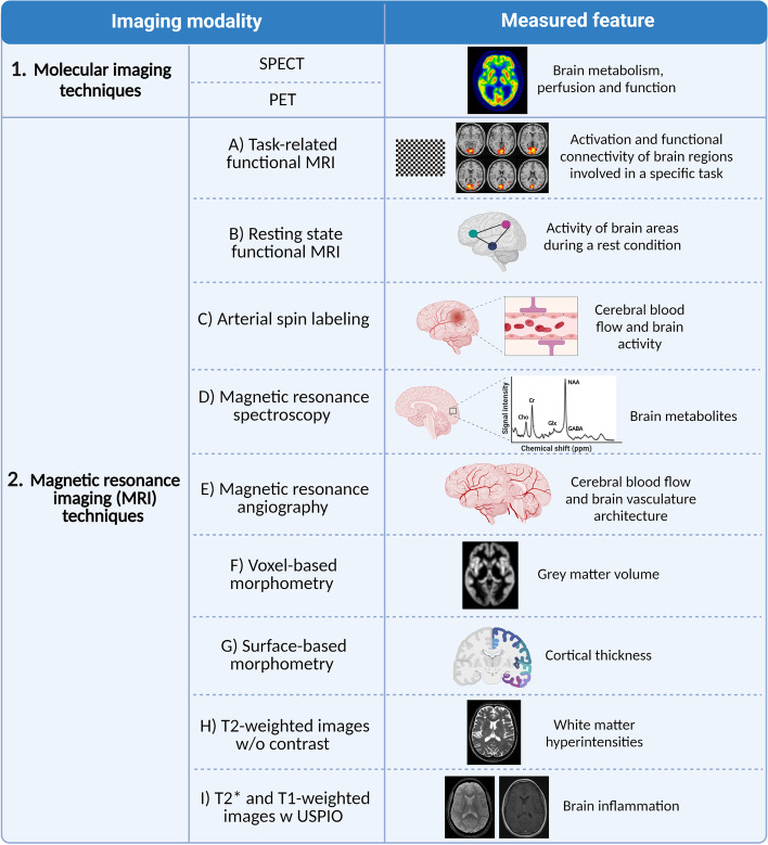
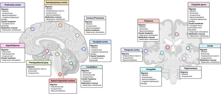
Main body: We performed a systematic literature search on PubMed and Embase databases for imaging studies investigating central and vascular effects of pharmacological and non-pharmacological treatments used to abort and prevent headache attacks. Sixty-three studies were included in the final qualitative analysis. Of these, 54 investigated migraine patients, 4 cluster headache patients and 5 patients with medication overuse headache. Most studies used functional magnetic resonance imaging (MRI) (n = 33) or molecular imaging (n = 14). Eleven studies employed structural MRI and a few used arterial spin labeling (n = 3), magnetic resonance spectroscopy (n = 3) or magnetic resonance angiography (n = 2). Different imaging modalities were combined in eight studies. Despite of the variety of imaging approaches and results, some findings were consistent. This systematic review suggests that triptans may cross the blood-brain barrier to some extent, though perhaps not sufficiently to alter the intracranial cerebral blood flow. Acupuncture in migraine, neuromodulation in migraine and cluster headache patients, and medication withdrawal in patients with medication overuse headache could promote headache improvement by reverting headache-affected pain processing brain areas. Yet, there is currently no clear evidence for where each treatment acts, and no firm imaging predictors of efficacy. This is mainly due to a scarcity of studies and heterogeneous treatment schemes, study designs, subjects, and imaging techniques. In addition, most studies used small sample sizes and inadequate statistical approaches, which precludes generalizable conclusions.
Conclusion: Several aspects of headache treatments remain to be elucidated using imaging approaches, such as how pharmacological preventive therapies work, whether treatment-related brain changes may influence therapy effectiveness, and imaging biomarkers of clinical response. In the future, well-designed studies with homogeneous study populations, adequate sample sizes and statistical approaches are needed.
Keywords: Biomarkers; Headache; Migraine; Neuroimaging; Secondary headaches.
© 2023. The Author(s).
Conflict of interest statement
The authors declare no competing interests.
Figures
Similar articles
-
Pharmacological interventions for acute attacks of vestibular migraine.Cochrane Database Syst Rev. 2023 Apr 12;4(4):CD015322. doi: 10.1002/14651858.CD015322.pub2. Cochrane Database Syst Rev. 2023. PMID: 37042545 Free PMC article.
-
Non-pharmacological interventions for prophylaxis of vestibular migraine.Cochrane Database Syst Rev. 2023 Apr 12;4(4):CD015321. doi: 10.1002/14651858.CD015321.pub2. Cochrane Database Syst Rev. 2023. PMID: 37042522 Free PMC article.
-
Preventive drug treatments for adults with chronic migraine: a systematic review with economic modelling.Health Technol Assess. 2024 Oct;28(63):1-329. doi: 10.3310/AYWA5297. Health Technol Assess. 2024. PMID: 39365169 Free PMC article.
-
Antidepressants for pain management in adults with chronic pain: a network meta-analysis.Health Technol Assess. 2024 Oct;28(62):1-155. doi: 10.3310/MKRT2948. Health Technol Assess. 2024. PMID: 39367772 Free PMC article.
-
Systemic pharmacological treatments for chronic plaque psoriasis: a network meta-analysis.Cochrane Database Syst Rev. 2021 Apr 19;4(4):CD011535. doi: 10.1002/14651858.CD011535.pub4. Cochrane Database Syst Rev. 2021. Update in: Cochrane Database Syst Rev. 2022 May 23;5:CD011535. doi: 10.1002/14651858.CD011535.pub5. PMID: 33871055 Free PMC article. Updated.
Cited by
-
Evaluating Headache and Facial Pain in a Headache Diagnostic Laboratory: Experiences from the Danish Headache Center.Diagnostics (Basel). 2023 Aug 14;13(16):2671. doi: 10.3390/diagnostics13162671. Diagnostics (Basel). 2023. PMID: 37627930 Free PMC article. Review.
-
Regional distribution of unbound eletriptan and sumatriptan in the CNS and PNS in rats: implications for a potential central action.J Headache Pain. 2024 Oct 30;25(1):187. doi: 10.1186/s10194-024-01894-0. J Headache Pain. 2024. PMID: 39478486 Free PMC article.
-
Differences in Cortical Morphology in People With and Without Migraine: A Registry for Migraine (REFORM) MRI Study.Neurology. 2024 May;102(9):e209305. doi: 10.1212/WNL.0000000000209305. Epub 2024 Apr 17. Neurology. 2024. PMID: 38630960 Free PMC article. Clinical Trial.
-
Functional connectivity of the visual cortex in chronic migraine before and after medication withdrawal therapy.Neuroimage Clin. 2023;40:103543. doi: 10.1016/j.nicl.2023.103543. Epub 2023 Nov 17. Neuroimage Clin. 2023. PMID: 37988998 Free PMC article.
References
Publication types
MeSH terms
LinkOut - more resources
Full Text Sources
Medical
