. 2023 May 23;13(1):95.
doi: 10.1186/s13578-023-01028-0.

Microglia degrade Tau oligomers deposit via purinergic P2Y12-associated podosome and filopodia formation and induce chemotaxis

Affiliations

Microglia degrade Tau oligomers deposit via purinergic P2Y12-associated podosome and filopodia formation and induce chemotaxis

Subashchandrabose Chinnathambi et al. Cell Biosci. .

Abstract

Background: Tau protein forms neurofibrillary tangles and becomes deposited in the brain during Alzheimer's disease (AD). Tau oligomers are the most reactive species, mediating neurotoxic and inflammatory activity. Microglia are the immune cells in the central nervous system, sense the extracellular Tau via various cell surface receptors. Purinergic P2Y12 receptor can directly interact with Tau oligomers and mediates microglial chemotaxis via actin remodeling. The disease-associated microglia are associated with impaired migration and express a reduced level of P2Y12, but elevate the level of reactive oxygen species and pro-inflammatory cytokines.

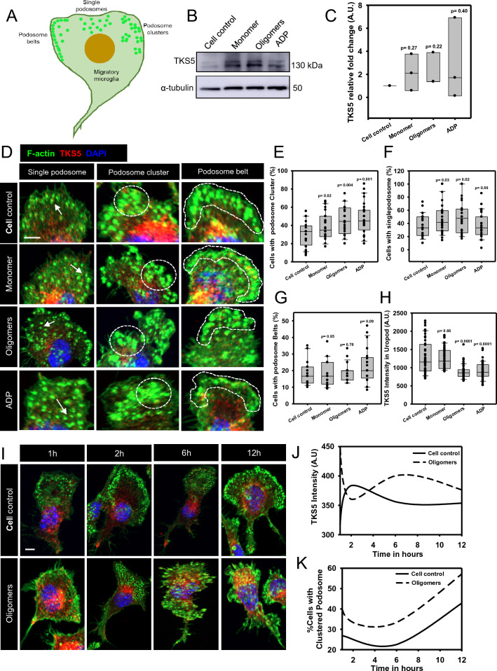
Results: Here, we studied the formation and organization of various actin microstructures such as-podosome, filopodia and uropod in colocalization with actin nucleator protein Arp2 and scaffold protein TKS5 in Tau-induced microglia by fluorescence microscopy. Further, the relevance of P2Y12 signaling either by activation or blockage was studied in terms of actin structure formations and Tau deposits degradation by N9 microglia. Extracellular Tau oligomers facilitate the microglial migration via Arp2-associated podosome and filopodia formation through the involvement of P2Y12 signaling. Similarly, Tau oligomers induce the TKS5-associated podosome clustering in microglial lamella in a time-dependent manner. Moreover, the P2Y12 was evidenced to localize with F-actin-rich podosome and filopodia during Tau-deposit degradation. The blockage of P2Y12 signaling resulted in decreased microglial migration and Tau-deposit degradation.

Conclusions: The P2Y12 signaling mediate the formation of migratory actin structures like- podosome and filopodia to exhibit chemotaxis and degrade Tau deposit. These beneficial roles of P2Y12 in microglial chemotaxis, actin network remodeling and Tau clearance can be intervened as a therapeutic target in AD.

Keywords: Filopodia; Microglia; Migration; P2Y12; Podosome; Tau oligomers.


Conflict of interest statement

The authors declared that there is no competing interests associated with this study.

Figures

Fig. 1
Fig. 1
Tau oligomers induce podosome and filopodia formation, orchestrated with Arp2. A Extracellular Tau oligomers-induced podosome accumulation more in microglial lamellipodia, than Tau monomers as observed by immunofluorescence (IF) assay. The microglial podosome was found to be colocalized with actin-nucleator protein Arp2 for rapid actin polymerization in response to Tau oligomers similar to ADP exposure in migratory microglia (scale bar 10 μm). B A simplistic illustration of podosome where actin cross-linking is mediated by actin nuclear Arp2/3 complex. The actin cores are surrounded by the vinculin ring and adhesion protein receptors. The attachment of actin core fibers with membrane receptors is driven by adaptor protein TKS5. C The quantification of microscopic images showed that the exposure of Tau oligomers has significantly induced the podosome+ microglia by 70% as compared to the monomer-exposed group (50%). (No. of experiment = 3) (n = 13). D Moreover, podosome-associated area has increased almost twice in Tau-exposed microglial cells as compared to cell control. (No. of experiment = 3) (n = 40). E Tau oligomers have increased the number of filopodia three times more compared to untreated control microglia. While, the Tau monomer and ADP exposure have induced the filopodia numbers twice than the control population, as seen by microscopic image quantification. (No. of experiment = 3) (n = 50). F Similar to podosome, Tau oligomers facilitated the localization of Arp2 in filopodia and branched uropod in microglia, for actin polymerization during migration (scale bar 10 μm). G The Arp2 level was reduced from microglial uropod upon Tau oligomers exposure, relating rapid actin dynamics towards frontal lamellipodia. (No. of experiment = 3) (n = 50). H, I. The Arp2 expression level remained unaltered in various Tau and ADP exposure by Western blot analysis and relative fold change calculation in N9 microglia. (n = 3). J The extracellular Tau oligomers have the potential to induce the accumulation of podosome and filopodia by localizing Arp2 at lamellipodia of microglia, which plays an essential role in migration and mechanosensing
Fig. 2
Fig. 2
Extracellular Tau modulates the formation of TKS5-mediated podosome clusters in microglia. A Microglia rearrange the podosome differently at frontal lamellipodia such as podosome belts, clustered podosome, and single podosome, for the adherence to the substratum by membrane-associated actin polymerization. B, C Western blot analysis and relative fold change showed no significant changes in the expression of TKS5 protein upon various Tau exposure and ADP treatment in microglia (n = 3). D IF study revealed that microglia form various rearrangements of podosome upon exposure to extracellular Tau species and ADP. Among all structures, the podosome clusters were more evident in oligomer-treated microglia than in monomer exposure (scale bar 10 μm). E The clustered podosome accumulated by 20% more in lamellipodia upon Tau oligomers exposure, similar to ADP and cell control. (No. of experiment = 3) (n = 28). F While, The amount of single podosome arrangement in microglia remains unaltered among various Tau-treated groups. (No. of experiment = 3) (n = 28) G The podosome belts containing cells remain unaltered in Tau-induced migratory microglia. (No. of experiment = 3) (n = 28). H Furthermore, the TKS5 intensity of uropod was decreased in Tau oligomer-induced microglia, which may signify the rapid actin and actin-associated protein turnover from the rear end towards lamella upon migration. (No. of experiment = 3) (n = 50). I, J The time-dependent IF study showed that the extracellular Tau oligomers induced the accumulation of clustered podosome in microglial lamellipodia (scale bar 10 μm). Then, the quantification of microscopic images revealed that the TKS5 intensity was increased during the oligomers exposure from 6 h, compared to cell control. (No. of experiment = 3) (n = 40). K Further, the clustered podosome-containing microglial population have increased in a time-dependent manner in Tau oligomers-exposed microglia than to untreated cells. (No. of experiment = 3) (n = 10)
Fig. 3
Fig. 3
P2Y12-associated podosome modulates microglial migration and invasion. A Purinergic P2Y12 receptors were colocalized with F-actin-rich podosome in lamellipodia during Tau-induced microglia migration. Similarly, the activation of P2Y12 signaling by ADP has also induced the colocalization of P2Y12 in podosome-rich microglial lamella, as seen by the IF study (scale bar 10 μm). B Fluorescence quantification of podosome-rich lamella near the membrane (box marked area) showed the colocalization of P2Y12 and F-actin at the distances of 14 μm, in Tau monomer, oligomers-exposed microglia. C, D The wound scratch assay showed that the extracellular Tau and ADP, together and separately induced the microglial migration as quantified by %wound closure at 24 h. The blockage of P2Y12 signaling by Clopidogrel has reduced the microglial migration. But, Tau oligomers restored the microglial migration even upon clopidogrel-induced blockage, as observed and quantified by phase contrast imaging. (No. of experiment = 3) (n = 12) (Scale bar 100 μm). E, F In trans-well migration assay, Tau oligomers have induced the microglial invasion more than Tau monomer treatment. P2Y12 activation by ADP has induced microglial trans-migration in both Tau monomer and oligomers exposure. The quantification of microscopic images revealed that Clopidogrel exposure has significantly reduced the microglial invasion, which was eventually restored by Tau oligomers exposure. These emphasize that Tau oligomers were better chemoattractants and can intervene P2Y12 signaling during migration/invasion. (No. of experiment = 2) (n = 12) (scale bar 100 μm)
Fig. 4
Fig. 4
Extracellular Tau facilitates P2Y12 localization in filopodia and uropod, which degrades Tau deposits. A P2Y12 was colocalized with F-actin in filopodia and branched uropod of migratory microglia. Extracellular Tau oligomers exposure induced the localization of P2Y12 more in microglial filopodia and uropod as compared to monomer-treated cells. (Scale bar 10 μm) B The P2Y12+ filopodia+ microglia has significantly increased up to 80% upon Tau oligomers exposure than monomer exposure at 50%. (No. of experiment = 3) (n = 22). C P2Y12 intensity has increased in uropod by Tau oligomers and ADP exposure in migratory microglia (No. of experiment = 3) (n = 50). D The coverslips were coated with Tau monomer and oligomers to mimic the scenario of Tau depositions. The N9 microglia were seeded onto the coated coverslip to determine the potency of Tau plaques degradation. Microglia degraded Tau monomeric deposits by 8 h and Tau oligomers deposits by 24 h after seeding, through the formation of the F-actin-rich P2Y12+ podosome. It was evident that the degradation of Tau monomers was faster than oligomeric deposits (scale bar 10 μm). E The percentage of microglia, which degraded Tau monomer- deposits, was 38%, while 30% of microglia degraded Tau oligomers deposits. (No. of experiment = 3) (n = 40). F The P2Y12-associated podosome in microglia degraded Tau monomer deposits more than Tau oligomers-deposits. (No. of experiment = 3) (n = 30). G Microglia was also found to degrade Tau deposits through filopodia formation, which was orchestrated with P2Y12 (scale bar 10 μm). It was evident for the first time that P2Y12+ filopodia could degrade Tau monomer and oligomers deposits, which connects the signaling of chemotaxis and matrix degradation. H Microglia degraded the Tau deposits more in the case of the Tau monomer than the Tau oligomers deposits, through the formation of P2Y12+ filopodia. (No. of experiment = 3) (n = 30). I Extracellular Tau oligomers have induced the P2Y12-driven chemotaxis, which leads to the substratum adhesion and Tau deposit degradation through the formation of podosome and filopodia in migratory microglia
Fig. 5
Fig. 5
Microglia degrades Tau deposits by actin remodeling, localized with TKS5 and Arp2. A Microglia degraded Tau deposits through the accumulation of podosome and filopodia, which were localized with Arp2 actin nucleator at the site of degradation (Scale bar 10 μm). The arrow indicates the degradation area. B The actin remodeling is mediated by Arp2, where filopodia contained more Arp2 than podosome, relating to rapid actin polymerization at the Tau deposits degradation site (No. of experiment = 3) (n = 30). C Similarly, the TKS5 adaptor protein became colocalized with podosome and filopodia at the site of Tau deposits degradation (scale bar 10 μm). The arrow indicates the degradation area. D. But, the colocalization of F-actin and TKS5 did not alter between podosome and filopodia-associated Tau deposits degradation. (No. of experiment = 3) (n = 30) E Tau fluorescence intensity was significantly reduced in the degradation area of Tau monomer and oligomers-deposits as compared to the non-degraded area. Moreover, Tau monomer was significantly degraded more than oligomers in microglia-mediated deposit degradation (No. of experiment = 3) (n = 100). F The quantification of relative degraded area/total cell area revealed that monomer deposits were better degraded by microglia as compared to Tau oligomers deposits. (No. of experiment = 3) (n = 45). Hence, It can be concluded that microglia prefer to degrade Tau monomer more than oligomers as deposits as emphasized by Time and area of degradation
Fig. 6
Fig. 6
P2Y12-activation and blockage influence Tau deposit degradation through the Arp2-mediated podosome and filopodia formation. A P2Y12 activation by ADP has induced more filopodia formation for Tau deposit degradation. While, microglia, which degrade Tau, deposits by P2Y12.+ podosome formation, were independent of P2Y12 signaling activation/blockage. Microglia have been shown to degrade Tau deposits as scattered spots at the cell surface upon Clopidogrel treatment. While upon ADP exposure, microglia degraded the Tau deposits throughout the cell surface (Scale bar 10 μm). B The percentage of cells with Tau deposit degradation has been reduced by Clopidogrel exposure. While the ADP-mediated P2Y12 activation showed unaltered Tau deposit degradation compared to the control. (No. of experiment = 3) (n = 33). C P2Y12 activation resulted in more Arp2-localized filopodia formation that leads to Tau degradation, while Clopidogrel treatment did not alter the membrane-associated actin and Tau deposit degradation (Scale bar 10 μm). D Similarly, TKS5 localization was reduced in podosome and filopodia during Tau deposit degradation upon P2Y12-activated microglia (Scale bar 10 μm). E P2Y12 activation leads to the Arp2-associated filopodia formation at the site of Tau deposit degradation. At the same time, the blockage of P2Y12 signaling by clopidogrel resulted in more accumulation of Arp2-orchestrated podosome and filopodia at the Tau degradation site. (No. of experiment = 3) (n = 25). F ADP-mediated P2Y12 activation lead to the reduced TKS5 colocalization at podosome and filopodia while P2Y12 blockage did not alter TKS5 localization in remodeled actin network. (No. of experiment = 3) (n = 25)
Fig. 7
Fig. 7
Microglia degrade extracellular Tau deposits by P2Y12-mediated podosome and filopodia formation. Extracellular Tau oligomers have induced Arp2 and TKS5-associated podosome clusters and filopodia formation during P2Y12-mediated microglial migration and invasion. Moreover, Tau monomer and oligomers deposits can be degraded by microglial filopodia and podosome formation. The blockage of P2Y12 signaling has reduced the microglial chemotaxis and P2Y12-associated filopodia formation and Tau-deposits degradation. Hence, P2Y12 signaling plays a dual role in extracellular Tau-induced microglial chemotaxis and the clearance of Tau deposits via podosome and filopodia-associated actin remodeling

Similar articles

Cited by

References

    1. Gorantla NV, Chinnathambi S. Tau protein squired by molecular chaperones during Alzheimer’s disease. J Mol Neurosci. 2018;66(3):356–368. doi: 10.1007/s12031-018-1174-3. - DOI - PubMed
    1. Das R, Chinnathambi S. Microglial priming of antigen presentation and adaptive stimulation in Alzheimer’s disease. Cell Mol Life Sci. 2019;76(19):3681–3694. doi: 10.1007/s00018-019-03132-2. - DOI - PMC - PubMed
    1. Sonawane SK, Chinnathambi S. Prion-like propagation of post-translationally modified tau in Alzheimer’s disease: a hypothesis. J Mol Neurosci. 2018;65(4):480–490. doi: 10.1007/s12031-018-1111-5. - DOI - PubMed
    1. Iqbal K, Gong C-X, Liu F. Hyperphosphorylation-induced tau oligomers. Front Neurol. 2013;4:112. doi: 10.3389/fneur.2013.00112. - DOI - PMC - PubMed
    1. Castillo-Carranza DL, et al. Specific targeting of tau oligomers in htau mice prevents cognitive impairment and tau toxicity following injection with brain-derived tau oligomeric seeds. J Alzheimers Dis. 2014;40(s1):S97–S111. doi: 10.3233/JAD-132477. - DOI - PubMed

LinkOut - more resources