

Identification of BRAF, CCND1, and MYC mutations in a patient with multiple primary malignant tumors: a case report and review of the literature
- PMID: 37221610
- PMCID: PMC10207802
- DOI: 10.1186/s12957-023-03036-3
Identification of BRAF, CCND1, and MYC mutations in a patient with multiple primary malignant tumors: a case report and review of the literature
Abstract
Background: Multiple primary malignant tumors (MPMTs), usually associated with worse malignant behavior and prognosis comparing to a single primary tumor, and have recently been found to have an increasing incidence globally. However, the pathogenesis of MPMTs remains to be clarified. Here, we report a unique case of the coexistence of malignant melanoma (MM), papillary thyroid carcinoma (PTC), and clear-cell renal cell carcinoma (ccRCC) along with our perceptions on its pathogenesis.
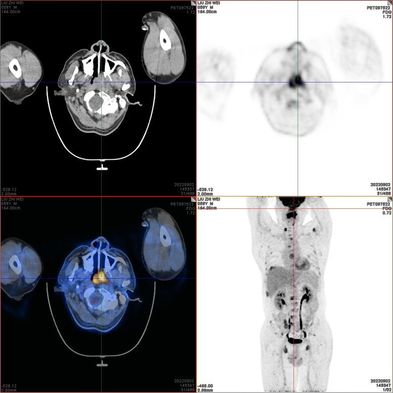
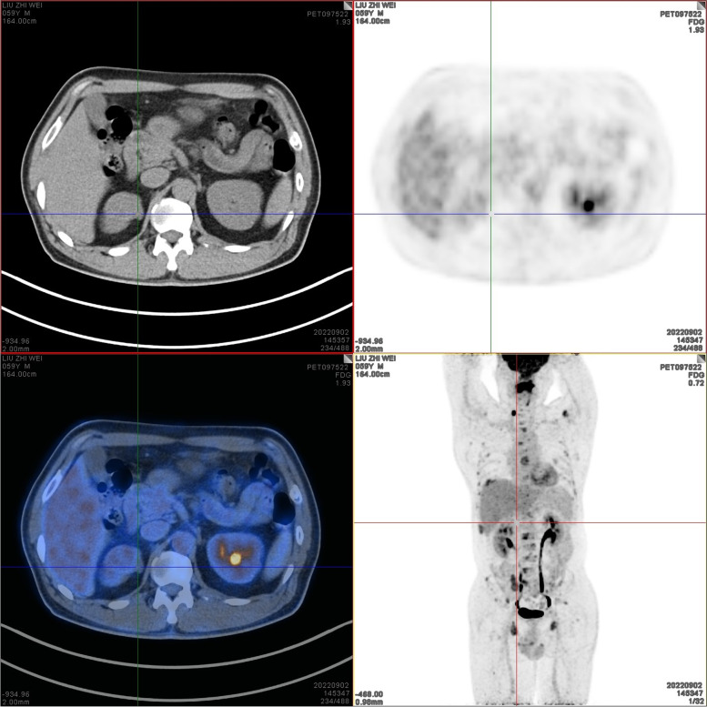
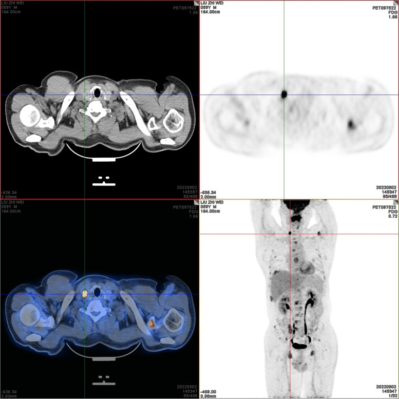
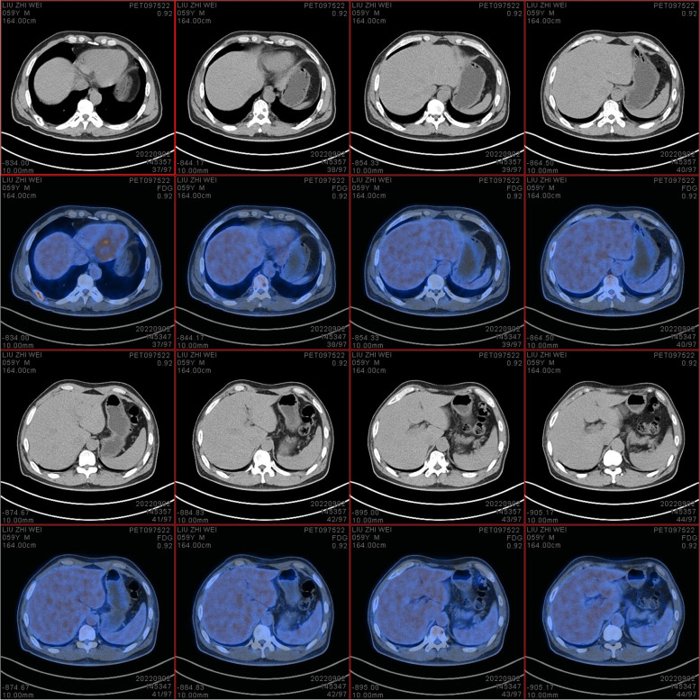
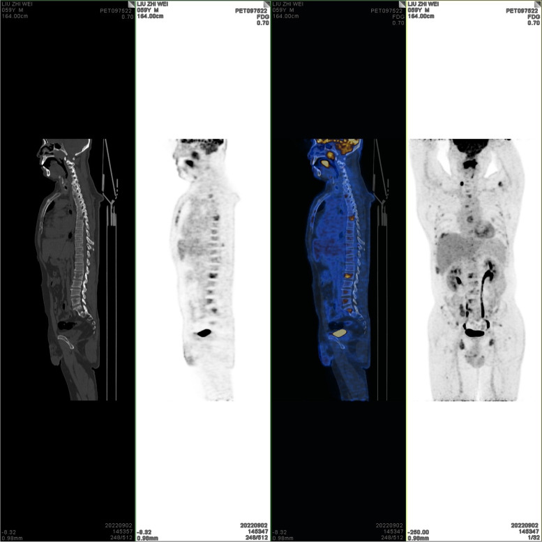
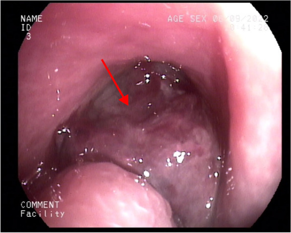
Case presentation: The case reported is of a 59-year-old male patient with unilateral nasal obstruction as well as a renal occupying lesion. Positron emission tomography-computed tomography (PET-CT) revealed a palpable mass of 32 × 30 mm on the posterior and left walls of the nasopharynx. In addition, an isodense nodule was observed in the right superior renal pole, approximately 25 mm in diameter, as well as a slightly hypodense shadow in the right leaf of the thyroid, approximately 13 mm in diameter. Nasal endoscopy and magnetic resonance imaging (MRI) confirmed the existence of a nasopharyngeal neoplasm. Afterward, biopsies of the nasopharyngeal neoplasm, thyroid gland and kidney were performed, and the patient was diagnosed with MM, PTC, and ccRCC according to the pathological and immunohistochemical results. Moreover, mutation of BRAFV600E was detected in bilateral thyroid tissues, and amplification of both CCND1 and MYC oncogenes were detected in the nasopharyngeal melanoma. After chemotherapy, the patient is now in good overall condition.
Conclusions: This is the first reported case of a patient with the co-existence of MM, PTC and ccRCC undergoing chemotherapy with a favorable prognosis. Herein, we suggest that such a combination may be non-random, as for mutation of BRAFV600E might account for the co-occurrence of PTC and MM, while mutations of CCND1 and MYC cause the coexistence of MM and ccRCC. This finding may provide valuable guidance on the diagnosis and treatment of such disease, as well as the prevention of developing a second or third tumor for patients with a single primary.
Keywords: Clear-cell renal cell carcinoma; Genetic linkage; Malignant melanoma; Multiple primary malignant tumors; Papillary thyroid carcinoma.
© 2023. The Author(s).
Conflict of interest statement
We certify that we have no affiliations with any organization or entity with any financial interest or non-financial interest.
Figures


Similar articles
-
Synchronous occurrence of medullary and papillary carcinoma of the thyroid in a patient with cutaneous melanoma: determination of BRAFV600E in peripheral blood and tissues. Report of a case and review of the literature.Endocr Pathol. 2014 Sep;25(3):324-31. doi: 10.1007/s12022-014-9303-1. Endocr Pathol. 2014. PMID: 24858900 Review.
-
Pancreatic metastasis arising from a BRAF(V600E)-positive papillary thyroid cancer: the role of endoscopic ultrasound-guided biopsy and response to sorafenib therapy.Thyroid. 2012 May;22(5):536-41. doi: 10.1089/thy.2011.0247. Epub 2012 Mar 21. Thyroid. 2012. PMID: 22435913
-
Tumor-to-tumor metastases: papillary thyroid carcinoma into a clear cell renal cell carcinoma.J Otolaryngol Head Neck Surg. 2017 Mar 1;46(1):17. doi: 10.1186/s40463-017-0193-3. J Otolaryngol Head Neck Surg. 2017. PMID: 28249616 Free PMC article.
-
Clinicopathological characteristics including BRAF V600E mutation status and PET/CT findings in papillary thyroid carcinoma.Clin Endocrinol (Oxf). 2017 Jul;87(1):73-79. doi: 10.1111/cen.13335. Epub 2017 Apr 18. Clin Endocrinol (Oxf). 2017. PMID: 28329426
-
BRAF gene mutations in synchronous papillary thyroid carcinoma and Langerhans cell histiocytosis co-existing in the thyroid gland: a case report and literature review.BMC Cancer. 2019 Feb 22;19(1):170. doi: 10.1186/s12885-019-5372-3. BMC Cancer. 2019. PMID: 30795755 Free PMC article. Review.
Cited by
-
Predicting CircRNA-Disease Associations Based on Heterogeneous Graph Neural Network and Knowledge Graph Attribute Mining Attention.Interdiscip Sci. 2025 Sep;17(3):586-597. doi: 10.1007/s12539-025-00706-6. Epub 2025 May 13. Interdiscip Sci. 2025. PMID: 40358837
-
Durable response to nivolumab rechallenge in a patient with metastatic clear cell renal cell carcinoma.IJU Case Rep. 2024 Apr 8;7(4):293-296. doi: 10.1002/iju5.12727. eCollection 2024 Jul. IJU Case Rep. 2024. PMID: 38966764 Free PMC article.
-
Molecular Basis of BRAF Inhibitor Resistance in Melanoma: A Systematic Review.Pharmaceuticals (Basel). 2025 Aug 21;18(8):1235. doi: 10.3390/ph18081235. Pharmaceuticals (Basel). 2025. PMID: 40872623 Free PMC article. Review.
-
Synchronous double primary small cell lung cancer and invasive ductal breast carcinoma: a case report.BMC Pulm Med. 2024 Feb 22;24(1):93. doi: 10.1186/s12890-024-02897-y. BMC Pulm Med. 2024. PMID: 38388422 Free PMC article.
References
-
- Warren SGO. Multiple primary malignant tumors: A survey of the literature and a statistical study. Am J Cancer. 1932;16:1358–1414.
-
- WHO/IARC International rules for multiple primary cancers. Asian Pac J Cancer Prev. 2005;6(1):104–6. - PubMed
Publication types
MeSH terms
Substances
LinkOut - more resources
Full Text Sources
Medical
Research Materials
