

Coping with abrasive food: diverging composition of radular teeth in two Porifera-consuming nudibranch species (Mollusca, Gastropoda)
- PMID: 37221862
- PMCID: PMC10206459
- DOI: 10.1098/rsif.2022.0927
Coping with abrasive food: diverging composition of radular teeth in two Porifera-consuming nudibranch species (Mollusca, Gastropoda)
Abstract
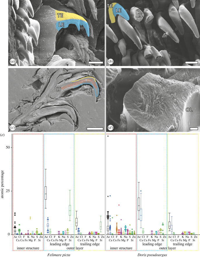
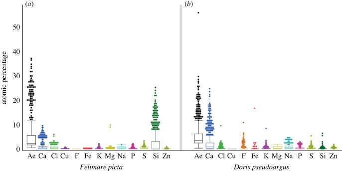
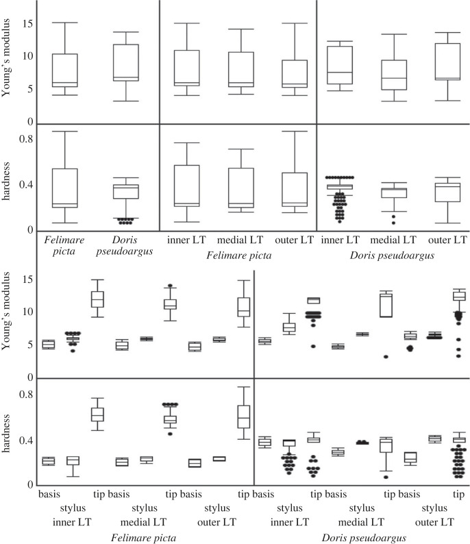
Molluscs forage with their radula, a chitinous membrane with teeth. Adaptations to hard or abrasive ingesta were well studied in Polyplacophora and Patellogastropoda, but for other taxa there are large gaps in knowledge. Here, we investigated the nudibranch gastropods Felimare picta and Doris pseudoargus, both of which feed on Porifera. Tooth morphologies were documented by scanning electron microscopy, and mechanical properties were tested by nanoindentation. We found that these parameters are rather similar in both species, indicating that teeth are similar in their function. To study the composition, teeth were visualized using confocal laser scanning microscopy (CLSM), to determine the degree of tanning, and analysed with energy-dispersive X-ray spectroscopy, to test the elemental composition. The emitted autofluorescence signal and the inorganic content differed between the species. This was especially prominent when studying the inner and outer tooth surfaces (leading and trailing edges). In F. picta, we detected high proportions of Si, whereas teeth of D. pseudoargus contained high amounts of Ca, which influenced the autofluorescence signal in CLSM. Employing nanoindentation, we determined high Young's modulus and hardness values for the leading edges of teeth, which relate to the Si and Ca content. This highlights that teeth with a similar morphology and mechanical properties can be mechanically enhanced via different chemical pathways in Nudibranchia.
Keywords: Nudibranchia; biomineralization; elemental composition; feeding; mechanical properties; mollusca.
Conflict of interest statement
The authors declare that they have no competing interests.
Figures


Similar articles
-
Elemental composition and material properties of radular teeth in the heterobranch snail Gastropteron rubrum (Mollusca, Gastropoda, Cephalaspidea) foraging on hard organisms.Ecol Evol. 2023 Aug 14;13(8):e10332. doi: 10.1002/ece3.10332. eCollection 2023 Aug. Ecol Evol. 2023. PMID: 37589038 Free PMC article.
-
Radular Tooth Coating in Members of Dendronotidae and Flabellinidae (Nudibranchia, Gastropoda, Mollusca).J Morphol. 2024 Sep;285(9):e21773. doi: 10.1002/jmor.21773. J Morphol. 2024. PMID: 39252400
-
Elemental analyses reveal distinct mineralization patterns in radular teeth of various molluscan taxa.Sci Rep. 2022 May 7;12(1):7499. doi: 10.1038/s41598-022-11026-w. Sci Rep. 2022. PMID: 35525838 Free PMC article.
-
Main patterns of radula formation and ontogeny in Gastropoda.J Morphol. 2023 Jan;284(1):e21538. doi: 10.1002/jmor.21538. J Morphol. 2023. PMID: 36426387 Review.
-
Attack on crypsis: Molecular and morphological study of Dendrodoris Ehrenberg, 1831 (Mollusca: Gastropoda: Nudibranchia) from the Mediterranean Sea and Northern Atlantic Ocean reinstates Dendrodoris temarana Pruvot-Fol, 1953.Zootaxa. 2022 May 5;5133(3):383-406. doi: 10.11646/zootaxa.5133.3.4. Zootaxa. 2022. PMID: 36101093 Review.
Cited by
-
Wear patterns of radular teeth in Loligo vulgaris (Cephalopoda; Mollusca) are related to their structure and mechanical properties.Interface Focus. 2024 Apr 12;14(2):20230082. doi: 10.1098/rsfs.2023.0082. eCollection 2024 Apr 15. Interface Focus. 2024. PMID: 38618237 Free PMC article.
-
Elemental composition and material properties of radular teeth in the heterobranch snail Gastropteron rubrum (Mollusca, Gastropoda, Cephalaspidea) foraging on hard organisms.Ecol Evol. 2023 Aug 14;13(8):e10332. doi: 10.1002/ece3.10332. eCollection 2023 Aug. Ecol Evol. 2023. PMID: 37589038 Free PMC article.
-
Mineral Composition of Skeletal Elements in Dorid Nudibranchia Onchidoris muricata (Gastropoda, Mollusca).Biomimetics (Basel). 2025 Mar 29;10(4):211. doi: 10.3390/biomimetics10040211. Biomimetics (Basel). 2025. PMID: 40277610 Free PMC article.
-
Silica-associated proteins from hexactinellid sponges support an alternative evolutionary scenario for biomineralization in Porifera.Nat Commun. 2024 Jan 7;15(1):181. doi: 10.1038/s41467-023-44226-7. Nat Commun. 2024. PMID: 38185711 Free PMC article.
-
Wear-coping mechanisms and functional morphology of the radular teeth of Vittina turrita (Neritimorpha, Gastropoda).J R Soc Interface. 2025 May;22(226):20250016. doi: 10.1098/rsif.2025.0016. Epub 2025 May 28. J R Soc Interface. 2025. PMID: 40432502 Free PMC article.
References
-
- Aboul-Ela IA. 1959. On the food of nudibranchs. Biol. Bull. 117, 439-442. (10.2307/1538855) - DOI
-
- Young D. 1969. The functional morphology of the feeding apparatus of some Indo-West-Pacific dorid nudibranchs. Malacologia 9, 421-446.
-
- McBeth J. 1971. Studies on the food of nudibranchs. Veliger 14, 158-161.
-
- McDonald GR, Nybakken JW. 1978. Additional notes on the food of some California nudibranchs with a summary of known food habits of California species. Veliger 21, 110-119.
-
- Cattaneo-Vietti R, Balduzzi A. 1991. Relationship between radular morphology and food in the Doridina (Mollusca: Nudibranchia). Malacologia 32, 211-217.
Publication types
MeSH terms
LinkOut - more resources
Full Text Sources
