Treatment effects and lip profile changes following premolars extraction treatment vs fixed functional treatment in Class II division 1 malocclusion: A randomized controlled clinical trial
- PMID: 37222338
- PMCID: PMC10202449
- DOI: 10.1590/2177-6709.28.2.e232140.oar
Treatment effects and lip profile changes following premolars extraction treatment vs fixed functional treatment in Class II division 1 malocclusion: A randomized controlled clinical trial
Abstract
Objective: The objective of this two-arm parallel randomized controlled trial was to evaluate the treatment effects and lip profile changes in skeletal Class II patients subjected to premolars extraction treatment versus fixed functional treatment.
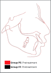
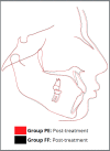
Methods: Forty six subjects fulfilling inclusion criteria were randomly distributed into Group PE (mean age 13.03±1.78 years) and Group FF (mean age 12.80±1.67 years) (n=23 each). Group PE was managed by therapeutic extraction of maxillary first premolars and mandibular second premolars, followed by mini-implant-supported space closure; and Group FF, by fixed functional appliance therapy. Skeletal, dental, and soft-tissue changes were analyzed using pre and post-treatment lateral cephalograms. Data obtained from this open label study was subjected to blind statistical analysis.
Results: Extraction treatment resulted in greater increase of nasolabial angle (NLA: 3.1 [95% CI 2.08, 4.19], p<0.001), significant improvement of upper lip (UL-E line: -2.91 [95% CI -3.54, -2.28], p<0.001, UL-S line: -2.50 [95% CI -2.76, -2.24], p<0.001, UL-SnPog': -2.32 [95% CI -2.90, -1.74], p<0.01) and lower lip position (LL-E line: -0.68 [95% CI -1.36, 0.00], p<0.01, LL-S line: -0.55 [95% CI -1.11, 0.02], p<0.01, and LL-SnPog': -0.64 [95% CI -1.20, -0.07], p<0.01), lip thickness (UL thickness: 2.27 [95% CI 1.79, 2.75], p<0.001; LL thickness: 0.41 [95% CI -0.16, 0.97], p<0.01), upper lip strain (UL strain: -2.68 [95% CI -3.32, -2.04], p<0.001) and soft tissue profile (N'-Sn-Pog': 2.68 [95% CI 1.87, 3.50], p<0.01). No significant difference was observed between the groups regarding skeletal changes in the maxilla and mandible, growth pattern, overjet, overbite, interincisal angle and soft tissue chin position (p>0.05). Premolar extraction treatment demonstrated significant intrusion-retraction of maxillary incisors, better maintenance of maxillary incisor inclination, and significant mandibular molar protraction; whereas functional treatment resulted in retrusive and intrusive effect on maxillary molars, marked proclination of mandibular anterior teeth, and significant extrusion of mandibular molars. Both treatment modalities had similar treatment duration. Implant failure was seen in 7.9% of cases, whereas failure of fixed functional appliance was observed in 9.09% of cases.
Conclusions: Premolar extraction therapy is a better treatment modality, compared to fixed functional appliance therapy for Class II patients with moderate skeletal discrepancy, increased overjet, protruded maxillary incisors and protruded lips, as it produces better dentoalveolar response and permits greater improvement of the soft tissue profile and lip relationship.
Objetivo:: O objetivo desse estudo randomizado controlado paralelo de dois braços foi avaliar os efeitos do tratamento e as mudanças no perfil labial em pacientes esqueléticos Classe II submetidos a tratamento com extração de pré-molares (EP) versus tratamento funcional fixo (FF).
Métodos:: Quarenta e seis indivíduos que preencheram os critérios de inclusão foram distribuídos aleatoriamente em Grupo EP (idade média 13,03±1,78 anos) e Grupo FF (idade média 12,80±1,67 anos) (n=23 cada). O grupo EP foi tratado com extração dos primeiros pré-molares superiores e segundos pré-molares inferiores, seguida de fechamento do espaço com ancoragem em mini-implantes; e o Grupo FF, com tratamento usando aparelhos funcionais fixos. As alterações esqueléticas, dentárias e de tecidos moles foram analisadas usando cefalogramas laterais pré e pós-tratamento. Os dados obtidos desse estudo aberto foram submetidos a análise estatística cega.
Resultados:: O tratamento com extrações resultou em maior aumento do ângulo nasolabial (ANL: 3,1 [IC 95% 2,08, 4,19], p<0,001), melhora significativa do lábio superior (Ls-Linha E: -2,91 [IC 95% -3,54, -2,28], p<0,001, Ls-Linha S: -2,50 [IC 95% -2,76, -2,24], p<0,001, Ls-SnPog’: -2,32 [IC 95% -2,90, -1,74], p<0,01) e posição do lábio inferior (Li-Linha E: -0,68 [IC 95% -1,36, 0,00], p<0,01, Li-Linha S: -0,55 [IC 95% -1,11, 0,02], p<0,01, e Li-SnPog’: -0,64 [IC 95% -1,20, -0,07], p<0,01), espessura dos lábios (espessura Ls: 2,27 [IC 95% 1,79, 2,75], p<0,001; espessura Li: 0,41 [IC 95% -0,16, 0,97], p<0,01), tensão do lábio superior (tensão Ls: -2,68 [IC 95% -3,32, -2,04], p<0,001) e perfil de tecidos moles (N’-Sn-Pog’: 2,68 [IC 95% 1,87, 3,50], p<0,01). Nenhuma diferença significativa foi observada entre os grupos quanto às alterações esqueléticas na maxila e mandíbula, padrão de crescimento, sobressaliência, sobremordida, ângulo interincisal e posição dos tecidos moles do mento (p>0,05). O tratamento com extração de pré-molares demonstrou significativa intrusão-retração dos incisivos superiores, melhor manutenção da inclinação dos incisivos superiores e protração significativa dos molares inferiores; enquanto o tratamento funcional resultou em efeito retrusivo e intrusivo nos molares superiores, proclinação acentuada dos dentes anteriores inferiores e extrusão significativa dos molares inferiores. Ambas as modalidades de tratamento tiveram duração de tratamento semelhante. A falha do mini-implante foi observada em 7,9% dos casos, enquanto a falha do aparelho funcional fixo foi observada em 9,09% dos casos.
Conclusões:: O tratamento com extração de pré-molares é uma modalidade de tratamento melhor do que os aparelhos funcionais fixos para pacientes Classe II com discrepância esquelética moderada, sobressaliência aumentada, incisivos superiores protruídos e lábios protruídos, pois produz melhor resposta dentoalveolar e permite maior melhora do perfil dos tecidos moles e relacionamento labial.
Conflict of interest statement
The authors report no commercial, proprietary or financial interest in the products or companies described in this article.
Figures


Similar articles
-
Treatment Effects and Lip Profile Changes Following Surgical Mandibular Advancement Versus Premolar Extractions in Class II Div 1 Malocclusion: A Randomized Controlled Trial.J Craniofac Surg. 2022 Jan-Feb 01;33(1):81-86. doi: 10.1097/SCS.0000000000007986. J Craniofac Surg. 2022. PMID: 34320575 Clinical Trial.
-
Mini-implants vs fixed functional appliances for treatment of young adult Class II female patients: a prospective clinical trial.Angle Orthod. 2012 Mar;82(2):294-303. doi: 10.2319/042811-302.1. Epub 2011 Aug 26. Angle Orthod. 2012. PMID: 21867432 Free PMC article. Clinical Trial.
-
Dentoskeletal morphology in adults with Class I, Class II Division 1, or Class II Division 2 malocclusion with increased overbite.Am J Orthod Dentofacial Orthop. 2019 Aug;156(2):248-256.e2. doi: 10.1016/j.ajodo.2019.03.006. Am J Orthod Dentofacial Orthop. 2019. PMID: 31375235
-
One phase or two phase orthodontic treatment for Class II division 1 malocclusion ?Evid Based Dent. 2019 Sep;20(3):72-73. doi: 10.1038/s41432-019-0049-y. Evid Based Dent. 2019. PMID: 31562403 Review.
-
The extraction of permanent second molars and its effect on the dentofacial complex of patients treated with the Tip-Edge appliance.Eur J Orthod. 2002 Oct;24(5):501-18. doi: 10.1093/ejo/24.5.501. Eur J Orthod. 2002. PMID: 12407946 Review.
References
-
- Proffit WR, Fields HW, Jr, Moray LJ. Prevalence of malocclusion and orthodontic treatment need in the United States estimates from the NHANES III survey. Int J Adult Orthodon Orthognath Surg. 1998;13(2):97–106. - PubMed
-
- O'Brien K, Macfarlane T, Wright J, Conboy F, Appelbe P, Birnie D, et al. Early treatment for Class II malocclusion and perceived improvements in facial profile. Am J Orthod Dentofacial Orthop. 2009;135(5):580–585. - PubMed
-
- Tadic N, Woods M. Contemporary Class II orthodontic and orthopaedic treatment a review. Aust Dent J. 2007;52(3):168–174. - PubMed
-
- Janson G, Mendes LM, Junqueira CH, Garib DG. Soft-tissue changes in Class II malocclusion patients treated with extractions a systematic review. Eur J Orthod. 2016;38(6):631–637. - PubMed
-
- Zierhut EC, Joondeph DR, Artun J, Little RM. Long-term profile changes associated with successfully treated extraction and nonextraction Class II Division 1 malocclusions. Angle Orthod. 2000;70(3):208–219. - PubMed
Publication types
MeSH terms
LinkOut - more resources
Full Text Sources

