Review
. 2023 May 19;47(3):fuad025.
doi: 10.1093/femsre/fuad025.

Cyclic-di-AMP signalling in lactic acid bacteria

Affiliations
Review

Cyclic-di-AMP signalling in lactic acid bacteria

Mark S Turner et al. FEMS Microbiol Rev. .

Abstract

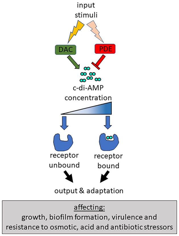
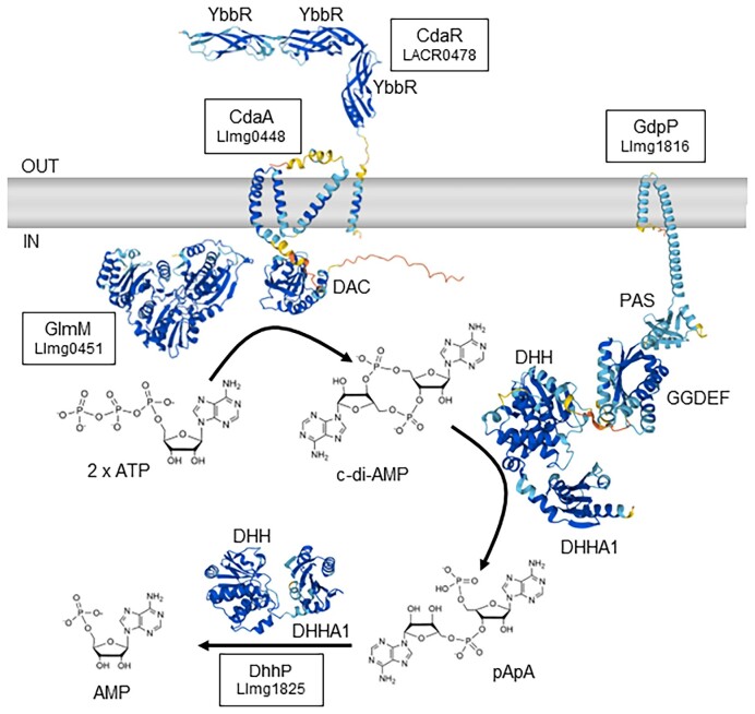
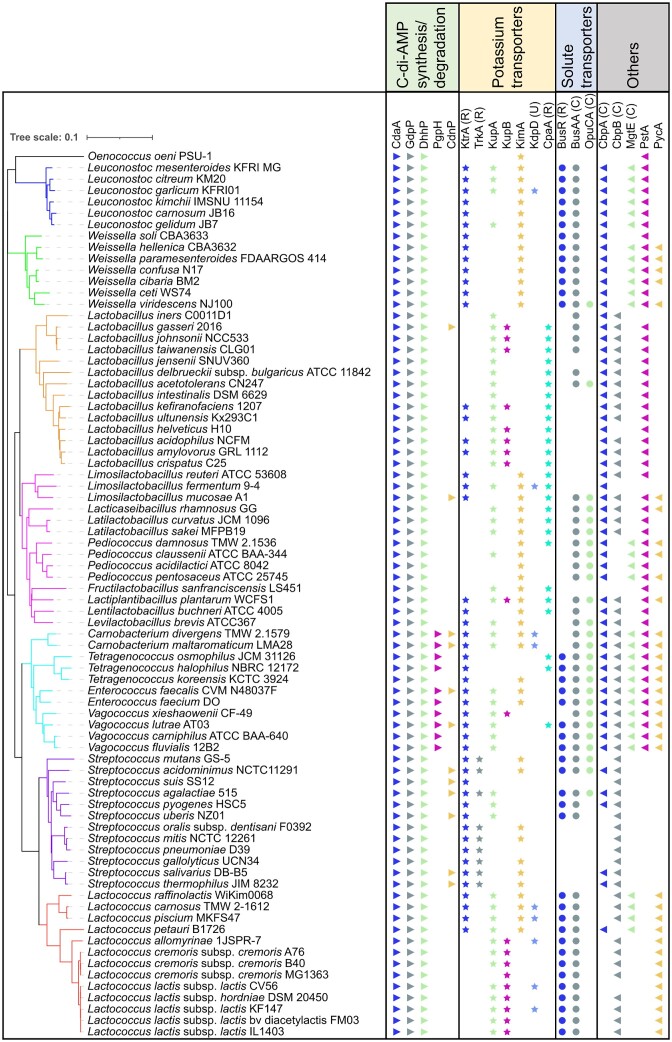
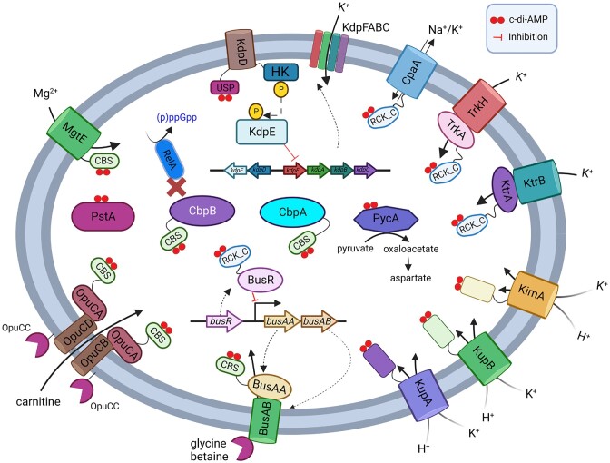
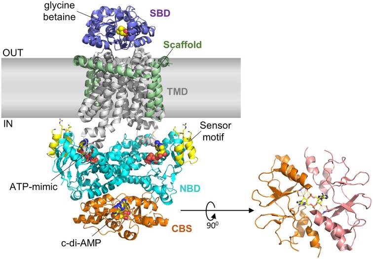
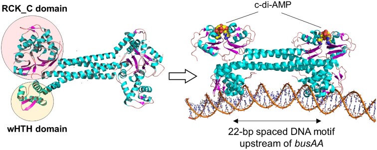
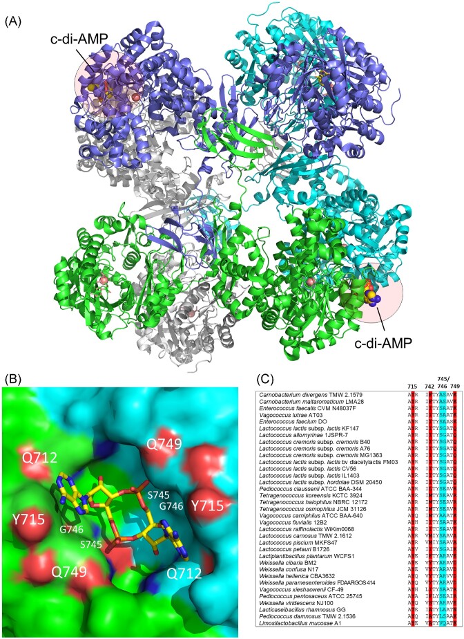
Cyclic dimeric adenosine monophosphate (cyclic-di-AMP) is a nucleotide second messenger present in Gram-positive bacteria, Gram-negative bacteria and some Archaea. The intracellular concentration of cyclic-di-AMP is adjusted in response to environmental and cellular cues, primarily through the activities of synthesis and degradation enzymes. It performs its role by binding to protein and riboswitch receptors, many of which contribute to osmoregulation. Imbalances in cyclic-di-AMP can lead to pleiotropic phenotypes, affecting aspects such as growth, biofilm formation, virulence, and resistance to osmotic, acid, and antibiotic stressors. This review focuses on cyclic-di-AMP signalling in lactic acid bacteria (LAB) incorporating recent experimental discoveries and presenting a genomic analysis of signalling components from a variety of LAB, including those found in food, and commensal, probiotic, and pathogenic species. All LAB possess enzymes for the synthesis and degradation of cyclic-di-AMP, but are highly variable with regards to the receptors they possess. Studies in Lactococcus and Streptococcus have revealed a conserved function for cyclic-di-AMP in inhibiting the transport of potassium and glycine betaine, either through direct binding to transporters or to a transcriptional regulator. Structural analysis of several cyclic-di-AMP receptors from LAB has also provided insights into how this nucleotide exerts its influence.

Keywords: compatible solutes; cyclic-di-AMP; lactic acid bacteria; osmotic stress; potassium.


Conflict of interest statement

None declared.

Figures

Figure 1.
Figure 1.
Overview of the c-di-AMP signalling system. The c-di-AMP synthesis and degradation enzymes respond to external or cellular stimuli resulting in changes in c-di-AMP levels. Upon reaching a certain intracellular concentration, c-di-AMP binds to receptors leading to activation or inhibition of activity and a subsequent cellular output and adaptation.
Figure 2.
Figure 2.
Synthesis and degradation of c-di-AMP with predicted structures of proteins. Uniprot accessions are from L. cremoris strains: A2RIF7 (CdaA; Llmg_0448), Q031P3 (CdaR; LACR_0478), A2RM60 (GdpP; Llmg_1816), A2RIG0 (GlmM; Llmg_0451), and A2RM69 (DhhP; Llmg_1825). AlphaFold predictions of protein tertiary structures and Rhea chemical structures are provided under a Creative Commons Attribution (CC BY 4.0) License (Jumper et al. , Bansal et al. , Varadi et al. 2022). Colour coding in protein structures indicates the confidence of the prediction by AlphaFold (dark blue = very high confidence; light blue = confident; yellow = low confidence; orange = very low confidence). Note that only monomers are shown for simplicity.
Figure 3.
Figure 3.
Presence and analysis of c-di-AMP signalling components in representative LAB. Representative taxa genomes of LAB were selected from the NCBI database. A phylogenetic tree was constructed using FastME 2.1.6.1 (Lefort et al. 2015), which was based on the Genome BLAST Distance Phylogeny (GBDP) distances. These distances were calculated from 16S rRNA gene sequences utilizing the TYGS platform (Meier-Kolthoff and Goker 2019). The iTOL software (Letunic and Bork 2021) was used to visualize the phylogenetic tree and receptors. For each strain, we searched for homologs of c-di-AMP synthesis and degradation enzymes, and their receptors using the custom BLAST function in Geneious Prime software version 2022.1.1 (Biomatters Ltd, New Zealand). This search was compared with published c-di-AMP receptors that originated from either LAB or other c-di-AMP-producing bacteria. Specific c-di-AMP binding domains are denoted as follows: RCK_C domain (R), CBS domain (C), and USP domain (U).
Figure 4.
Figure 4.
Overview of c-di-AMP receptors found in LAB with binding domains and functions indicated. Created with BioRender.com.
Figure 5.
Figure 5.
Structure of the L. lactis BusA (OpuA) glycine betaine transporter complex obtained by cryo-EM studies. Shown here is the c-di-AMP-inhibited conformation of the ATPase BusAA with c-di-AMP bound at the interface of the tandem CBS domains and the ATP mimic AMP–PNP bound in the NBD domain (Protein Data Bank 7AHH) (Sikkema et al. 2020). In BusAB, glycine betaine bound in the solute-binding protein (SBD), which is fused to the TMD. Highlighted here are also the stabilizing scaffold motif located on the periphery of the TMD of BusAB and the ionic strength sensor motif in BusAA with the functionally important Lys16, Arg17, and Lys19 shown in stick representation.
Figure 6.
Figure 6.
Binding of c-di-AMP promotes BusR-DNA interaction based on the structural studies of S. agalactiae BusR. The binding of c-di-AMP to the RCK_C domain of the transcriptional repressor BusR induces a conformational change to release the wHTH domain into a protruding and DNA-interacting position. The c-di-AMP bound BusR subsequently binds to a 22-bp sequence in the promoter region of busAA to repress the transcription of the busAA-AB genes and hence expression levels of the glycine betaine transporter (Protein Data Bank 7B5Y; Bandera et al. 2021).
Figure 7.
Figure 7.
Structure of c-di-AMP bound PC from L. lactis (A). The c-di-AMP binding pocket (B) and an alignment of key residues in the c-di-AMP binding pocket of PC in LAB (blue highlighted amino acids should have small side chains in order to not block the c-di-AMP pocket) (C). In the close-up view of the c-di-AMP binding site at the dimeric interface (B), the three key residues (Q712, Y715, and Q749) that are directly involved in c-di-AMP binding are highlighted. The two residues with short side chains (S745, G746) located deeper in the pocket and permit c-di-AMP binding are also highlighted (Protein Data Bank 5VYZ; Choi et al. 2017).

Similar articles

Cited by

References

    1. Andrade WA, Firon A, Schmidt Tet al. . Group B Streptococcus degrades cyclic-di-AMP to modulate STING-dependent type I interferon production. Cell Host Microbe. 2016;20:49–59. - PMC - PubMed
    1. Bai Y, Yang J, Eisele LEet al. . Two DHH subfamily 1 proteins in Streptococcus pneumoniae possess cyclic di-AMP phosphodiesterase activity and affect bacterial growth and virulence. J Bacteriol. 2013;195:5123–32. - PMC - PubMed
    1. Bai Y, Yang J, Zarrella TMet al. . Cyclic di-AMP impairs potassium uptake mediated by a cyclic di-AMP binding protein in Streptococcus pneumoniae. J Bacteriol. 2014;196:614–23. - PMC - PubMed
    1. Bandera AM, Bartho J, Lammens Ket al. . BusR senses bipartite DNA binding motifs by a unique molecular ruler architecture. Nucleic Acids Res. 2021;49:10166–77. - PMC - PubMed
    1. Bansal P, Morgat A, Axelsen KBet al. . Rhea, the reaction knowledgebase in 2022. Nucleic Acids Res. 2022;50:D693–700. - PMC - PubMed

Publication types

MeSH terms