. 2023 Jun 15;11(3):e0536122.
doi: 10.1128/spectrum.05361-22. Epub 2023 May 24.

A C. albicans TRAPP Complex-Associated Gene Contributes to Cell Wall Integrity, Hyphal and Biofilm Formation, and Tissue Invasion

Affiliations

A C. albicans TRAPP Complex-Associated Gene Contributes to Cell Wall Integrity, Hyphal and Biofilm Formation, and Tissue Invasion

Dakota Ma et al. Microbiol Spectr. .

Abstract

While endocytic and secretory pathways are well-studied cellular processes in the model yeast Saccharomyces cerevisiae, they remain understudied in the opportunistic fungal pathogen Candida albicans. We previously found that null mutants of C. albicans homologs of the S. cerevisiae early endocytosis genes ENT2 and END3 not only exhibited delayed endocytosis but also had defects in cell wall integrity, filamentation, biofilm formation, extracellular protease activity, and tissue invasion in an in vitro model. In this study, we focused on a potential C. albicans homolog to S. cerevisiae TCA17, which was discovered in our whole-genome bioinformatics approach aimed at identifying genes involved in endocytosis. In S. cerevisiae, TCA17 encodes a transport protein particle (TRAPP) complex-associated protein. Using a reverse genetics approach with CRISPR-Cas9-mediated gene deletion, we analyzed the function of the TCA17 homolog in C. albicans. Although the C. albicans tca17Δ/Δ null mutant did not have defects in endocytosis, it displayed an enlarged cell and vacuole morphology, impaired filamentation, and reduced biofilm formation. Moreover, the mutant exhibited altered sensitivity to cell wall stressors and antifungal agents. When assayed using an in vitro keratinocyte infection model, virulence properties were also diminished. Our findings indicate that C. albicans TCA17 may be involved in secretion-related vesicle transport and plays a role in cell wall and vacuolar integrity, hyphal and biofilm formation, and virulence. IMPORTANCE The fungal pathogen Candida albicans causes serious opportunistic infections in immunocompromised patients and has become a major cause of hospital-acquired bloodstream infections, catheter-associated infections, and invasive disease. However, due to a limited understanding of Candida molecular pathogenesis, clinical approaches for the prevention, diagnosis, and treatment of invasive candidiasis need significant improvement. In this study, we focus on identifying and characterizing a gene potentially involved in the C. albicans secretory pathway, as intracellular transport is critical for C. albicans virulence. We specifically investigated the role of this gene in filamentation, biofilm formation, and tissue invasion. Ultimately, these findings advance our current understanding of C. albicans biology and may have implications for the diagnosis and treatment of candidiasis.

Keywords: Candida albicans; TRAPP complex; biofilm; filamentation; pathogenesis; secretion; trafficking; virulence.


Conflict of interest statement

The authors declare no conflict of interest.

Figures
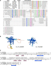
FIG 1
FIG 1
Alignment of protein sequences between C. albicans C4_06460C and homologs in other organisms. (A) Protein alignment between C. albicans C4_06460C, S. cerevisiae YEL048C (TCA17), and homologs in seven species of Candida. Regions of conservation are identified, specifically between residues 26 and 42 and residues 74 and 90 of the C. albicans product. Several key conserved residues are illustrated within boxes, including prolines (P27 and P36; orange), lysines (K75, K138, and K173; blue), and an asparagine-proline motif (NP; purple). (B) AlphaFold models of the putative protein structure of C. albicans C4_06460C and S. cerevisiae YEL048C. Notably, the program identified in both proteins three alpha-helical regions and five beta-sheet stretches with “high confidence” (dark blue color), which are found in a similar tertiary arrangement. In addition, C. albicans has loops that less rigorously conform to helical algorithms (light blue) as well as regions that are unstructured (orange and yellow). (C) The protein alignment extends the homology to human TRAPP2CL. The conserved residues are boxed as above. The regions of highest amino acid homology coincide with the secondary structures identified by AlphaFold in all three homologs. The boxes below the alignments delimit those structures as found in C. albicans.
FIG 2
FIG 2
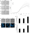
The C. albicans tca17Δ/Δ null mutant (KO) shows impaired growth and cell morphology. (A) Growth on yeast extract peptone dextrose (YPD) plates at 30°C, 37°C, and 42°C. (B) Growth curve in liquid YNB at 30°C. OD600 values were recorded every 30 min for 16 h. Experiments were conducted in triplicate, with three replicates per strain. Error bars indicate the 95% confidence interval of OD600 values at each time point for each strain. (C) Yeast cell morphology was visualized under differential interference contrast (DIC) and fluorescence microscopy with Calcofluor white (CW) staining. Scale bar, 10 μm. (D) Comparison of cell length between wild-type (WT), knock-in (KI), and KO strains. Statistical significance was determined with a Student’s t test (WT/KO, P < 0.0001; KI/KO, P < 0.0001; WT/KI, P = 0.14). The length of the KO cell is significantly (***) longer than the length of the WT and KI cells. (E) Comparison of cell area between WT, KI, and KO strains. Statistical significance was determined with a Student’s t test (WT/KO, P < 0.0001; KI/KO, P < 0.0001; WT/KI, P = 0.96).
FIG 3
FIG 3
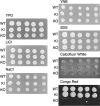
The C. albicans tca17Δ/Δ mutant strain has a decreased tolerance to the cell wall stressor Congo red. The tca17Δ/Δ null mutant (KO), wild-type (WT), and knock-in (KI) strains were grown for 48 h on YPD plates under cationic cell stress conditions (500 mM NaCl or 100 mM LiCl) and on YNB plates containing cell wall stressors SDS (0.05%), Calcofluor white (50 μg/mL), and Congo red (140 μg/mL). The images shown are representative of three different experiments.
FIG 4
FIG 4
The C. albicans tca17Δ/Δ mutant strain (KO) has increased sensitivity to the antifungal drug caspofungin. The tca17Δ/Δ mutant (KO), wild-type (WT), and knock-in (KI) strains were grown for 48 h on YPD plates containing antifungal drugs at various concentrations as follows: amphotericin B at 0.11 μg/mL, 0.33 μg/mL, and 1.0 μg/mL (A), caspofungin at 0.025 μg/mL, 0.05 μg/mL, and 0.1 μg/mL (B), and fluconazole at 1.0 μg/mL, 2.0 μg/mL, and 4.0 μg/mL (C). The images shown in A to C are representative of three different experiments.
FIG 5
FIG 5
Membrane-related endocytosis is intact in the C. albicans tca17Δ/Δ mutant (KO) compared to the wild-type (WT) and knock-in (KI) strains. Membrane-related endocytosis was tracked over time using the lipophilic dye FM4-64 (red) and visualized under DIC and fluorescence microscopy. The images shown are representative of three different experiments. Scale bar, 10 μm.
FIG 6
FIG 6
The C. albicans tca17Δ/Δ mutant strain is defective in filamentation and biofilm formation. (A) Agar plate assays of C. albicans hyphae formation. Wild-type (WT), knock-in (KI), and knockout (KO) strains were spotted on filamentation medium (RPMI 1640, M199, FCS, and Spider) and incubated at 37°C for 72 h. (B) Filaments visualized under differential interference contrast (DIC) and fluorescence microscopy with Calcofluor white (CW) staining. Filamentation was induced by growing the strains in liquid RPMI 1640 overnight at 37°C. The images shown are representative of three different experiments. Scale bar, 10 μm. (C) Biofilm metabolic activity relative to the WT using an XTT reduction assay. The activity of WT biofilms was arbitrarily set as 100%. Error bars indicate standard deviation. Each individual data point was run in triplicate and averaged. Statistical significance was determined with a Student’s t test (WT/KO, P < 0.0001; KI/KO, P < 0.0001; WT/KI, P = 0.07). (D) Visualization of biofilm growth for 48 h on a plastic surface in RPMI 1640 medium under a DIC filter. Scale bar, 50 μm.
FIG 7
FIG 7
The dissolution of cell-cell adhesions in human VK-2 cells is impaired in the presence of the C. albicans tca17Δ/Δ null mutant (KO). (A) Human VK-2 cells were infected with wild-type (WT), knock-in (KI), and knockout (KO) strains and incubated for 6 and 24 h. E-cadherin was fluorescently labeled using an Alexa Fluor 488-tagged antibody. DAPI dye was used to label the nucleus. Scale bar, 10 μm. (B) Western blotting of E-cadherin in C. albicans-infected VK-2 cells. Tubulin is used as a loading control. (C) The C. albicans strains were further tested in a protease agar plate assay for their ability to lyse BSA after overnight incubation at 30°C. The images shown in A to C are representative of three different experiments.
FIG 8
FIG 8
The C. albicans tca17Δ/Δ mutant has a reduced ability to kill human VK-2 cells in vitro. (A) Proportion of live (white bars) and dead (black bars) VK-2 cells 6 h after C. albicans infection. (B) Proportion of live (white bars) and dead (black bars) VK-2 cells 24 h after C. albicans infection. Error bars in A and B are the standard deviations of three different data sets. Each individual data point was run in triplicate and averaged. Statistical significance between the cells infected with the wild-type (WT), the complemented knock-in (KI), and the knockout (KO) strains was determined by Student’s t test; (*, P < 0.05).

References

    1. Wächtler B, Citiulo F, Jablonowski N, Förster S, Dalle F, Schaller M, Wilson D, Hube B. 2012. Candida albicans-epithelial interactions: dissecting the roles of active penetration, induced endocytosis and host factors on the infection process. PLoS One 7:e36952. doi:10.1371/journal.pone.0036952. - DOI - PMC - PubMed
    1. Schaller M, Korting HC, Schäfer W, Bastert J, Chen W, Hube B. 1999. Secreted aspartic proteinase (Sap) activity contributes to tissue damage in a model of human oral candidosis. Mol Microbiol 34:169–180. doi:10.1046/j.1365-2958.1999.01590.x. - DOI - PubMed
    1. Ibrahim AS, Filler SG, Sanglard D, Edwards JE, Hube B. 1998. Secreted aspartyl proteinases and interactions of Candida albicans with human endothelial cells. Infect Immun 66:3003–3005. doi:10.1128/IAI.66.6.3003-3005.1998. - DOI - PMC - PubMed
    1. Navarro-García F, Sánchez M, Nombela C, Pla J. 2001. Virulence genes in the pathogenic yeast Candida albicans. FEMS Microbiol Rev 25:245–268. doi:10.1111/j.1574-6976.2001.tb00577.x. - DOI - PubMed
    1. Braun BR, Head WS, Wang MX, Johnson AD. 2000. Identification and characterization of TUP1-regulated genes in Candida albicans. Genetics 156:31–44. doi:10.1093/genetics/156.1.31. - DOI - PMC - PubMed

Publication types

Substances

LinkOut - more resources