. 2023 Jun 28;89(6):e0210322.
doi: 10.1128/aem.02103-22. Epub 2023 May 24.

PBP2b Mutations Improve the Growth of Phage-Resistant Lactococcus cremoris Lacking Polysaccharide Pellicle

Affiliations

PBP2b Mutations Improve the Growth of Phage-Resistant Lactococcus cremoris Lacking Polysaccharide Pellicle

Hugo Guérin et al. Appl Environ Microbiol. .

Abstract

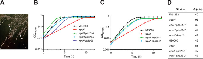
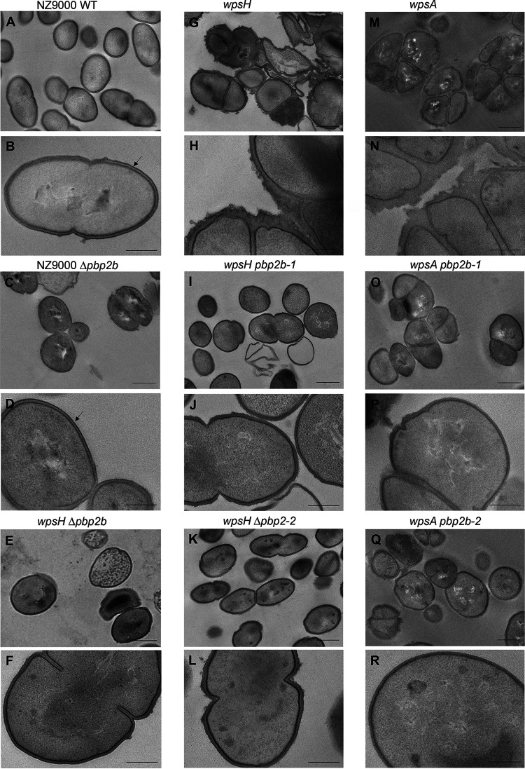
Lactococcus lactis and Lactococcus cremoris are Gram-positive lactic acid bacteria widely used as starter in milk fermentations. Lactococcal cells are covered with a polysaccharide pellicle (PSP) that was previously shown to act as the receptor for numerous bacteriophages of the Caudoviricetes class. Thus, mutant strains lacking PSP are phage resistant. However, because PSP is a key cell wall component, PSP-negative mutants exhibit dramatic alterations of cell shape and severe growth defects, which limit their technological value. In the present study, we isolated spontaneous mutants with improved growth, from L. cremoris PSP-negative mutants. These mutants grow at rates similar to the wild-type strain, and based on transmission electron microscopy analysis, they exhibit improved cell morphology compared to their parental PSP-negative mutants. In addition, the selected mutants maintain their phage resistance. Whole-genome sequencing of several such mutants showed that they carried a mutation in pbp2b, a gene encoding a penicillin-binding protein involved in peptidoglycan biosynthesis. Our results indicate that lowering or turning off PBP2b activity suppresses the requirement for PSP and ameliorates substantially bacterial fitness and morphology. IMPORTANCE Lactococcus lactis and Lactococcus cremoris are widely used in the dairy industry as a starter culture. As such, they are consistently challenged by bacteriophage infections which may result in reduced or failed milk acidification with associated economic losses. Bacteriophage infection starts with the recognition of a receptor at the cell surface, which was shown to be a cell wall polysaccharide (the polysaccharide pellicle [PSP]) for the majority of lactococcal phages. Lactococcal mutants devoid of PSP exhibit phage resistance but also reduced fitness, since their morphology and division are severely impaired. Here, we isolated spontaneous, food-grade non-PSP-producing L. cremoris mutants resistant to bacteriophage infection with a restored fitness. This study provides an approach to isolate non-GMO phage-resistant L. cremoris and L. lactis strains, which can be applied to strains with technological functionalities. Also, our results highlight for the first time the link between peptidoglycan and cell wall polysaccharide biosynthesis.

Keywords: Lactococcus; bacteriophage resistance; cell wall; lactic acid bacteria; penicillin-binding proteins; polysaccharide pellicle.


Conflict of interest statement

The authors declare no conflict of interest.

Figures

FIG 1
FIG 1
(A) Isolation of suppressor mutants from a L. cremoris PSP-negative strain on solid GM17 medium. Larger colonies (indicated by white arrows) arise spontaneously among the small colonies formed by VES5748 (wpsH) PSP-negative strain. (B and C) Growth in liquid GM17 medium of MG1363, wpsH mutant, and its pbp2b derivatives (B) and of NZ9000, wpsA mutant, and its pbp2b derivatives (C). Each point represents the mean value of three biological replicates. The standard deviations for each value were very low and could not be clearly represented on the corresponding curves. (D) Generation time (G) determined in the exponential growth phase for each strain.
FIG 2
FIG 2
3D homology model of PBP2b. The pedestal domain is shown in teal, and transpeptidase domain is shown in blue. Amino acid substitutions found in the four wpsH pbp2b mutants are displayed as orange spheres. The catalytic amino acid residues are highlighted as yellow spheres. Inserted at the upper right is a closeup view of the G292V mutation and its interaction sphere, with a highlight on the salt bridge between D276 and K298 and on the interaction between K298 and P524. Inserted at the lower right is a closer view of the accessible surface of the active site that contains the three other substitutions.
FIG 3
FIG 3
SEC-HPLC analysis of CWPS extracted from L. cremoris NZ9000, wpsH and wpsA mutants, and their respective pbp2b mutant derivatives. Peaks containing rhamnan and PSP oligosaccharides are indicated. *, Nonpolysaccharidic compounds.
FIG 4
FIG 4
TEM images of L. cremoris NZ9000, PSP-negative mutants, and their pbp2b derivatives. Black arrows indicate the outer electron dense layer attributed to PSP. Scale bars, 500 nm (A, C, E, G, I, K, M, O, and Q) and 200 nm (B, D, F, H, J, L, N, P, and R). (A and B) NZ9000 WT strain, showing typical ovoid shape, septation at midcell and PSP layer at the cell surface. (C and D) NZ9000 Δpbp2b (VES7552). (E and F) wpsH Δpbp2b (VES7552). (G and H) wpsH (VES5748). (I and J) wpsH pbp2b-1 (VES7484). (K and L) wpsH pbp2b-2 (VES7485). (M and N) wpsA (VES7810). (O and P) wpsA pbp2b-1 (VES7806). (Q and R) wpsA pbp2b-2 (VES7808).
FIG 5
FIG 5
Cell length to width ratios measured on SEM images of each indicated strain. A whiskers plot and string chart are shown. Each dot indicates a measured cell.
FIG 6
FIG 6
PG analysis of L. cremoris NZ9000, wpsH and wpsA mutants, and their respective pbp2b derivatives. (A) Representative muropeptide profiles obtained after PG digestion with mutanolysin and separation by RP-UHPLC for NZ9000 and wpsH pbp2b-2 strains. Identification of peaks containing monomers, dimers, or oligomers is based on mass spectrometry analysis and previous muropeptide identification (39). Monomers correspond to disaccharide peptides, dimers to two cross-linked monomers, and oligomers to three or more cross-linked monomers. (B) Relative abundances (%) of different muropeptide species for NZ9000, the wpsH mutant and its pbp2b derivatives (upper panel), and the wpsA mutant and its pbp2b derivatives (lower panel). Each value is the mean of three biological replicates. Errors bars represent ± the standard deviations of the mean. Abundance of each muropeptide species in mutant strains was compared to that of WT using a two-way analysis of variance, followed by Dunnett’s multiple comparison with a 95% confidence interval. The P values of significant differences are indicated by asterisks (*, P < 0.05; **, P < 0.01).

Similar articles

Cited by

References

    1. Ortiz Charneco G, de Waal PP, van Rijswijck IMH, van Peij N, van Sinderen D, Mahony J. 2023. Bacteriophages in the dairy industry: a problem solved? Annu Rev Food Sci Technol 14:367–385. doi:10.1146/annurev-food-060721-015928. - DOI - PubMed
    1. Garneau JE, Moineau S. 2011. Bacteriophages of lactic acid bacteria and their impact on milk fermentations. Microb Cell Fact 10(Suppl 1):S20. doi:10.1186/1475-2859-10-S1-S20. - DOI - PMC - PubMed
    1. Deveau H, Labrie SJ, Chopin MC, Moineau S. 2006. Biodiversity and classification of lactococcal phages. Appl Environ Microbiol 72:4338–4346. doi:10.1128/AEM.02517-05. - DOI - PMC - PubMed
    1. Spinelli S, Veesler D, Bebeacua C, Cambillau C. 2014. Structures and host-adhesion mechanisms of lactococcal siphophages. Front Microbiol 5:3. doi:10.3389/fmicb.2014.00003. - DOI - PMC - PubMed
    1. Mahony J, Cambillau C, van Sinderen D. 2017. Host recognition by lactic acid bacterial phages. FEMS Microbiol Rev 41:S16–S26. doi:10.1093/femsre/fux019. - DOI - PubMed

Supplementary concepts