

Flatfoot over the centuries: the background of current conservative and operative treatments
- PMID: 37222816
- PMCID: PMC10439239
- DOI: 10.1007/s00264-023-05837-3
Flatfoot over the centuries: the background of current conservative and operative treatments
Abstract
Purpose: Although flatfoot is a widespread human condition, historical medical texts and ancient illustrations on this deformity are extremely rare. Nowadays, doubts regarding its management remain unsolved. This historical review aims to identify the presence of pes planus since the prehistoric era and examine the treatments proposed over the centuries up to the present.
Method: For this propose, we performed an extensive electronic search of the relevant literature, complemented by a manual search of additional sources from archaeological to artistic, literary, historical, and scientific accounts, describing flatfoot and its treatment in different eras.
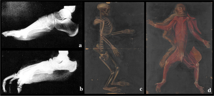
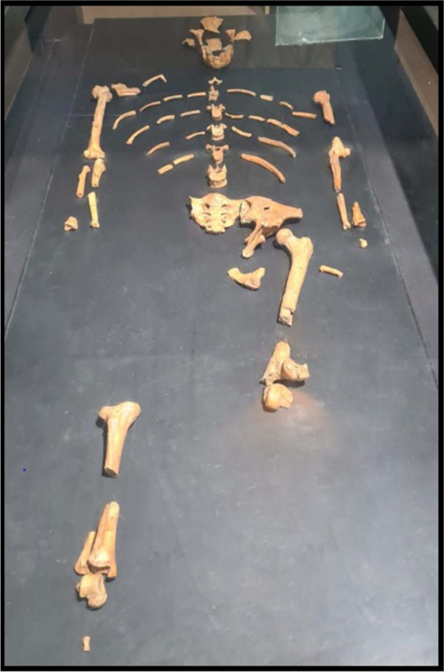
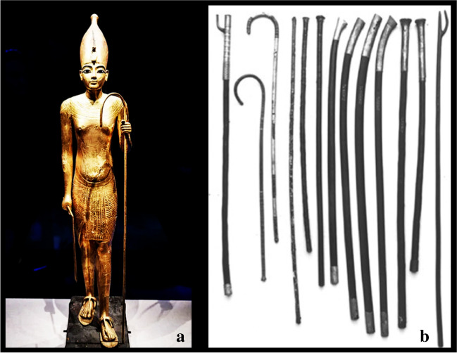
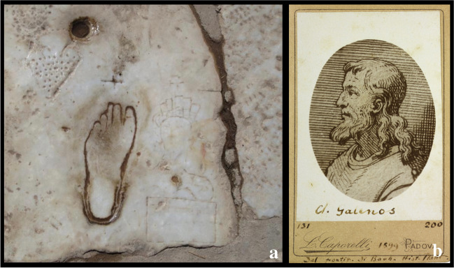
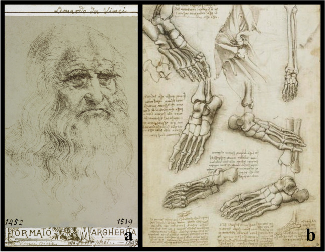
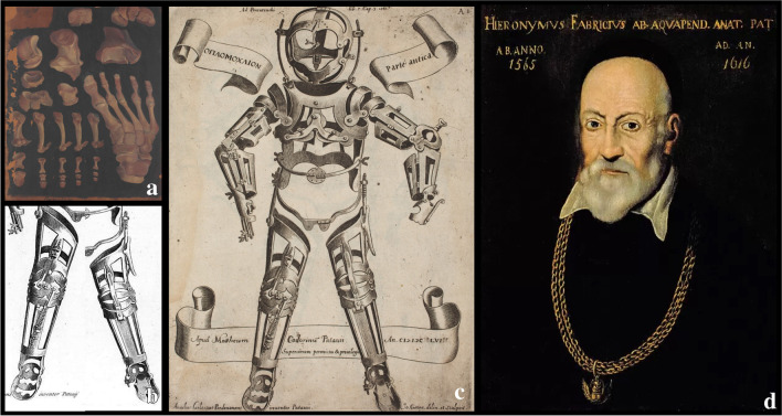
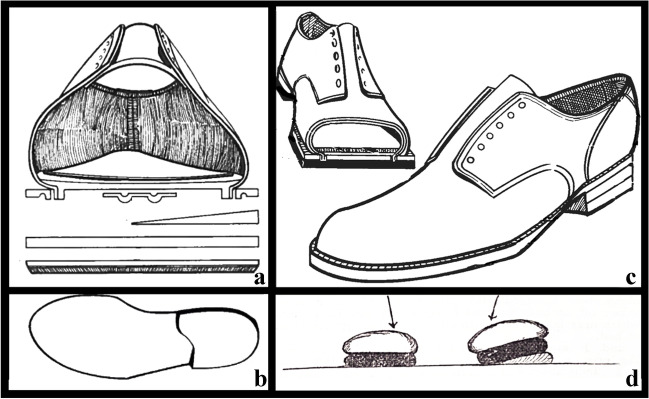
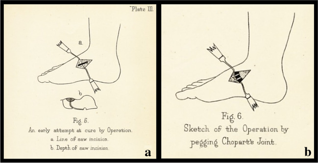
Results: Flatfoot accompanied the evolutionary timeline of human species: from Lucy Australopithecus to Homo Sapiens. It was described among various diseases suffered by Tutankhamun (1343-1324 B.C.), while the first anatomical description dates to Emperor Trajan (53-117 A.D.) and the medical studies of Galen (129-201 A.D.). It was also represented in the anatomical drawings of Leonardo da Vinci (1452-1519) and Girolamo Fabrici d'Acquapendente (1533-1619). Historically, the conservative treatment by insoles was the only one proposed until the nineteenth century. Since then, the most popular surgical procedures performed for correction have been osteotomies, arthrodesis, arthrorisis, and tendon lengthening and transfer.
Conclusion: During the centuries, conservative therapeutic strategies have not radically changed in their substance, while operative ones have become the protagonists during the twentieth century up to the present. Nevertheless, after more than 2000 years of history, there is no consensus regarding the best indication for the flatfoot and if it really needs to be treated.
Keywords: Arthrodesis, Arthrorisis; Flatfoot; Foot deformities; Insoles; Osteotomies; Pes planus; Tendon lengthening; Tendon transfer.
© 2023. The Author(s).
Conflict of interest statement
The authors declare no competing interests.
Figures
References
-
- Krogman WM (1951) The scars of human evolution. Sci Am 185(6): 54–57. JSTOR. http://www.jstor.org/stable/24950554. Accessed 2 Sept 2022
-
- Helfet AJ (1980) Disorders of the foot. Lippincott Williams & Wilkins, Philadelphia
-
- Standring S (2017) Anatomia Del Gray 41 Ed.: 2 Volumi. Edra
-
- Aiello L, Dean C (2002) Chapter twenty-three - the hominoid foot, Editor: Leslie Aiello, Christopher Dean, An Introduction to Human Evolutionary Anatomy, Academic Press, Pages 507–538, ISBN 9780120455911. 10.1016/B978-0-08-057100-3.50027-4
Publication types
MeSH terms
LinkOut - more resources
Full Text Sources
