

Microglia proliferation underlies synaptic dysfunction in the prefrontal cortex: implications for the pathogenesis of HIV-1-associated neurocognitive and affective alterations
- PMID: 37222970
- PMCID: PMC10629500
- DOI: 10.1007/s13365-023-01147-x
Microglia proliferation underlies synaptic dysfunction in the prefrontal cortex: implications for the pathogenesis of HIV-1-associated neurocognitive and affective alterations
Abstract
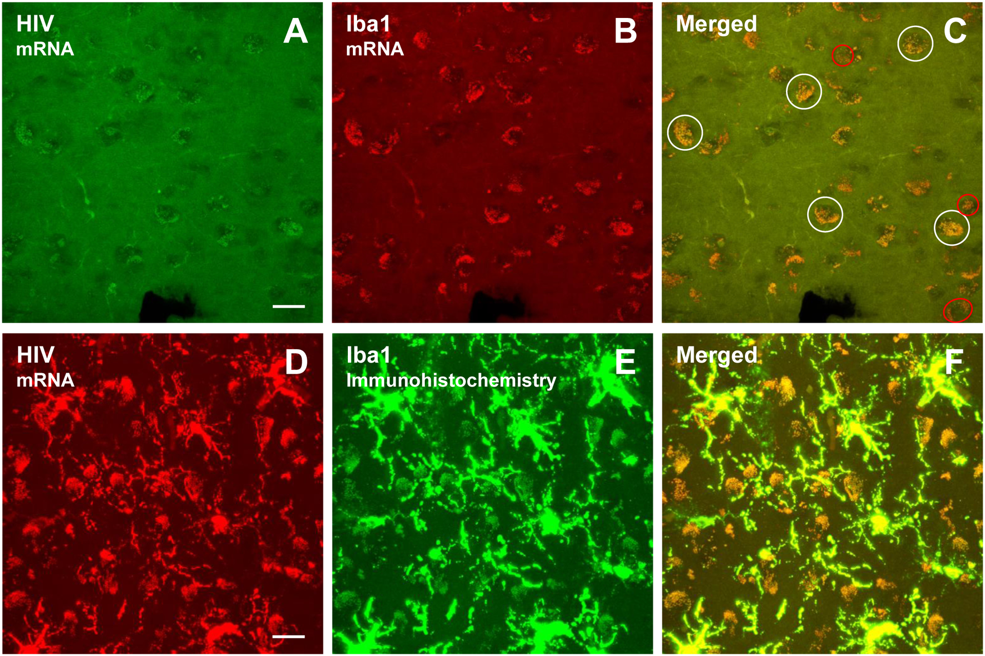
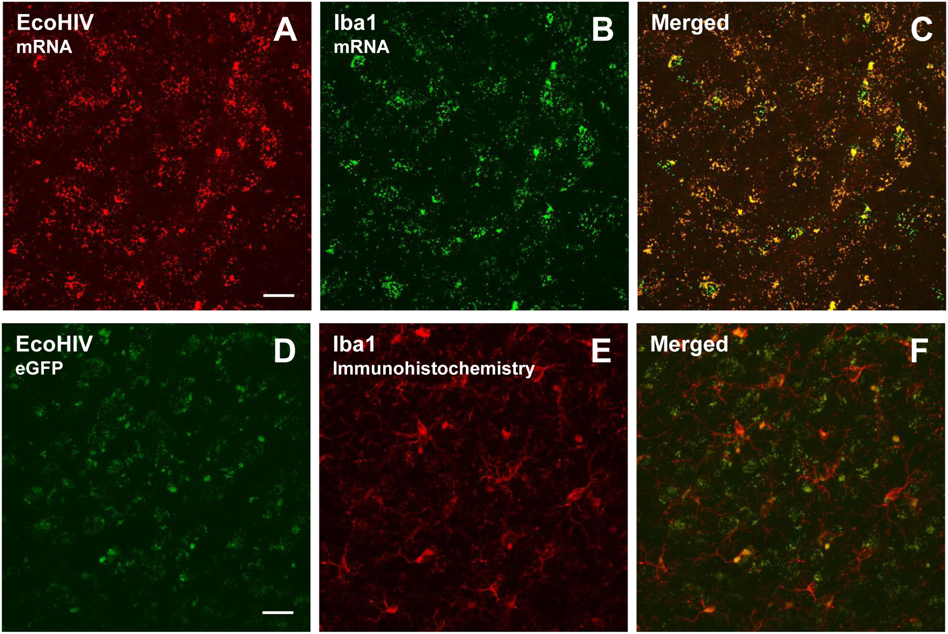
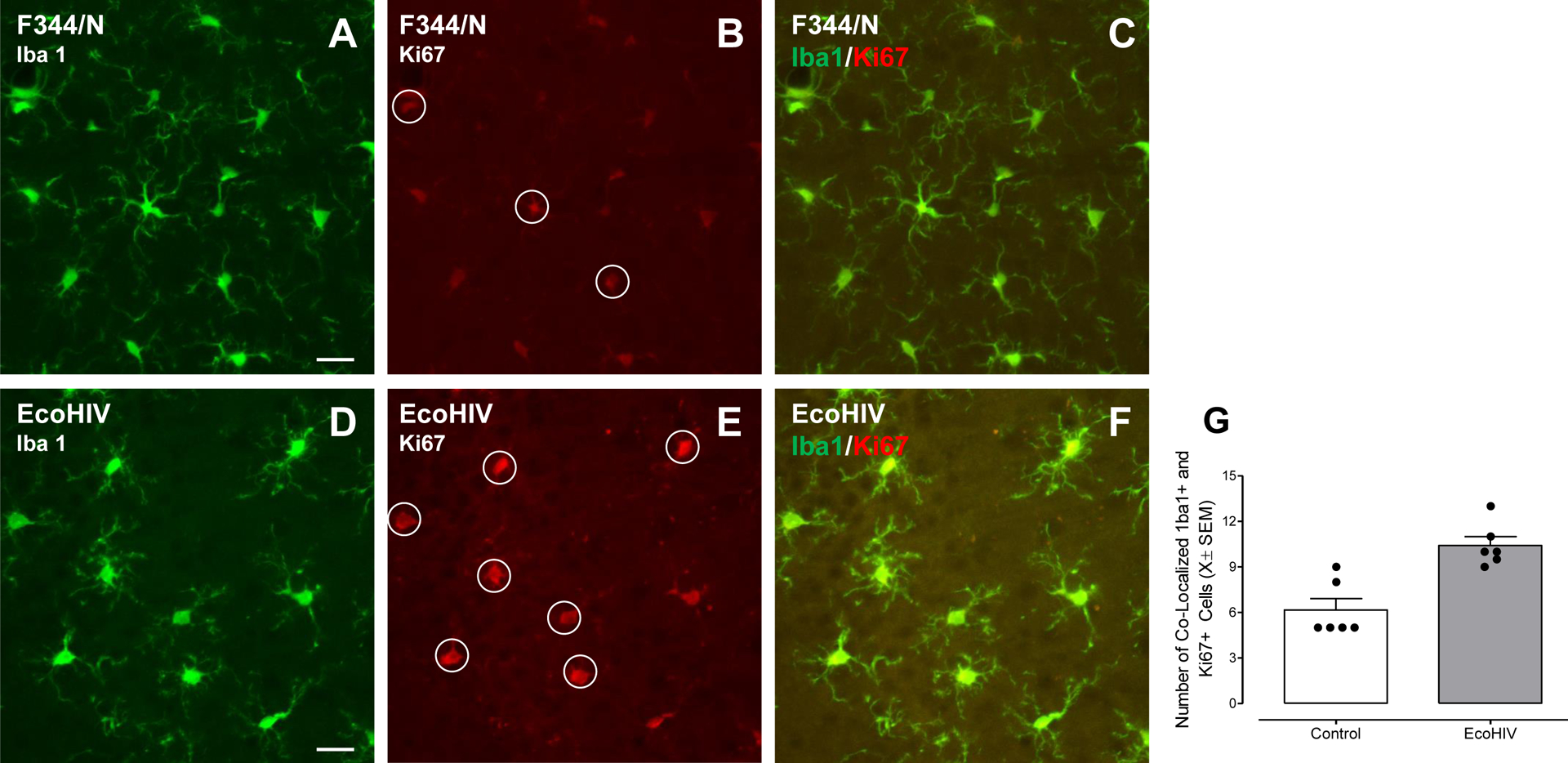
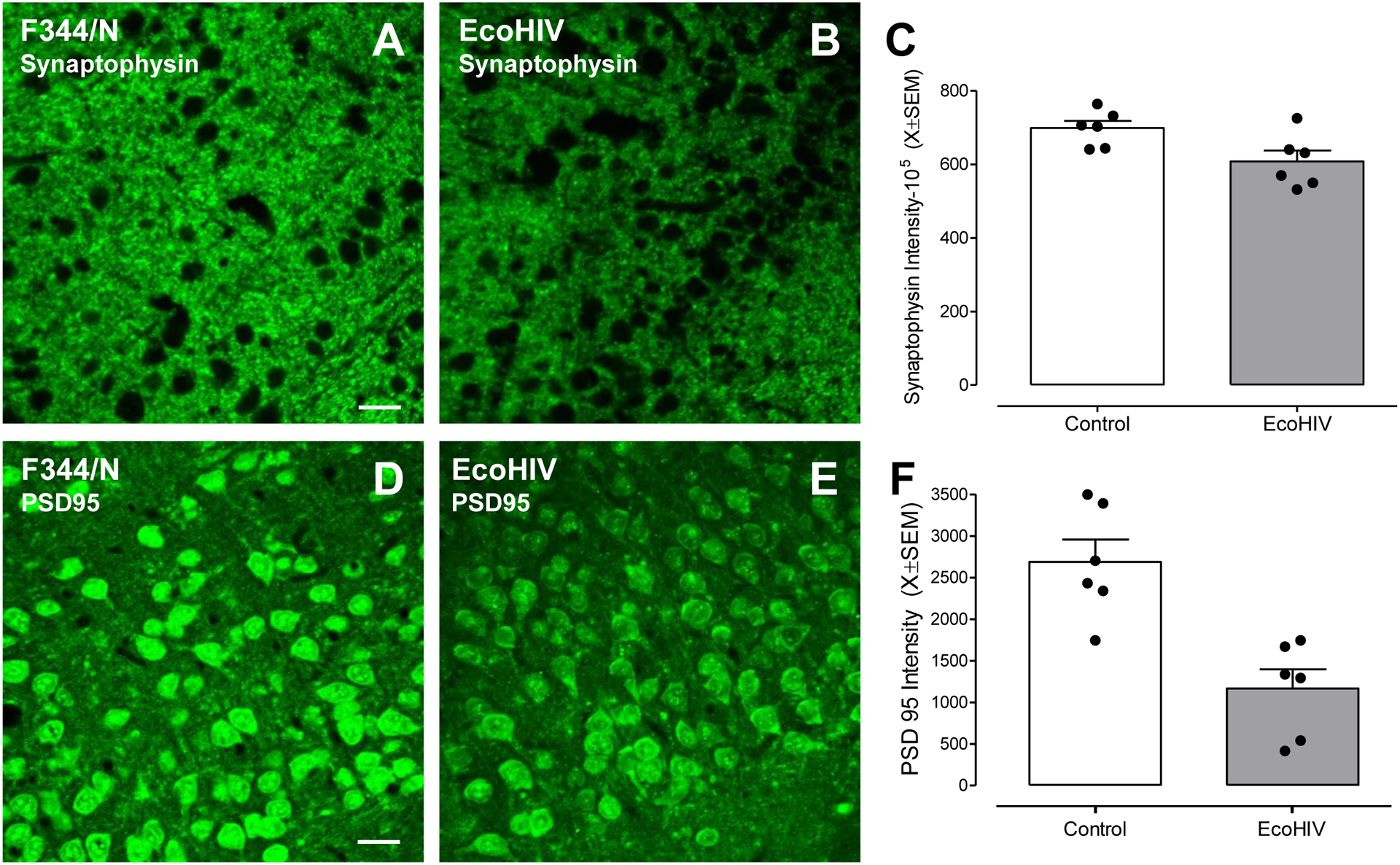
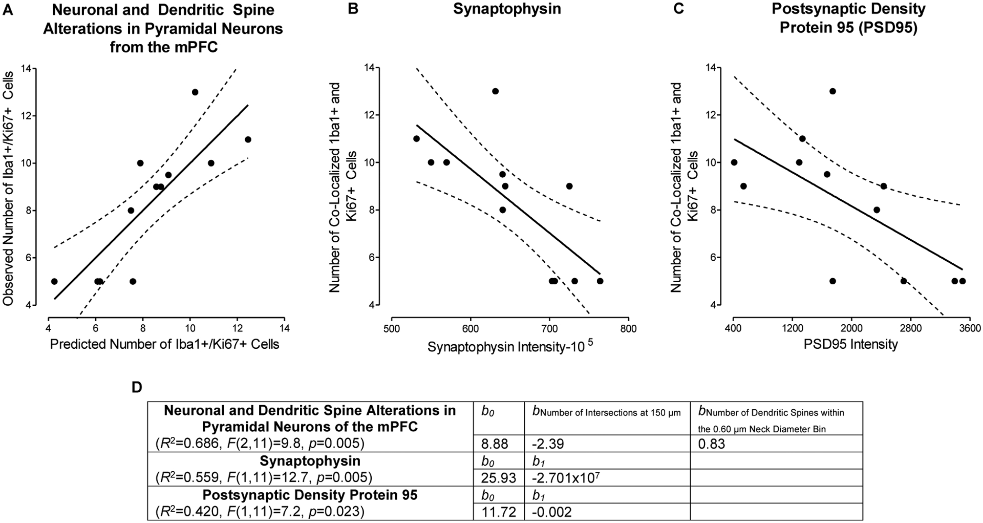
Microglia, which are productively infected by HIV-1, are critical for brain development and maturation, as well as synaptic plasticity. The pathophysiology of HIV-infected microglia and their role in the pathogenesis of HIV-1-associated neurocognitive and affective alterations, however, remains understudied. Three complementary aims were undertaken to critically address this knowledge gap. First, the expression of HIV-1 mRNA in the dorsolateral prefrontal cortex of postmortem HIV-1 seropositive individuals with HAND was investigated. Utilization of immunostaining and/or RNAscope multiplex fluorescent assays revealed prominent HIV-1 mRNA in microglia of postmortem HIV-1 seropositive individuals with HAND. Second, measures of microglia proliferation and neuronal damage were evaluated in chimeric HIV (EcoHIV) rats. Eight weeks after EcoHIV inoculation, enhanced microglial proliferation was observed in the medial prefrontal cortex (mPFC) of EcoHIV rats, evidenced by an increased number of cells co-localized with both Iba1 + and Ki67 + relative to control animals. Neuronal damage in EcoHIV infected rats was evidenced by pronounced decreases in both synaptophysin and postsynaptic density protein 95 (PSD-95), markers of presynaptic and postsynaptic damage, respectively. Third, regression analyses were conducted to evaluate whether microglia proliferation mechanistically underlies neuronal damage in EcoHIV and control animals. Indeed, microglia proliferation accounted for 42-68.6% of the variance in synaptic dysfunction. Collectively, microglia proliferation induced by chronic HIV-1 viral protein exposure may underlie the profound synaptodendritic alterations in HIV-1. Understanding how microglia are involved in the pathogenesis of HAND and HIV-1-associated affective disorders affords a key target for the development of novel therapeutics.
Keywords: Dendritic Spines; EcoHIV; Proliferation.
© 2023. The Author(s) under exclusive licence to The Journal of NeuroVirology, Inc.
Conflict of interest statement
CONFLICT OF INTEREST
The authors declare that they have no conflict of interest.
Figures
Update of
-
Microglia Proliferation Underlies Synaptic Dysfunction in the Prefrontal Cortex: Implications for the Pathogenesis of HIV-1-Associated Neurocognitive and Affective Alterations.bioRxiv [Preprint]. 2023 Jan 21:2023.01.20.524942. doi: 10.1101/2023.01.20.524942. bioRxiv. 2023. Update in: J Neurovirol. 2023 Aug;29(4):460-471. doi: 10.1007/s13365-023-01147-x. PMID: 36711456 Free PMC article. Updated. Preprint.
Similar articles
-
Microglia Proliferation Underlies Synaptic Dysfunction in the Prefrontal Cortex: Implications for the Pathogenesis of HIV-1-Associated Neurocognitive and Affective Alterations.bioRxiv [Preprint]. 2023 Jan 21:2023.01.20.524942. doi: 10.1101/2023.01.20.524942. bioRxiv. 2023. Update in: J Neurovirol. 2023 Aug;29(4):460-471. doi: 10.1007/s13365-023-01147-x. PMID: 36711456 Free PMC article. Updated. Preprint.
-
Microglial HIV-1 Expression: Role in HIV-1 Associated Neurocognitive Disorders.Viruses. 2021 May 17;13(5):924. doi: 10.3390/v13050924. Viruses. 2021. PMID: 34067600 Free PMC article.
-
A Rat Model of EcoHIV Brain Infection.J Vis Exp. 2021 Jan 21;(167):10.3791/62137. doi: 10.3791/62137. J Vis Exp. 2021. PMID: 33554966 Free PMC article.
-
HIV-Associated Apathy/Depression and Neurocognitive Impairments Reflect Persistent Dopamine Deficits.Cells. 2021 Aug 21;10(8):2158. doi: 10.3390/cells10082158. Cells. 2021. PMID: 34440928 Free PMC article. Review.
-
Opioid and chemokine regulation of cortical synaptodendritic damage in HIV-associated neurocognitive disorders.Brain Res. 2019 Nov 15;1723:146409. doi: 10.1016/j.brainres.2019.146409. Epub 2019 Aug 26. Brain Res. 2019. PMID: 31465771 Free PMC article. Review.
Cited by
-
Identification of EcoHIV-Infected Cells in Microglia-Manipulated Transgenic Mice.J Vis Exp. 2024 Dec 20;(214):10.3791/67150. doi: 10.3791/67150. J Vis Exp. 2024. PMID: 39760359
-
Perturbation of 3D nuclear architecture, epigenomic aging and dysregulation, and cannabinoid synaptopathy reconfigures conceptualization of cannabinoid pathophysiology: part 2-Metabolome, immunome, synaptome.Front Psychiatry. 2023 Oct 3;14:1182536. doi: 10.3389/fpsyt.2023.1182536. eCollection 2023. Front Psychiatry. 2023. PMID: 37854446 Free PMC article. Review.
-
HIV-1 mRNA Knockdown with CRISPR/Cas9 Enhances Neurocognitive Function.Res Sq [Preprint]. 2023 Oct 16:rs.3.rs-3266933. doi: 10.21203/rs.3.rs-3266933/v1. Res Sq. 2023. Update in: J Neurovirol. 2024 Feb;30(1):71-85. doi: 10.1007/s13365-024-01193-z. PMID: 37886577 Free PMC article. Updated. Preprint.
-
HIV-1 mRNA knockdown with CRISPR/CAS9 enhances neurocognitive function.J Neurovirol. 2024 Feb;30(1):71-85. doi: 10.1007/s13365-024-01193-z. Epub 2024 Feb 14. J Neurovirol. 2024. PMID: 38355914 Free PMC article.
References
-
- Akiyama H, Nishimura T, Kondo H, Ikeda K, Hayashi Y, McGeer PL (1994) Expression of the receptor for macrophage colony stimulating factor by brain microglia and its upregulation in brains of patients with Alzheimer’s disease and amyotrophic lateral sclerosis. Brain Res. 639: 171–174. doi: 10.1016/0006-8993(94)91779-5. - DOI - PubMed
-
- Antonucci F, Turola E, Riganti L, Caleo M, Gabrielli M, Perrotta C, Novellino L, Clementi E, Giussani P, Viani P, Matteoli M, Verderio C (2012) Microvesicles released from microglia stimulate synaptic activity via enhanced sphingolipid metabolism. EMBO J. 31: 1231–1240. doi: 10.1038/emboj.2011.489. - DOI - PMC - PubMed
Publication types
MeSH terms
Substances
Grants and funding
LinkOut - more resources
Full Text Sources
Medical
Miscellaneous
