

Rhizopus homothallicus, an emerging pathogen causing cavitary lung lesions
- PMID: 37223060
- PMCID: PMC10202401
- DOI: 10.1099/acmi.0.000526.v3
Rhizopus homothallicus, an emerging pathogen causing cavitary lung lesions
Abstract
Introduction: Rhizopus homothallicus is an emerging pathogen that causes pulmonary mucormycosis.
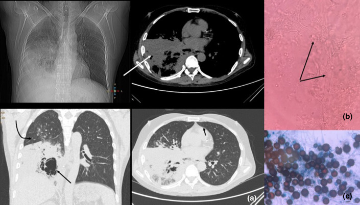
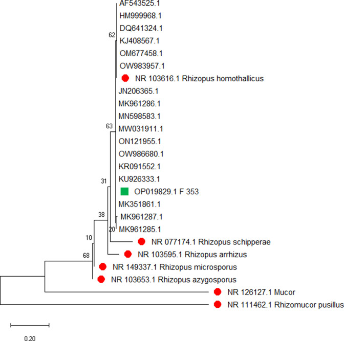
Case presentation: We report a case of pneumonia caused by R. homothallicus in a 54-year-old type 2 diabetic patient. The organism was isolated from bronchoalveolar lavage fluid and preliminarily identified by fungal morphology and finally by sequencing of the internal transcribed spacer region.
Conclusion: Mucormycosis may be associated with cavitary lung lesions against a backdrop of poorly controlled diabetes or other immunosuppressed states. Pulmonary mucormycosis may have variable clinical and radiological presentations. Therefore, strong clinical suspicion and prompt management can address the high fatality associated with the disease.
Keywords: Rhizopus homothallicus; cavitary; mucormycosis; pulmonary.
© 2023 The Authors.
Conflict of interest statement
The authors declare that there are no conflicts of interest.
Figures
Similar articles
-
Fatal Pulmonary Mucormycosis due to Rhizopus homothallicus.Mycopathologia. 2017 Oct;182(9-10):907-913. doi: 10.1007/s11046-017-0151-7. Epub 2017 Jun 3. Mycopathologia. 2017. PMID: 28580534
-
Clinical and Mycologic Characteristics of Emerging Mucormycosis Agent Rhizopus homothallicus.Emerg Infect Dis. 2023 Jul;29(7):1313-1322. doi: 10.3201/eid2907.221491. Emerg Infect Dis. 2023. PMID: 37347535 Free PMC article.
-
Rhizopus homothallicus Causing Invasive Infections: Series of Three Cases from a Single Centre in North India.Mycopathologia. 2017 Oct;182(9-10):921-926. doi: 10.1007/s11046-017-0153-5. Epub 2017 Jun 16. Mycopathologia. 2017. PMID: 28623532
-
Mucormycosis in India: unique features.Mycoses. 2014 Dec;57 Suppl 3:85-90. doi: 10.1111/myc.12243. Epub 2014 Sep 3. Mycoses. 2014. PMID: 25187095 Review.
-
Primary Cutaneous Mucormycosis Caused by Rhizopus oryzae: A Case Report and Review of Literature.Mycopathologia. 2017 Apr;182(3-4):387-392. doi: 10.1007/s11046-016-0084-6. Epub 2016 Nov 3. Mycopathologia. 2017. PMID: 27807669 Review.
Cited by
-
Clinical characteristics, outcome, and factors associated with mortality of pulmonary mucormycosis: a retrospective single-center study from Pakistan.Ther Adv Infect Dis. 2024 May 6;11:20499361241251744. doi: 10.1177/20499361241251744. eCollection 2024 Jan-Dec. Ther Adv Infect Dis. 2024. PMID: 38716078 Free PMC article.
References
Publication types
LinkOut - more resources
Full Text Sources