

Review
doi: 10.2147/IJWH.S351095.
eCollection 2023.
Prevalence, Impact, and Diagnostic Challenges of Benign Breast Disease: A Narrative Review
Affiliations
- PMID: 37223067
- PMCID: PMC10202205
- DOI: 10.2147/IJWH.S351095
Item in Clipboard
Review
Prevalence, Impact, and Diagnostic Challenges of Benign Breast Disease: A Narrative Review
Int J Womens Health.
.
Abstract
Benign breast diseases, which are commonly seen in clinical practice, have various clinical presentations and implications, as well as management strategies. This article describes common benign breast lesions, presentations of these lesions, and typical radiographic and histologic findings. Also included in this review are the most recent data and guideline-based recommendations for the management of benign breast diseases at diagnosis, including surgical referral, medical management, and ongoing surveillance.
Keywords: breast biopsy; imaging; management; surveillance.
© 2023 Fraker et al.
Conflict of interest statement
The authors declare that they have no competing interests.
Figures

Sonographic image showing a fibroepithelial lesion with borderline findings. Biopsy revealed phyllodes tumor.

Sonographic image showing multiple papillomas.

Sonographic image showing a single papilloma with cystic spaces.

Magnetic resonance image showing enhancement in the area of pseudoangiomatous stromal hyperplasia.
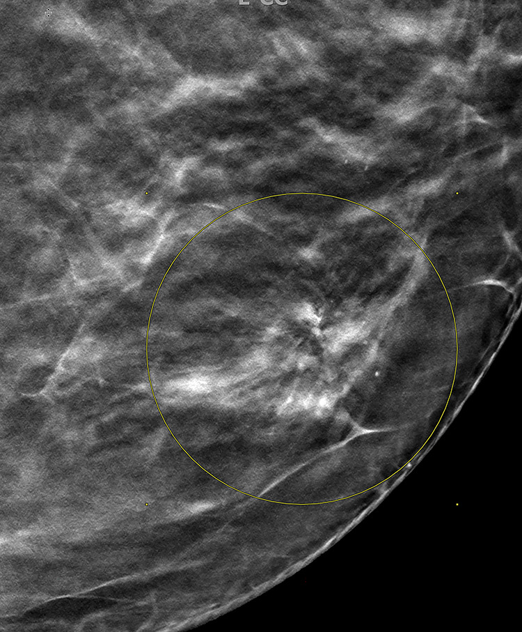
Mammographic image showing distortion in an area of radial scar. Yellow circle on figure outlines area of architectural distortion on mammography, biopsy revealed radial scar on pathology.

Mammographic image showing calcifications. Biopsy revealed flat epithelial atypia and atypical ductal hyperplasia. Yellow and white circles on figure outline areas of calcifications on mammography, biopsies revealed flat epithelial atypia and atypical ductal hyperplasia.
References
Publication types
LinkOut - more resources
Full Text Sources
