. 2021 Apr 12:2:uqab002.
doi: 10.1093/femsml/uqab002. eCollection 2021.

The composition and function of Enterococcus faecalis membrane vesicles

Affiliations

The composition and function of Enterococcus faecalis membrane vesicles

Irina Afonina et al. Microlife. .

Abstract

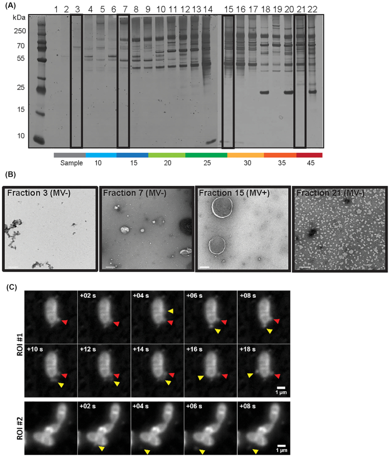
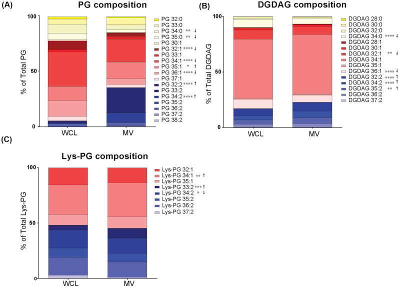
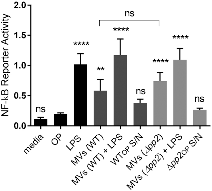
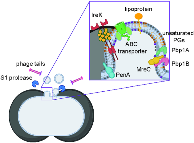
Membrane vesicles (MVs) contribute to various biological processes in bacteria, including virulence factor delivery, antimicrobial resistance, host immune evasion and cross-species communication. MVs are frequently released from the surface of both Gram-negative and Gram-positive bacteria during growth. In some Gram-positive bacteria, genes affecting MV biogenesis have been identified, but the mechanism of MV formation is unknown. In Enterococcus faecalis, a causative agent of life-threatening bacteraemia and endocarditis, neither mechanisms of MV formation nor their role in virulence has been examined. Since MVs of many bacterial species are implicated in host-pathogen interactions, biofilm formation, horizontal gene transfer, and virulence factor secretion in other species, we sought to identify, describe and functionally characterize MVs from E. faecalis. Here, we show that E. faecalis releases MVs that possess unique lipid and protein profiles, distinct from the intact cell membrane and are enriched in lipoproteins. MVs of E. faecalis are specifically enriched in unsaturated lipids that might provide membrane flexibility to enable MV formation, providing the first insights into the mechanism of MV formation in this Gram-positive organism.

Keywords: Enterococcus faecalis; NF-kB signaling; horizontal gene transfer; lipidomics; membrane vesicles; proteomics.


Conflict of interest statement

None declared.

Figures

Figure 1.
Figure 1.
Enterococcus faecalis produces MVs ranging from 50–400 nm in size. (A) 22 fractions consisting of 200 μL each were collected from the top of a 4.4 mL OptiPrep gradient, separated by SDS-PAGE, and silver stained. (B) Selected fractions (# 3, 7, 15, 21 indicated in the boxes in panel A) were negatively stained and viewed by TEM. Scale bar is 200 nm. (C) In-situ imaged live E. faecalis on the agar pad. Panels of consecutive frames (+0.2 s) from two distinct areas on the pad. The existing vesicle is indicated with a red arrow, the new vesicle—with a yellow arrow. Scale bar 1 μL.
Figure 2.
Figure 2.
MVs are enriched in polyunsaturated PG species. Constituent distribution of individual lipid species from whole cell lysate (WCL) or MVs within the analysed lipid classes (A), phosphatidylglycerol (PG), (B), diglucosyl-diacylglycerol (DGDAG), and (C), lysyl-phosphatidylglycerol (Lys-PG). Each stack represents the mean from 6 biological replicates. *, P ≤ 0.05; **, P ≤ 0.01; ****, P ≤ 0.0001; Fisher's LSD test for one-way ANOVA.
Figure 3.
Figure 3.
Phage tails co-purify with MVs, but phage tail production does not contribute to MV abundance. (A) TEM on assembled phage tails that are present within purified MVs. Scale bar is 100 nm. (B) TEM image of negatively stained E. faecalis, where MVs are associated with the cells surface. Scale bar is 100 nm. (C) The concentration of MVs in WT and the Δpp2 mutant as determined by Nanosight from three independent experiments. Statistical analysis was performed by the unpaired t-test. ns: P > 0.05.
Figure 4.
Figure 4.
Plasmid DNA co-purifies with MVs but is not transferred to E. faecalis or E. colicells. (A) DNA concentration was measured by Qubit in intact MV samples and in MVs lysed by boiling. Data shown from three independent experiments. Statistical analysis was performed by the unpaired t-test using GraphPad. ****, P < 0.0001. (B) Agarose gel showing PCR product amplified with plasmid-specific primers on crude and purified MV fractions of WT and WT pGCP123. The expected plasmid PCR product is 240 bp. (C) Agarose gel showing PCR product amplified with plasmid-specific primers on intact and lysed MVs from WT pGCP123, subjected to DNAse treatment or treatment with inactivated DNAse prior to PCR. DNAse treated pGCP123 serves as a control. (D) The number of transformants following E. faecalis and E. coli incubation with MVs extracted from WT or WT pGCP123 determined by CFU enumeration on non-selective BHI or selective media with kanamycin. Statistical analysis was performed by the one-way ANOVA using one-way ANOVA test with Tukey's multiple comparison test. ****, P < 0.0001.
Figure 5.
Figure 5.
MVs but not phage tails activate NF-kB pathway in macrophages. RAW-blue cells derived from RAW267.4 macrophages were stimulated with lipopolysaccharide (LPS) at 100 ng/mL (positive control), OptiPrep (OP) (negative control), MV-free concentrated supernatant in OptiPrep from WT and Δpp2 (WTOP S/N and Δpp2OP S/N) (secondary controls), MVs derived from WT and Δpp2 at 1000 particles/macrophage, and LPS + MVs. Six hours after stimulation, the NF-kB response was measured by secreted embryonic alkaline phosphatase reporter activity, transcribed from the plasmid under NF-kB inducible promoter. Statistical analysis was performed by the one-way ANOVA using one-way ANOVA test with Tukey's multiple comparison test. ****, P < 0.0001, ** P < 0.001, ns: P > 0.05 among all of the conditions as compared to OptiPrep negative control.
Figure 6.
Figure 6.
Septal model for MV formation in E. faecalis. Cartoon depiction for MV formation from the septal region, where peptidoglycan is thinnest, during the cell divison. Enterococcus faecalisMVs are enriched in more flexible unsaturated PGs, and have a high abundance of septal proteins—Pbp1A, Pbp1B, MreC, PenA, that are involved in cell division, and IreK kinase, that is bound to un-crosslinked peptidoglycan through PASTA domains. Phage tails and S1 extracellular protease co-purify with MV fraction.

References

    1. Beilharz K, Nováková L, Fadda Det al. Control of cell division in Streptococcus pneumoniae by the conserved Ser/Thr protein kinase StkP. Proc Natl Acad Sci. 2012;109:E905– 13. - PMC - PubMed
    1. Beveridge TJ. Structures of gram-negative cell walls and their derived membrane vesicles. J Bacteriol. 1999;181:4725– 33. - PMC - PubMed
    1. Biagini M, Garibaldi M, Aprea Set al. The human pathogen Streptococcus pyogenes releases lipoproteins as Lipoprotein-rich Membrane Vesicles. Mol Cell Proteomics. 2015;2138–49. - PMC - PubMed
    1. Bligh EG, Dyer WJ. A rapid method of total lipid extraction and purification. Can J Biochem Physiol. 1959;37:911– 7. - PubMed
    1. Boes A, Olatunji S, Breukink Eet al. Regulation of the peptidoglycan polymerase activity of PBP1b by antagonist actions of the core divisome proteins FtsBLQ and FtsN. mBio. 2019;10:e01912–18. - PMC - PubMed

LinkOut - more resources