. 2023 Apr 24:6:0124.
doi: 10.34133/research.0124. eCollection 2023.

SARS-CoV-2 RBD and Its Variants Can Induce Platelet Activation and Clearance: Implications for Antibody Therapy and Vaccinations against COVID-19

Affiliations

SARS-CoV-2 RBD and Its Variants Can Induce Platelet Activation and Clearance: Implications for Antibody Therapy and Vaccinations against COVID-19

Xiaoying Ma et al. Research (Wash D C). .

Abstract

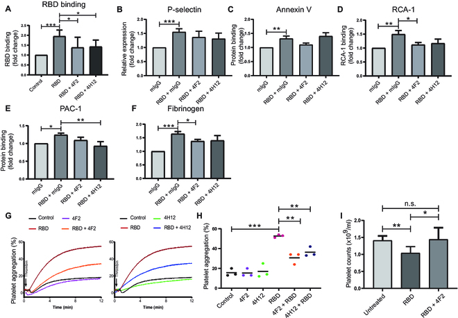
The COVID-19 pandemic caused by SARS-CoV-2 virus is an ongoing global health burden. Severe cases of COVID-19 and the rare cases of COVID-19 vaccine-induced-thrombotic-thrombocytopenia (VITT) are both associated with thrombosis and thrombocytopenia; however, the underlying mechanisms remain inadequately understood. Both infection and vaccination utilize the spike protein receptor-binding domain (RBD) of SARS-CoV-2. We found that intravenous injection of recombinant RBD caused significant platelet clearance in mice. Further investigation revealed the RBD could bind platelets, cause platelet activation, and potentiate platelet aggregation, which was exacerbated in the Delta and Kappa variants. The RBD-platelet interaction was partially dependent on the β3 integrin as binding was significantly reduced in β3-/- mice. Furthermore, RBD binding to human and mouse platelets was significantly reduced with related αIIbβ3 antagonists and mutation of the RGD (arginine-glycine-aspartate) integrin binding motif to RGE (arginine-glycine-glutamate). We developed anti-RBD polyclonal and several monoclonal antibodies (mAbs) and identified 4F2 and 4H12 for their potent dual inhibition of RBD-induced platelet activation, aggregation, and clearance in vivo, and SARS-CoV-2 infection and replication in Vero E6 cells. Our data show that the RBD can bind platelets partially though αIIbβ3 and induce platelet activation and clearance, which may contribute to thrombosis and thrombocytopenia observed in COVID-19 and VITT. Our newly developed mAbs 4F2 and 4H12 have potential not only for diagnosis of SARS-CoV-2 virus antigen but also importantly for therapy against COVID-19.


Figures

Fig. 1.
Fig. 1.
The RBD-rFc induces platelet clearance in vivo. (A) CD1 mouse platelets were counted at different time intervals (0, 1, 3, 8, 24, and 48 h) after I.V. injection with either a PBS control or RBD-rFc (PBS, n = 8; 0.25 μg/g RBD-rFc, n = 4; 0.5 μg/g RBD-rFc, n = 4; 1 μg/g RBD-rFc, n = 11). (B) A total of 2 × 10⁸ Far Red-labeled platelets were incubated with 2.5 μg/ml RBD-rFc for 30 min and then I.V. injected into CD1 mice. Circulating platelets were quantified at different time intervals (0, 1, 3, and 24 h). All the data were expressed as means ± SEM. Two-way ANOVA followed by Tukey’s multiple comparisons test was applied to evaluate the difference between RBD group and control group. *P < 0.05, **P < 0.01, ***P < 0.001, versus control at the same time point. *P < 0.05, **P < 0.01, versus baseline.
Fig. 2.
Fig. 2.
The RBD can bind to platelets partially via integrin αIIbβ3. The binding of RBD (10 μg/ml) to healthy human donor (A to C) and mouse (E to H) washed platelets were analyzed by flow cytometry using Alexa Fluor 647-labeled anti-RBD mAb 4H12. The binding of RBD-rFC to human (D) and mouse (I) platelets was analyzed by flow cytometry using FITC-labeled anti-rabbit Fc antibody. A total of 5 × 10⁵ platelets were incubated with wild-type RBD-rFc or RBD-RGE-rFc (RBD with RGE mutation). (G) RBD binding to integrin β3 knockout, and wild-type BALB/c mice platelets were analyzed by flow cytometry using Alexa Fluor 647-labeled anti-RBD mAb 4H12. All the data are displayed as means ± SEM. Two-way ANOVA Tukey’s multiple comparisons test was applied to evaluate the difference between groups. *P < 0.05, **P < 0.01, ***P < 0.0001. (J) Direct binding analysis of labeled RBD as a function of integrin αIIbβ3 concentration in vitro utilizing normalized mean fluorescence anisotropy of Alexa Fluor 488 in binding buffer. (K) Direct binding kinetics characterization of spike-αIIbβ3 using BLI in binding buffer (20 mM tris [pH 7.4], 137 mM NaCl, 1 mM MgCl2, 1 mM MnCl2, 1 mM CaCl2, and 50% glycerol) plus 1X Octet Kinetics Buffer in a concentration range from 68 to 512 nM (red sensograms) at 25 °C. Mean control sensogram (black) is 2% BSA in binding buffer.
Fig. 3.
Fig. 3.
The RBD induces human platelet activation and potentiates platelet aggregation in vitro. (A to E) Human platelet activation (P-selection expression; PAC-1, fibrinogen binding), apoptosis (Annexin V binding) and desialylation (RCA-1 binding) were detected via flow cytometry. RBD without tag: 50 μg/ml, n = 3. All flow cytometry data are expressed as fold change from control group. (F to H) Human gel-filtered platelet (preincubated with 200 μg/ml RBD-rFc) aggregation was stimulated by 0.02 U/ml thrombin, or 2 μM ADP with fibrinogen, or 2 μg/ml collagen. n = 5 to 9. All the data were expressed as means ± SEM (***P < 0.001, **P < 0.01, *P < 0.05).
Fig. 4.
Fig. 4.
The κ, δ, and δ+ variants have enhanced platelet binding, activation, and potentiation of platelet aggregation. (A) The binding of RBD-rFc variants (200 μg/ml) to human healthy donors’ platelets was analyzed by flow cytometry using FITC-labeled anti-rabbit IgG (Fc specific). (B to F) Human platelet activation (P-selection expression/PAC-1 or fibrinogen binding), apoptosis (Annexin V binding), and desialylation (RCA-1 binding) were detected via flow cytometry, n = 5 to 10. (G to I) Human gel-filtered platelet (preincubated with 200 μg/ml RBD-rFc or RBD-rFc variants) aggregation was stimulated by 0.02 U/ml thrombin, or 2 μM ADP with addition of fibrinogen or 2 μg/ml collagen. All the data were expressed as means ± SEM. *P < 0.05, **P < 0.01, ***P < 0.001.
Fig. 5.
Fig. 5.
The novel anti-RBD mAbs 4F2 and 4H12 inhibit RBD-induced platelet activation and RBD-potentiated platelet aggregation. (A) The inhibitive effect of anti-RBD mAbs (50 μg/ml) on RBD-rFc (200 μg/ml) binding to human washed platelets was analyzed by flow cytometry using FITC-labeled anti-rabbit IgG (Fc specific). (B to F) The inhibitive effect of anti-RBD mAbs 4F2 and 4H12 (50 μg/ml) on RBD-induced human platelet activation (P-selection expression/PAC-1 or fibrinogen binding), apoptosis (Annexin V binding), and desialylation (RCA-1 binding) was detected via flow cytometry. (G and H) Human gel-filtered platelet (preincubated with rFC control, 200 μg/ml RBD-rFc, or 50 μg/ml 4F2 + 200 μg/ml RBD-rFc) aggregation was stimulated by 0.02 U/ml thrombin. (I) CD1 mouse platelets were counted at 0 h (Untreated) and 3 h after I.V. injection with RBD-rFc or RBD-rFc + 4F2 (RBD-rFc incubated with 4F2 before injection, n = 3). RBD-rFc: 1 μg/g, 4F2: 1 μg/g. All data are expressed as means ± SEM. n.s. denotes no significance. *P < 0.05, **P < 0.01, ***P < 0.001.
Fig. 6.
Fig. 6.
Anti-RBD mAbs 4F2 and 4H12 inhibit SARS-CoV-2 infection in a dose-dependent manner. (A) Different concentrations of anti-RBD mAb 4F2 or 4H12 were mixed with MOI 2 of SARS-CoV-2 for 30 min and then added to the culture medium of Vero E6 cells (the inoculum was not removed). Vero E6 cells were recovered 15 h postinfection and viral RNA was assayed by RT-qPCR. Dotted horizontal lines indicate the limit of detection (LOD). (B) Virus progeny was evaluated for viable virus in a TCID50 assay. (C) The addition of 4F2 or 4H12 was not toxic to the Vero E6 cells. (D) Detection of SARS-CoV-2 entry proteins and viral proteins in cells lysate by western blot. Glyceraldehyde-3-phosphate dehydrogenase was used as a loading control. (E to G) Densitometric analysis of protein bands. Data are represented as means ± SEM. *P < 0.05, **P < 0.01, versus SARS-CoV-2 group.

References

    1. Gu SX, Tyagi T, Jain K, Gu VW, Lee SH, Hwa JM, Kwan JM, Krause DS, Lee AI, Halene S, et al. . Thrombocytopathy and endotheliopathy: Crucial contributors to COVID-19 thromboinflammation. Nat Rev Cardiol. 2021;18(3):194–209. - PMC - PubMed
    1. Helms J, Tacquard C, Severac F, Leonard-Lorant I, Ohana M, Delabranche X, Merdji H, Clere-Jehl R, Schenck M, Gandet FF, et al. . High risk of thrombosis in patients with severe SARS-CoV-2 infection: A multicenter prospective cohort study. Intensive Care Med. 2020;46(6):1089–1098. - PMC - PubMed
    1. Liao D, Zhou F, Luo L, Xu M, Wang H, Xia J, Gao Y, Cai L, Wang Z, Yin P, et al. . Haematological characteristics and risk factors in the classification and prognosis evaluation of COVID-19: A retrospective cohort study. Lancet Haematol. 2020;7(9):e671–e678. - PMC - PubMed
    1. Ciceri F, Beretta L, Scandroglio AM, Colombo S, Landoni G, Ruggeri A, Peccatori J, D’Angelo A, De Cobelli F, Rovere-Querini P, et al. . Microvascular COVID-19 lung vessels obstructive thromboinflammatory syndrome (MicroCLOTS): An atypical acute respiratory distress syndrome working hypothesis. Crit Care Resusc. 2020;22(2):95–97. - PMC - PubMed
    1. Boeckh-Behrens T, Golkowski D, Ikenberg B, Schlegel J, Protzer U, Schulz C, Novotny J, Kreiser K, Zimmer C, Hemmer B, et al. . COVID-19-associated large vessel stroke in a 28-year-old patient : NETs and platelets possible key players in acute thrombus formation. Clin Neuroradiol. 2021;31(2):511–514. - PMC - PubMed