. 2023 May 8:6:0130.
doi: 10.34133/research.0130. eCollection 2023.

Transcranial Magneto-Acoustic Stimulation Attenuates Synaptic Plasticity Impairment through the Activation of Piezo1 in Alzheimer's Disease Mouse Model

Affiliations

Transcranial Magneto-Acoustic Stimulation Attenuates Synaptic Plasticity Impairment through the Activation of Piezo1 in Alzheimer's Disease Mouse Model

Fangxuan Chu et al. Research (Wash D C). .

Abstract

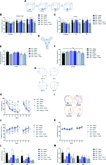
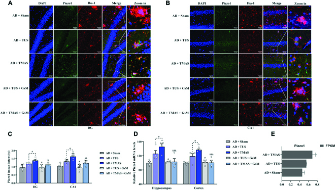
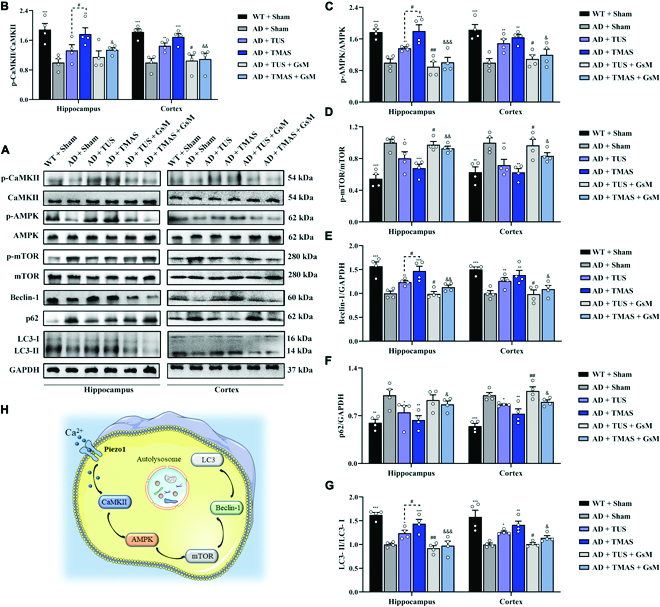
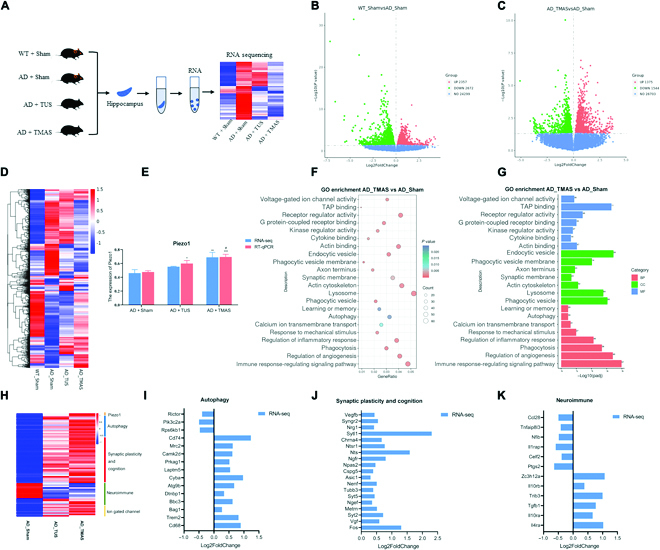
The neuropathological features of Alzheimer's disease include amyloid plaques. Rapidly emerging evidence suggests that Piezo1, a mechanosensitive cation channel, plays a critical role in transforming ultrasound-related mechanical stimuli through its trimeric propeller-like structure, but the importance of Piezo1-mediated mechanotransduction in brain functions is less appreciated. However, apart from mechanical stimulation, Piezo1 channels are strongly modulated by voltage. We assume that Piezo1 may play a role in converting mechanical and electrical signals, which could induce the phagocytosis and degradation of Aβ, and the combined effect of mechanical and electrical stimulation is superior to single mechanical stimulation. Hence, we design a transcranial magneto-acoustic stimulation (TMAS) system, based on transcranial ultrasound stimulation (TUS) within a magnetic field that combines a magneto-acoustic coupling effect electric field and the mechanical force of ultrasound, and applied it to test the above hypothesis in 5xFAD mice. Behavioral tests, in vivo electrophysiological recordings, Golgi-Cox staining, enzyme-linked immunosorbent assay, immunofluorescence, immunohistochemistry, real-time quantitative PCR, Western blotting, RNA sequencing, and cerebral blood flow monitoring were used to assess whether TMAS can alleviate the symptoms of AD mouse model by activating Piezo1. TMAS treatment enhanced autophagy to promote the phagocytosis and degradation of β-amyloid through the activation of microglial Piezo1 and alleviated neuroinflammation, synaptic plasticity impairment, and neural oscillation abnormalities in 5xFAD mice, showing a stronger effect than ultrasound. However, inhibition of Piezo1 with an antagonist, GsMTx-4, prevented these beneficial effects of TMAS. This research indicates that Piezo1 can transform TMAS-related mechanical and electrical stimuli into biochemical signals and identifies that the favorable effects of TMAS on synaptic plasticity in 5xFAD mice are mediated by Piezo1.


Figures

Fig. 1.
Fig. 1.
Description of the experimental processes and instruction of the treatment procedure. (A) Timeline of the experiment. (B) Schematic of the stimulation area and GsMTx-4 injection area [71]. (C) TMAS and TUS systems. (D) Schematic of the stimulation parameters.
Fig. 2.
Fig. 2.
TMAS treatment alleviates cognitive disorder in 5xFAD mice. (A) Protocol of the NOR test. (B and C) The RI based on the exploration duration and visit frequency. (D) Protocol of the Y-maze test. (E) The spontaneous alternation percentages in the Y-maze test. (F) The number of arm entries in the Y-maze test. (G) Experimental protocol of the MWM test. (H) Escape latencies in the IT and RT phases. (I) Illustration of the search strategies. (J and K) Cognitive scores and swimming speed in the IT and RT phases. (L and M) The number of platform crossings and quadrant occupancy time on day 6. n = 8 per group. *P < 0.05, **P < 0.01, ***P < 0.001 vs. the AD + Sham group. #P < 0.05, ##P < 0.01 vs. the AD + TUS group. &P < 0.05, &&P < 0.01, &&&P < 0.001 vs. the AD + TMAS group. n.s., not significant; SET, spatial exploration test; RET, reversal exploration test.
Fig. 3.
Fig. 3.
TMAS treatment ameliorates synaptic plasticity damage in 5xFAD mice. (A) Experimental protocol of in vivo electrophysiological recordings and positioning of stimulating and recording electrodes in hippocampal perforant pathway (PP)-DG circuit. (B) The fEPSP curve before and after LTP induction. (C) Changes in fEPSP slope from the PP to the DG. (D and E) Average fEPSP slope between the last 15 min during LTP and DEP. (F) Micrographs of Golgi–Cox-stained dendritic spines in the hippocampal area. Scale bar, 250 μm. (G) Representative picture of dendritic spines in the DG and CA1 region. The different types of dendritic spines are indicated by the black arrows. (H) The density of dendritic spines. (I) The proportions of different spine types. n = 8 per group. ***P < 0.001 vs. the AD + Sham group. #P < 0.05, ##P < 0.01, ###P < 0.001 vs. the AD + TUS group. &&&P < 0.001 vs. the AD + TMAS group. TBS, theta burst stimulation.
Fig. 4.
Fig. 4.
TMAS treatment activates Piezo1 in brain microglia. (A and B) Immunofluorescence images of Piezo1 and Iba-1 in the DG and CA1 regions. Scale bar, 50 μm. (C) The average fluorescence intensity of Piezo1. (D) The mRNA levels of piezo1 in hippocampal and cortical tissues. (E) RNA-seq analysis of Piezo1 in the hippocampus. n = 4 per group. *P < 0.05, ***P < 0.001 vs. the AD + Sham group. #P < 0.05 vs. the AD + TUS group. &P < 0.05, &&P < 0.01, &&&P < 0.001 vs. the AD + TMAS group.
Fig. 5.
Fig. 5.
TMAS treatment promotes microglial migration and phagocytosis of Aβ through the activation of Piezo1. (A) Microglial migration was evaluated by the Transwell assays. Scale bar, 100 μm. (B) Graph showing microglial migration. (C) Representative immunofluorescence image of Aβ (red) and Piezo1 (green) in BV2 cells. Scale bar, 20 μm. (D and E) The average fluorescence intensity of Piezo1 and Aβ in BV2 cells. (F) Immunofluorescence images of Aβ and Iba1 in the brains of mice. (G) Immunofluorescence images of Iba1, CD68, and Aβ in the brains of mice. (H) Number of microglia in the vicinity of Aβ plaques. (I) The mean fluorescence intensity of CD68. n = 4 per group. *P < 0.05, **P < 0.01, ***P < 0.001 vs. the Aβ group and AD + Sham group. #P < 0.05, ##P < 0.01 vs. the TUS + Aβ group and AD + TUS group. &P < 0.05, &&P < 0.01 vs. the TMAS + Aβ group and AD + TMAS group.
Fig. 6.
Fig. 6.
TMAS treatment activates autophagy via the CaMKII/AMPK/mTOR signaling pathway. (A) Immunoblotting of p-CaMKII, CaMKII, p-AMPK, AMPK, p-mTOR, mTOR, Beclin-1, p62, and LC3 in the hippocampus and cortex. (B to G) Quantitative analysis of p-CaMKII/CaMKII (B), p-AMPK/AMPK (C), p-mTOR/mTOR (D), Beclin-1/glyceraldehyde-3-phosphate dehydrogenase (GAPDH) (E), p62/GAPDH (F), and LC3-II/LC3-I (G) levels in the hippocampus and cortex. (H) Schematic diagram of CaMKII/AMPK/mTOR signaling pathway-mediated activation of Piezo1 channels induced by TMAS. n = 4 per group. *P < 0.05, **P < 0.01, ***P < 0.001 vs. the AD + Sham group. #P < 0.05, ##P < 0.01 vs. the AD + TUS group. &P < 0.05, &&P < 0.01, &&&P < 0.001 vs. the AD + TMAS group.
Fig. 7.
Fig. 7.
TMAS treatment alleviates the neuroinflammation in 5xFAD mice. (A) Immunohistochemical of Iba1 in the brains of mice. Scale bar, 50 μm. (B) Immunoblotting of iNOS, COX-2, IL-1β, IL-6, TNF-α, IL-10, and IL-4 in the hippocampus and cortex. (C and D) TMAS inhibited the production and release of proinflammatory factors in the hippocampi and cortices of 5xFAD mice. (E and F) TMAS increased the protein levels of IL-4 and IL-10 in the brain of 5xFAD mice. (G and H) TMAS inhibited the mRNA expression of proinflammatory factors and increased the mRNA expression of anti-inflammatory factors in the hippocampi and cortices of 5xFAD mice. (I) Fluorescence images of CD86 and Iba1 in the DG area. Scale bar, 50 μm. n = 4 per group. *P < 0.05, **P < 0.01, ***P < 0.001 vs. the AD + Sham group. #P < 0.05 vs. the AD + TUS group. &P < 0.05, &&P < 0.01, &&&P < 0.001 vs. the AD + TMAS group.
Fig. 8.
Fig. 8.
Transcriptome analysis of hippocampal tissue reveals that TMAS exerts its beneficial effect by activating Piezo1 in 5xFAD mice. (A) Schematic of RNA-seq analysis of hippocampal tissue from the 4 groups. (B and C) Differentially expressed genes compared to 5xFAD mice (log2 fold change > 0, P < 0.05). (D) Heatmap shows differentially expressed genes of 4 groups (log2 fold change > 0, P < 0.05). (E) The bar graph shows the expression of Piezo1, as determined by RT-qPCR and RNA-seq (P < 0.05). (F and G) GO enrichment analysis of differentially expressed genes in the brains of AD mice after TMAS treatment (log2 fold change > 0, P < 0.05). (H) Heatmap showing the relative gene expression of Piezo1- and autophagy-, synaptic plasticity-, and neuroimmunity-related genes in the AD, TUS, and TMAS-treated groups (log2 fold change > 0, P < 0.05). (I to K) The fold change in the expression of genes related to autophagy, synaptic plasticity, and neuroimmunity after TMAS treatment, as determined by RNA-seq (log2 fold change > 0, P < 0.05). n = 5 per group. BP, biological process; CC, cellular component; MF, molecular function.

References

    1. Wang H, Xu X, Pan YC, Yan Y, Hu XY, Chen R, Ravoo BJ, Guo DS, Zhang T. Recognition and removal of amyloid-β by a heteromultivalent macrocyclic coassembly: A potential strategy for the treatment of Alzheimer’s disease. Adv Mater. 2021;33(4):Article 2006483. - PubMed
    1. Hansen DV, Hanson JE, Sheng M. Microglia in Alzheimer’s disease. J Cell Biol. 2018;217(2):459–472. - PMC - PubMed
    1. Eimer WA, Vassar R. Neuron loss in the 5XFAD mouse model of Alzheimer’s disease correlates with intraneuronal Aβ42 accumulation and Caspase-3 activation. Mol Neurodegener. 2013;8:2. - PMC - PubMed
    1. Wang S, Li K, Zhao S, Zhang X, Yang Z, Zhang J, Zhang T. Early-stage dysfunction of hippocampal theta and gamma oscillations and its modulation of neural network in a transgenic 5xFAD mouse model. Neurobiol Aging. 2020;94:121–129. - PubMed
    1. Ivkovic S, Major T, Mitic M, Loncarevic-Vasiljkovic N, Jovic M, Adzic M. Fatty acids as biomodulators of Piezo1 mediated glial mechanosensitivity in Alzheimer’s disease. Life Sci. 2022;297:Article 120470. - PubMed

LinkOut - more resources