Review
. 2022 Jan 6;10(2):480-494.
doi: 10.1016/j.gendis.2021.12.006. eCollection 2023 Mar.

Mechanisms of NLRP3 inflammasome-mediated hepatic stellate cell activation: Therapeutic potential for liver fibrosis

Affiliations
Review

Mechanisms of NLRP3 inflammasome-mediated hepatic stellate cell activation: Therapeutic potential for liver fibrosis

Harsh Vardhan Charan et al. Genes Dis. .

Abstract

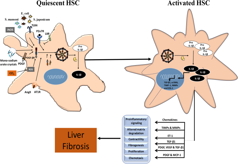
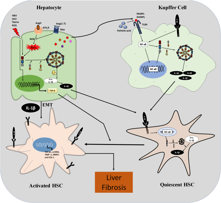
The liver injury leads to an inflammatory response, which causes the activation of hepatic stellate cells (HSCs) that further secrete ECM proteins and play an important role in liver fibrosis. Moreover, the inflammatory response is a driving force for fibrogenesis, which is triggered by many types of injuries. Exaggerated inflammatory immune responses are mediated by cytoplasmic protein complexes known as inflammasomes, which are involved in many chronic liver diseases. Inflammasomes are pattern recognition receptors (PRRs) that can sense any microbial motifs known as pathogen-associated molecular patterns (PAMPs), and host- or environmental-derived stress signals known as damage-associated molecular patterns (DAMPs). The inflammasomes cause caspase-mediated proteolytic cleavage of pro-IL-1β and pro-IL-18 into active IL-1β and IL-18. In this review, we provide a comprehensive summary of the important roles of NLRP3 inflammasome in the pathogenesis of liver fibrosis with an emphasis on several direct and indirect pathways responsible for the NLRP3 inflammasome-mediated HSCs activation and fibrogenesis. In addition, we discuss the general pharmacological and genetics strategies for the inhibition of NLRP3 inflammasome activation and its downstream signaling with examples of emerging pharmacotherapeutics, targeting the NLRP3 inflammasome signaling as well as a possible way to develop effective and safer NLRP3 inflammasome inhibitors.

Keywords: Hepatic stellate cells; Liver fibrosis; NLRP3 activation; NLRP3 inflammasome; NLRP3 inhibitors.


Figures

Figure 1
Figure 1
Mechanisms of NLRP3 inflammasome activation. NLRP3 is usually auto-repressed under physiological conditions but gets activated by undergoing oligomerization with ASC and pro-caspase-1 and releases active caspase-1, which is responsible for the conversion of pro-IL-1β (inactive) to IL-1β (active). The activation of NLRP3 inflammasome occurred mainly via 5 major upstream signals. 1) Potassium efflux: activation of the purinergic receptor via ATP causes potassium efflux that results in activation of NLRP3 complex; 2) calcium flux: activation of the calcium-sensing receptor causes phospholipase C mediated release of calcium ions from endoplasmic reticulum that results in NLRP3 oligomerization; 3) chloride efflux: efflux of chloride ions via chloride intracellular channel proteins CLIC1 and CLIC4 also acts as a signal for NLRP3 oligomerization; 4) lysosomal disruption: rupture of lysosomes due to large particulate materials also results in cathepsin B-mediated NLRP3 activation; and 5) reactive oxygen species (ROS): various ligands like imiquimod can induce mitochondrial stress resulting in the production of ROS that further cause the activation of NLRP3 inflammasome. ASC: adaptor protein apoptosis speck-like protein with a CARD domain; ATP: adenosine triphosphate; CASR: calcium-sensing receptor; P2x7R: P2x purinoceptor 7; PLC: phospholipase C; TWIK2: two-pore domain weak inwardly rectifying K+ channel 2.
Figure 2
Figure 2
NLRP3 inflammasome-mediated HSC activation and liver fibrogenesis. NLRP3 inflammasome is present in the cytoplasm of HSCs and is activated by various agents like PAMPs (S. mansoni, E. coli, S. japonicum); DAMPs (toxic agent-associated damage to cells), or through receptors like purinergic receptors (P2x7R), angiotensin II receptors (AT1R) and growth factor receptors (PDGF-βR). The activation of the NLRP3 inflammasome, in turn, activated the HSC (myofibroblast). Activated HSC further secretes various factors that result in an aggravated inflammatory response, chemotaxis of immune cells, altered matrix degradation, contractility and proliferation of myofibroblasts, and fibrogenesis. AngII: angiotensin II; AT1R: angiotensin II type 1 receptor; COL-1: collagen type 1; CCl4: carbon tetrachloride; ET-1: endothelin 1; iNOS: inducible nitric oxide synthase; LXR: liver X receptor; MCP-1: monocyte chemoattractant protein 1; MMP: matrix metalloproteinases; P2x7R: P2x purinoceptor 7; PDGF: platelet-derived growth factor; PDGF-βR: platelet-derived growth factor-β receptors; ROS: reactive oxygen species; α-SMA: α-smooth muscle actin; TIMP: tissue inhibitors of matrix metalloproteinases; TLR4: toll-like receptor 4; TGF-β1: transforming growth factor β1; VEGF: vascular endothelial growth factor.
Figure 3
Figure 3
Activation of NLRP3 inflammasome in the hepatocyte and Kupffer cells leading to HSCs activation and liver fibrogenesis. Damage to the hepatocytes due to various DAMPs or PAMPs results in the activation of the NLRP3 inflammasomes. Also, angiotensin II (AngII) acts on the angiotensin-II type 1 receptor (ATII1R) present on the surface of hepatocytes and results in the NADPH oxidase derived H2O2 mediated NLRP3 inflammasome activation. The activated NLRP3 inflammasome causes the activation and release of IL-1β, which subsequently activates HSCs. Additionally, IL-1β and NOX-derived H2O2-activated NLRP3 inflammasome cause the epithelial–mesenchymal transition (EMT), i.e., the conversion of hepatocytes to myofibroblasts. Damaged hepatocytes also release TGF-β, which acts as a pro-fibrogenic cytokine and converts quiescent HSCs to activated HSCs. Besides, damaged hepatocytes also release DAMPs that are recognized by toll-like receptors (TLR) present on Kupffer cells and activate the NLRP3 inflammasomes. TLR4 receptors on Kupffer cells are responsible for the recognition of various PAMPs and DAMPs and activate the NF-κB-mediated expression of the NLRP3, ASC, and pro-caspase 1, thereby the activation of NLRP3 inflammasome. NLRP3-mediated the release of IL-1β from Kupffer cells binds to its receptors present on the HSCs, which resulted in NF-κB-mediated NLRP3 activation, and ultimately activation of the HSCs (myofibroblast phenotype). These activated HSCs further release various pro-inflammatory mediators, pro-fibrogenic factors resulting in liver fibrosis. AngII: angiotensin II; Ang (1–7): angiotensin (1–7); ATII1R: angiotensin-II type 1 receptor; CCl4: carbon tetrachloride; CDCA: chenodeoxycholic acid; EMT: epithelial–mesenchymal transition; H2O2: hydrogen peroxide; HBV: hepatitis B virus; HCV: hepatitis C virus; LPS: lipopolysaccharides; Mas: Mas receptor; NOX: NADPH oxidase; ROS: reactive oxygen species; TGF-β: transforming growth factor-β; TLR4: Toll-like receptor 4.

References

    1. Higashi T., Friedman S.L., Hoshida Y. Hepatic stellate cells as key target in liver fibrosis. Adv Drug Deliv Rev. 2017;121:27–42. - PMC - PubMed
    1. Friedman S.L. Mechanisms of disease: mechanisms of hepatic fibrosis and therapeutic implications. Nat Clin Pract Gastroenterol Hepatol. 2004;1(2):98–105. - PubMed
    1. Bataller R., Brenner D.A. Liver fibrosis. J Clin Invest. 2005;115(2):209–218. - PMC - PubMed
    1. Mokdad A.A., Lopez A.D., Shahraz S., et al. Liver cirrhosis mortality in 187 countries between 1980 and 2010: a systematic analysis. BMC Med. 2014;12:145. - PMC - PubMed
    1. World Health Organization . 2018. The top 10 causes of death: leading causes of death by economy income group.http://www.who.int/en/news-room/fact-sheets/detail/the-top-10-causes-of-...

LinkOut - more resources