

Matrilin-2 within a three-dimensional lysine-modified chitosan porous scaffold enhances Schwann cell migration and axonal outgrowth for peripheral nerve regeneration
- PMID: 37223659
- PMCID: PMC10201561
- DOI: 10.3389/fbioe.2023.1142610
Matrilin-2 within a three-dimensional lysine-modified chitosan porous scaffold enhances Schwann cell migration and axonal outgrowth for peripheral nerve regeneration
Abstract
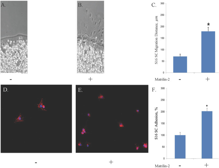
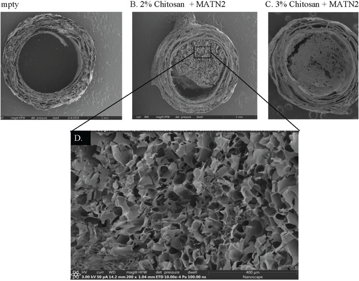
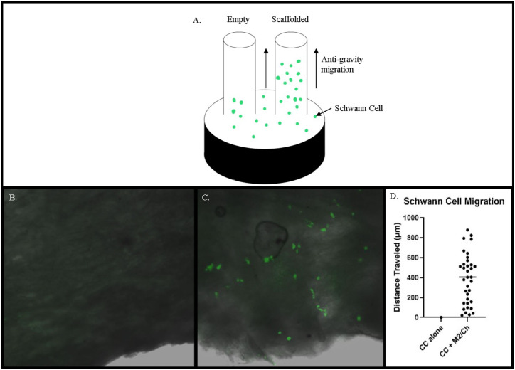
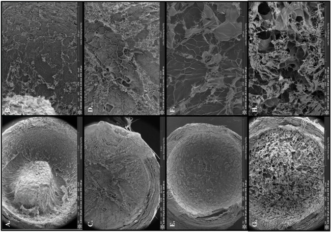
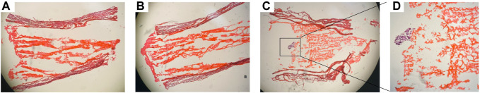
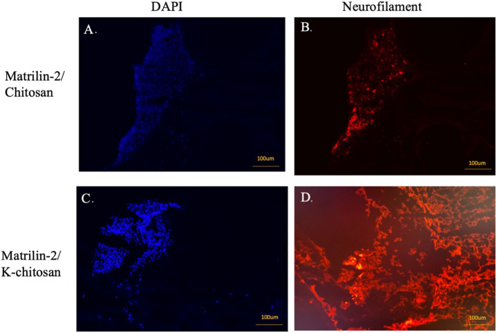
Background: Matrilin-2 is a key extracellular matrix protein involved in peripheral nerve regeneration. We sought to develop a biomimetic scaffold to enhance peripheral nerve regeneration by incorporating matrilin-2 within a chitosan-derived porous scaffold. We hypothesized that the use of such a novel biomaterial delivers microenvironmental cues to facilitate Schwann cell (SC) migration and enhance axonal outgrowth during peripheral nerve regeneration. Materials and Methods: The effect of matrilin-2 on SC migration was evaluated with agarose drop migration assay on matrilin-2 coated dishes. SC adhesion was determined with SCs cultured atop tissue culture dishes coated with matrilin-2. Various formulations of chitosan vs matrilin-2 in scaffold constructs were examined with scanning electron microscopy. The effect of the matrilin-2/chitosan scaffold on SC migration in the collagen conduits was determined by capillary migration assays. Neuronal adhesion and axonal outgrowth were evaluated with three-dimensional (3D) organotypic assay of dorsal root ganglions (DRG). DRG axonal outgrowth within the scaffolds was determined by immunofluorescence staining of neurofilaments. Results: Matrilin-2 induced SC migration and enhanced its adhesion. A formulation of 2% chitosan with matrilin-2 demonstrated an optimal 3D porous architecture for SC interaction. Matrilin-2/chitosan scaffold enabled SCs to migrate against gravity within conduits. Chemical modification of chitosan with lysine (K-chitosan) further improved DRG adhesion and axonal outgrowth than the matrilin-2/chitosan scaffold without lysine modification. Conclusion: We developed a matrilin-2/K-chitosan scaffold to mimic extracellular matrix cues and provide a porous matrix to enhance peripheral nerve regeneration. Taking advantage of matrilin-2's capability to stimulate SC migration and adhesion, we formulated a porous matrilin-2/chitosan scaffold to support axongal outgrowth. Chemical modification of chitosan with lysine further improved matrilin-2 bioactivity in the 3D scaffold. The 3D porous matrilin-2/K-chitosan scaffolds have high potential for enhancing nerve repair by stimulating SC migration, neuronal adhesion, and axonal outgrowth.
Keywords: axon; chitosan; lysine; matrilin-2; nerve regeneration; porous; scaffold; schwann cell.
Copyright © 2023 Li, Vorrius, Ge, Qiao, Zhu, Katarincic and Chen.
Conflict of interest statement
The authors declare that the research was conducted in the absence of any commercial or financial relationships that could be construed as a potential conflict of interest.
Figures
Similar articles
-
Matrilin-2 with a K-Chitosan Scaffold Enhances Functional Recovery and Nerve Regeneration in a Segmental Rat Sciatic Nerve Injury Model.Pharmaceuticals (Basel). 2025 May 6;18(5):686. doi: 10.3390/ph18050686. Pharmaceuticals (Basel). 2025. PMID: 40430505 Free PMC article.
-
A compound scaffold with uniform longitudinally oriented guidance cues and a porous sheath promotes peripheral nerve regeneration in vivo.Acta Biomater. 2018 Mar 1;68:223-236. doi: 10.1016/j.actbio.2017.12.010. Epub 2017 Dec 20. Acta Biomater. 2018. PMID: 29274478
-
The extracellular-matrix protein matrilin 2 participates in peripheral nerve regeneration.J Cell Sci. 2009 Apr 1;122(Pt 7):995-1004. doi: 10.1242/jcs.040378. J Cell Sci. 2009. PMID: 19295126
-
Unlocking nerve regeneration: electrical stimulation and bioscaffolds to enhance peripheral nerve regeneration.Front Neurosci. 2025 May 16;19:1594435. doi: 10.3389/fnins.2025.1594435. eCollection 2025. Front Neurosci. 2025. PMID: 40454247 Free PMC article. Review.
-
Regulation of nanomaterials in peripheral nerve regeneration from a microenvironmental perspective: biological effects and mechanisms.Mater Today Bio. 2025 Apr 29;32:101808. doi: 10.1016/j.mtbio.2025.101808. eCollection 2025 Jun. Mater Today Bio. 2025. PMID: 40453817 Free PMC article. Review.
Cited by
-
Harnessing three-dimensional porous chitosan microsphere embedded with adipose-derived stem cells to promote nerve regeneration.Stem Cell Res Ther. 2024 Jun 1;15(1):158. doi: 10.1186/s13287-024-03753-w. Stem Cell Res Ther. 2024. PMID: 38824568 Free PMC article.
-
Peripheral Nerve Regeneration Reimagined: Cutting-Edge Biomaterials and Biotechnological Innovations.Bioengineering (Basel). 2025 Aug 11;12(8):864. doi: 10.3390/bioengineering12080864. Bioengineering (Basel). 2025. PMID: 40868377 Free PMC article. Review.
-
Matrilin-2 with a K-Chitosan Scaffold Enhances Functional Recovery and Nerve Regeneration in a Segmental Rat Sciatic Nerve Injury Model.Pharmaceuticals (Basel). 2025 May 6;18(5):686. doi: 10.3390/ph18050686. Pharmaceuticals (Basel). 2025. PMID: 40430505 Free PMC article.
References
Grants and funding
LinkOut - more resources
Full Text Sources