

Characterization of membrane vesicles in Alteromonas macleodii indicates potential roles in their copiotrophic lifestyle
- PMID: 37223730
- PMCID: PMC10117737
- DOI: 10.1093/femsml/uqac025
Characterization of membrane vesicles in Alteromonas macleodii indicates potential roles in their copiotrophic lifestyle
Abstract
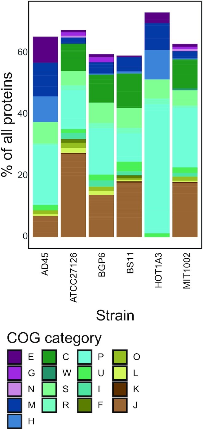
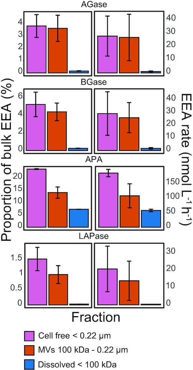
Bacterial membrane vesicles (MVs) are abundant in the oceans, but their potential functional roles remain unclear. In this study we characterized MV production and protein content of six strains of Alteromonas macleodii, a cosmopolitan marine bacterium. Alteromonas macleodii strains varied in their MV production rates, with some releasing up to 30 MVs per cell per generation. Microscopy imaging revealed heterogenous MV morphologies, including some MVs aggregated within larger membrane structures. Proteomic characterization revealed that A. macleodii MVs are rich in membrane proteins related to iron and phosphate uptake, as well as proteins with potential functions in biofilm formation. Furthermore, MVs harbored ectoenzymes, such as aminopeptidases and alkaline phosphatases, which comprised up to 20% of the total extracellular enzymatic activity. Our results suggest that A. macleodii MVs may support its growth through generation of extracellular 'hotspots' that facilitate access to essential substrates. This study provides an important basis to decipher the ecological relevance of MVs in heterotrophic marine bacteria.
Keywords: EVs; extracellular enzymes; iron uptake; marine bacteria; membrane transporters; moonlighting proteins.
© The Author(s) 2022. Published by Oxford University Press on behalf of FEMS.
Conflict of interest statement
The authors declare no competing interests.
Figures


Similar articles
-
Characterization of membrane vesicles secreted by seaweed associated bacterium Alteromonas macleodii KS62.Biochem Biophys Res Commun. 2019 Jun 25;514(2):422-427. doi: 10.1016/j.bbrc.2019.04.148. Epub 2019 Apr 30. Biochem Biophys Res Commun. 2019. PMID: 31053303
-
Development of a real-time quantitative PCR assay for detection and quantification of the marine bacterium Alteromonas macleodii from coastal environments.J Microbiol Methods. 2023 Jan;204:106629. doi: 10.1016/j.mimet.2022.106629. Epub 2022 Nov 29. J Microbiol Methods. 2023. PMID: 36460091
-
Different utilization of alginate and other algal polysaccharides by marine Alteromonas macleodii ecotypes.Environ Microbiol. 2015 Oct;17(10):3857-68. doi: 10.1111/1462-2920.12862. Epub 2015 May 8. Environ Microbiol. 2015. PMID: 25847866
-
Bacterial membrane vesicles, an overlooked environmental colloid: Biology, environmental perspectives and applications.Adv Colloid Interface Sci. 2015 Dec;226(Pt A):65-77. doi: 10.1016/j.cis.2015.08.013. Epub 2015 Sep 4. Adv Colloid Interface Sci. 2015. PMID: 26422802 Review.
-
Biofilm and bacterial membrane vesicles: recent advances.Expert Opin Ther Pat. 2024 Jun;34(6):475-491. doi: 10.1080/13543776.2024.2338101. Epub 2024 Apr 5. Expert Opin Ther Pat. 2024. PMID: 38578180 Review.
Cited by
-
Extracellular Vesicles and Bacteriophages: New Directions in Environmental Biocolloid Research.Environ Sci Technol. 2023 Nov 7;57(44):16728-16742. doi: 10.1021/acs.est.3c05041. Epub 2023 Oct 29. Environ Sci Technol. 2023. PMID: 37898880 Free PMC article. Review.
-
Membrane vesicles can contribute to cellulose degradation by Teredinibacter turnerae, a cultivable intracellular endosymbiont of shipworms.bioRxiv [Preprint]. 2024 Sep 8:2024.03.27.587001. doi: 10.1101/2024.03.27.587001. bioRxiv. 2024. Update in: Microb Biotechnol. 2024 Dec;17(12):e70064. doi: 10.1111/1751-7915.70064. PMID: 38585906 Free PMC article. Updated. Preprint.
-
Characterization of Spirulina-derived extracellular vesicles and their potential as a vaccine adjuvant.J Extracell Biol. 2024 Dec 12;3(12):e70025. doi: 10.1002/jex2.70025. eCollection 2024 Dec. J Extracell Biol. 2024. PMID: 39676887 Free PMC article.
-
Extracellular vesicle formation in Euryarchaeota is driven by a small GTPase.Proc Natl Acad Sci U S A. 2024 Mar 5;121(10):e2311321121. doi: 10.1073/pnas.2311321121. Epub 2024 Feb 26. Proc Natl Acad Sci U S A. 2024. PMID: 38408251 Free PMC article.
-
Membrane Vesicles Can Contribute to Cellulose Degradation by Teredinibacter turnerae, a Cultivable Intracellular Endosymbiont of Shipworms.Microb Biotechnol. 2024 Dec;17(12):e70064. doi: 10.1111/1751-7915.70064. Microb Biotechnol. 2024. PMID: 39659293 Free PMC article.
References
-
- Arnosti C, Wietz M, Brinkhoff Tet al. . The biogeochemistry of marine polysaccharides: sources, inventories, and bacterial drivers of the carbohydrate cycle. Ann Rev Marine Sci. 2021;13:81–108. - PubMed
-
- Arnosti C. Microbial extracellular enzymes and the marine carbon cycle. Ann Rev Marine Sci. 2011;3:401–25. - PubMed
-
- Baltar F, De Corte D, Thomson Bet al. . Teasing apart the different size pools of extracellular enzymatic activity in the ocean. Sci Total Environ. 2019;660:690–6. - PubMed
-
- Barrett AJ, Rawlings ND, Fred Woessner J. Handbook of Proteolytic Enzymes. Academic Press, 2012.
-
- Baumann P, Baumann L, Bowditch RDet al. . Taxonomy of Alteromonas: A. nigrifaciens sp. nov., nom. rev.; A. macleodii; and A. haloplanktis. Int J Syst Evol Microbiol. 1984;34:145–9.
Grants and funding
LinkOut - more resources
Full Text Sources