Review
. 2023 Apr 17:4:uqad015.
doi: 10.1093/femsml/uqad015. eCollection 2023.

Recent advances and perspectives in nucleotide second messenger signaling in bacteria

Affiliations
Review

Recent advances and perspectives in nucleotide second messenger signaling in bacteria

Regine Hengge et al. Microlife. .

Abstract

Nucleotide second messengers act as intracellular 'secondary' signals that represent environmental or cellular cues, i.e. the 'primary' signals. As such, they are linking sensory input with regulatory output in all living cells. The amazing physiological versatility, the mechanistic diversity of second messenger synthesis, degradation, and action as well as the high level of integration of second messenger pathways and networks in prokaryotes has only recently become apparent. In these networks, specific second messengers play conserved general roles. Thus, (p)ppGpp coordinates growth and survival in response to nutrient availability and various stresses, while c-di-GMP is the nucleotide signaling molecule to orchestrate bacterial adhesion and multicellularity. c-di-AMP links osmotic balance and metabolism and that it does so even in Archaea may suggest a very early evolutionary origin of second messenger signaling. Many of the enzymes that make or break second messengers show complex sensory domain architectures, which allow multisignal integration. The multiplicity of c-di-GMP-related enzymes in many species has led to the discovery that bacterial cells are even able to use the same freely diffusible second messenger in local signaling pathways that can act in parallel without cross-talking. On the other hand, signaling pathways operating with different nucleotides can intersect in elaborate signaling networks. Apart from the small number of common signaling nucleotides that bacteria use for controlling their cellular "business," diverse nucleotides were recently found to play very specific roles in phage defense. Furthermore, these systems represent the phylogenetic ancestors of cyclic nucleotide-activated immune signaling in eukaryotes.

Keywords: Ap4A; CBASS; biofilm; c-di-AMP; c-di-GMP; cGAMP; ppGpp.


Conflict of interest statement

None declared.

Figures
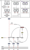
Figure 1.
Figure 1.
Model of (p)ppGpp regulation of PurR and global (p)ppGpp regulation of purine nucleotide synthesis in B. subtilis. (A) Model of evolution of PurR from PRT enzymes, with the active site of the latter becoming an effector binding pocket. (B) GTP and ATP synthesis in B. subtilis is regulated at multiple points by (p)ppGpp. This figure was previously published (Anderson et al. 2022) under the CC Attribution License.
Figure 2.
Figure 2.
(p)ppGpp interferes with the post- and cotranslational SRP-dependent membrane targeting pathway. In unstressed cells, SRP (Ffh protein in blue and the RNA in gray) can recognize a signal peptide (SP, pink) cotranslationally or post-translationally. Binding of GTP (green) then allows the formation of the SRP–FtsY targeting complex, which leads to GTP hydrolysis and transfer of the protein to the SecYEG translocon (light green). Under stringent conditions, (p)ppGpp (red) binds to SRP and prevents the formation of the SRP–FtsY targeting complex. This figure was previously published (Czech et al. 2022) under the CC Attribution License.
Figure 3.
Figure 3.
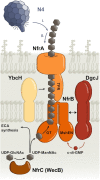
Model of the Nfr/DgcJ system and its role in locally c-di-GMP-activated exopolysaccharide production and bacteriophage N4 adsorption. DgcJ and NfrB colocalize via a direct protein–protein interaction. The C-terminal MshEN domain of NfrB binds c-di-GMP specifically produced by DgcJ, leading to an allosteric activation of the N-terminal GT domain of NfrB. WecB converts UDP-GlcNAc into UDP-ManNAc, which is used for the biosynthesis of the enterobaterial common antigen (ECA). In addition, the GT domain of NfrB uses UDP-ManNAc as a substrate to produce a putative ManNAc-polymer, which is secreted via the outer membrane protein NfrA. YbcH is a periplasmatic protein, which may play an auxiliary role, but is not essential for polysaccharide secretion. Phage N4 binds the exopolysaccharide secreted by the Nfr system as an initial receptor (I.) before interacting with NfrA (II.), which leads to the irriversible adsorption of the phage. This figure was previously published (Junkermeier and Hengge 2021) under the CC Attribution License.
Figure 4.
Figure 4.
Locally acting c-di-GMP control modules and global control of the cellular c-di-GMP level. If a strongly expressed master PDE keeps the cellular c-di-GMP pool low, local c-di-GMP production can become mandatory to activate specific effector/target systems. However, locally produced c-di-GMP would not significantly contribute to the global pool due to constant drainage of the latter by the master PDE. On the other hand, a strong DGC, which is expressed or activated under some particular conditions, could drive up cellular c-di-GMP to concentrations that can activate some effectors directly (depending on their KD), thus making local c-di-GMP production at these systems dispensable. Production of c-di-GMP is symbolized by gray arrows, degradation of c-di-GMP by gray lollypop symbols. Red arrows and lollypop symbols stand for activation and inhibition, respectively, by c-di-GMP. Bolts indicate signal input or regulatory output. Eff/target, effector/target component(s). This figure was previously published by R.H. (Hengge 2021) and is used here with permission.
Figure 5.
Figure 5.
The PDE PdeB is crucial to generate heterogeneity in Shewanella populations. Variation in protein copy number, late appearance at the cell pole and the following activation by interaction with HubP lead to high degree of variability in c-di-GMP levels. A plasmid-based reporter system (Zhou et al. 2016) was introduced in Shewanella for visualization of c-di-GMP content in vivo. The wild type (upper panel) shows much greater heterogeneity in c-di-GMP levels than a mutant lacking PdeB (lower panel). The scale bar equals 10 µm. This figure was kindly provided by Kai Thormann.
Figure 6.
Figure 6.
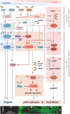
Regulatory network controlling the growth-vs.-survival balance and the integrated transition to adhesive multicellularity of E. coli. Signaling and regulatory proteins are shown by ovoid symbols (blue color indicates components that support growth and flagella-based activities, red color represents factors that drive maintenance, resilience, and multicellularity, and purple color designates components deployed by the flagellar control cascade, which, however, counteract flagellar activity and promote multicellularity). Transcriptional hubs are highlighted in light green. At the bottom, a vertical cryosection through a macrocolony biofilm of E. coli is shown that was rotated clockwise by 90o to align it roughly with the spatial distribution of the activities of the relevant network components. Extracellular matrix components, i.e. pEtN-cellulose and amyloid curli fibers, which are major targets of the control network, were stained with the fluorescent dye Thioflavin-S in order to visualize the morphologically different zones of the matrix architecture. At the top, the direction of nutrient and oxygen gradients in the biofilm is indicated. This figure was previously published by R.H. (Hengge 2020) and is used here with permission.
Figure 7.
Figure 7.
Glycogen granules in prespore chains of S. venezuelae. Transmission electron micrographs of wild type S. venezuelae stained for α-glucans. Colonies were grown for 5 days on maltose/yeast-extract/malt-extract (MYM) agar at 30°C. This figure was kindly provided by Natalia Tschowri.
Figure 8.
Figure 8.
Structure and function of dinucleases. (A) PDE with EAL or HD-GYP domains hydrolyze cyclic dinucleotides like c-di-GMP to linear molecules. Dinucleases of the Orn- and NrnC-type degrade these linear dinucleotides to mononucleotides. (B) Structure of substrate-bound Orn from V. cholera (left) and nano-RNAse C (NrnC) of Bartonella henselae (right). This figure was kindly provided by Holger Sondermann.
Figure 9.
Figure 9.
Different mechanisms to achieve potassium-dependent regulation of pyruvate carboxylation in B. subtilis and L. monocytogenes. Regulation is achieved indirectly via DarB-mediated control of the stringent response (expression of the pycA gene) and the DarB–PycA protein–protein interaction in B. subtilis. In contrast, c-di-AMP directly binds PycA in L. monocytogenes. Nevertheless, the regulatory logic and outcome is the same in both organisms. This figure was kindly provided by Jörg Stülke.

Similar articles

Cited by

References

    1. Abidi W, Zouhir S, Caleechurn Met al. Architecture and regulation of an enterobacterial cellulose secretion system. Sci Adv. 2021;7:eabd8049. - PMC - PubMed
    1. Abidi W, Torres-Sánchez L, Siroy Aet al. Weaving of bacterial cellulose by the Bcs secretion systems. FEMS Microbiol Rev. 2022;46:fuab051. - PMC - PubMed
    1. Aguilar C, Vlamakis H, Losick Ret al. Thinking about Bacillus subtilis as a multicellular organism. Curr Opin Microbiol. 2007;10:638–43. - PMC - PubMed
    1. Ahmad S, Wang B, Walker MDet al. An interbacterial toxin inhibits target cell growth by synthesizing (p)ppApp. Nature. 2019;575:674–8. - PMC - PubMed
    1. Al-Bassam MM, Haist J, Neumann SAet al. Expression patterns, genomic conservation and input into developmental regulation of the GGDEF/EAL/HD-GYP domain proteins in Streptomyces. Front Microbiol. 2018;9:2524. - PMC - PubMed

LinkOut - more resources