

Current and emerging trends in techniques for plant pathogen detection
- PMID: 37223788
- PMCID: PMC10200959
- DOI: 10.3389/fpls.2023.1120968
Current and emerging trends in techniques for plant pathogen detection
Abstract
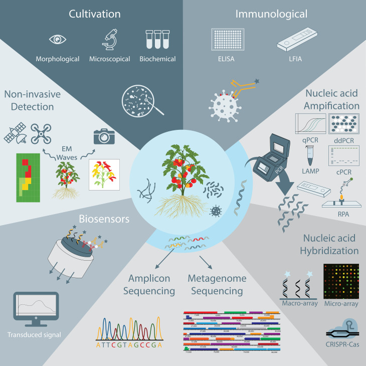
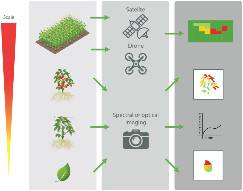
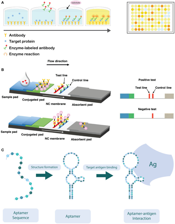
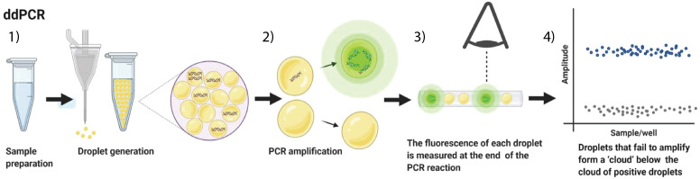
Plant pathogenic microorganisms cause substantial yield losses in several economically important crops, resulting in economic and social adversity. The spread of such plant pathogens and the emergence of new diseases is facilitated by human practices such as monoculture farming and global trade. Therefore, the early detection and identification of pathogens is of utmost importance to reduce the associated agricultural losses. In this review, techniques that are currently available to detect plant pathogens are discussed, including culture-based, PCR-based, sequencing-based, and immunology-based techniques. Their working principles are explained, followed by an overview of the main advantages and disadvantages, and examples of their use in plant pathogen detection. In addition to the more conventional and commonly used techniques, we also point to some recent evolutions in the field of plant pathogen detection. The potential use of point-of-care devices, including biosensors, have gained in popularity. These devices can provide fast analysis, are easy to use, and most importantly can be used for on-site diagnosis, allowing the farmers to take rapid disease management decisions.
Keywords: PCR-based detection; agriculture; biosensors; cultivation-based detection; immunologica detection; pathogen detection; plant pathogens; sequencing-based detection.
Copyright © 2023 Venbrux, Crauwels and Rediers.
Conflict of interest statement
The authors declare that the research was conducted in the absence of any commercial or financial relationships that could be construed as a potential conflict of interest.
Figures
Similar articles
-
Biosensors for plant pathogen detection.Biosens Bioelectron. 2017 Jul 15;93:72-86. doi: 10.1016/j.bios.2016.09.091. Epub 2016 Sep 28. Biosens Bioelectron. 2017. PMID: 27818053 Review.
-
A review of recent advances in plant-pathogen detection systems.Heliyon. 2022 Nov 28;8(12):e11855. doi: 10.1016/j.heliyon.2022.e11855. eCollection 2022 Dec. Heliyon. 2022. PMID: 36466579 Free PMC article. Review.
-
Novel plant disease detection techniques-a brief review.Mol Biol Rep. 2023 Nov;50(11):9677-9690. doi: 10.1007/s11033-023-08838-y. Epub 2023 Oct 12. Mol Biol Rep. 2023. PMID: 37823933 Review.
-
Enhancement of Plant Productivity in the Post-Genomics Era.Curr Genomics. 2016 Aug;17(4):295-6. doi: 10.2174/138920291704160607182507. Curr Genomics. 2016. PMID: 27499678 Free PMC article.
-
Fungal disease detection in plants: Traditional assays, novel diagnostic techniques and biosensors.Biosens Bioelectron. 2017 Jan 15;87:708-723. doi: 10.1016/j.bios.2016.09.032. Epub 2016 Sep 12. Biosens Bioelectron. 2017. PMID: 27649327 Review.
Cited by
-
Quantitative detection of the maize phytocytokine Zip1 utilizing ELISA.J Exp Bot. 2025 Jan 10;76(2):299-311. doi: 10.1093/jxb/erae423. J Exp Bot. 2025. PMID: 39673776 Free PMC article.
-
Portable solutions for plant pathogen diagnostics: development, usage, and future potential.Front Microbiol. 2025 Jan 31;16:1516723. doi: 10.3389/fmicb.2025.1516723. eCollection 2025. Front Microbiol. 2025. PMID: 39959158 Free PMC article. Review.
-
Soil Microbial Communities in Lemon Orchards Affected by Citrus Mal Secco Disease.Genes (Basel). 2024 Jun 21;15(7):824. doi: 10.3390/genes15070824. Genes (Basel). 2024. PMID: 39062603 Free PMC article.
-
Towards Pathogen-Free Coconut Germplasm Exchange.Plants (Basel). 2024 Jun 30;13(13):1809. doi: 10.3390/plants13131809. Plants (Basel). 2024. PMID: 38999649 Free PMC article. Review.
-
Pathogenic fungi (Sordariomycetes) associated with annual and perennial crops in Northern Thailand.MycoKeys. 2025 May 9;117:191-265. doi: 10.3897/mycokeys.117.137112. eCollection 2025. MycoKeys. 2025. PMID: 40386458 Free PMC article.
References
-
- Abdelfattah A., Malacrinò A., Wisniewski M., Cacciola S. O., Schena L. (2018). Metabarcoding: A powerful tool to investigate microbial communities and shape future plant protection strategies. Biol. Control 120, 1–10. doi: 10.1016/j.biocontrol.2017.07.009 - DOI
-
- Adams I., Fox A. (2016). “Diagnosis of plant viruses using next-generation sequencing and metagenomic analysis,” in Current research topics in plant virology. Eds. Wang A., Zhou X. (Springer International Publishing; ). doi: 10.1007/978-3-319-32919-2_14 - DOI
-
- Adkar-Purushothama C. R., Quaglino F., Casati P., Bianco P. A. (2010). Reverse transcription-duplex-polymerase chain reaction for simultaneous detection of citrus tristeza virus and ‘Candidatus liberibacter’ from citrus plants. J. Plant Dis. Prot. 117 (6), 241–243. doi: 10.1007/BF03356367 - DOI
Publication types
LinkOut - more resources
Full Text Sources