

Pyroptosis, apoptosis, and autophagy are involved in infection induced by two clinical Klebsiella pneumoniae isolates with different virulence
- PMID: 37223846
- PMCID: PMC10200925
- DOI: 10.3389/fcimb.2023.1165609
Pyroptosis, apoptosis, and autophagy are involved in infection induced by two clinical Klebsiella pneumoniae isolates with different virulence
Abstract
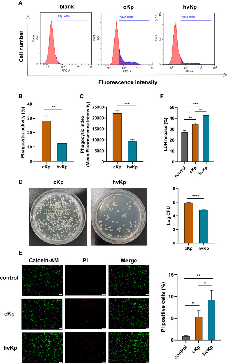
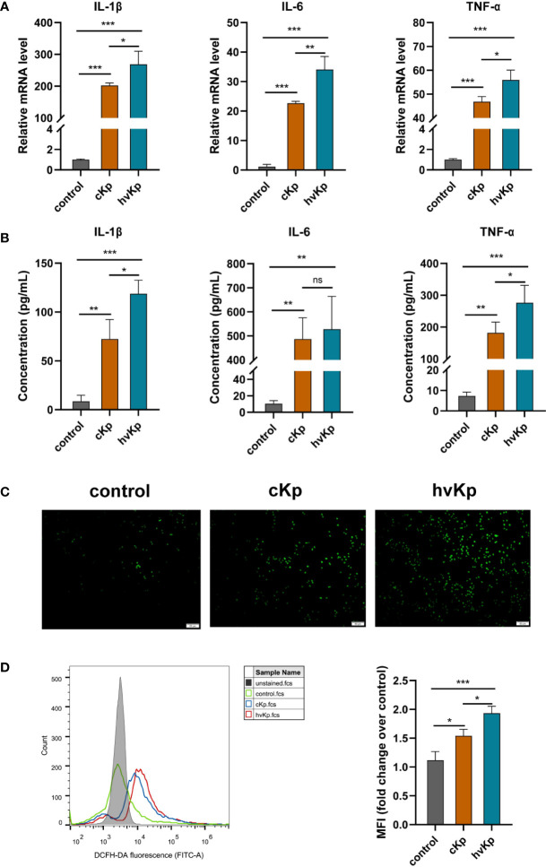
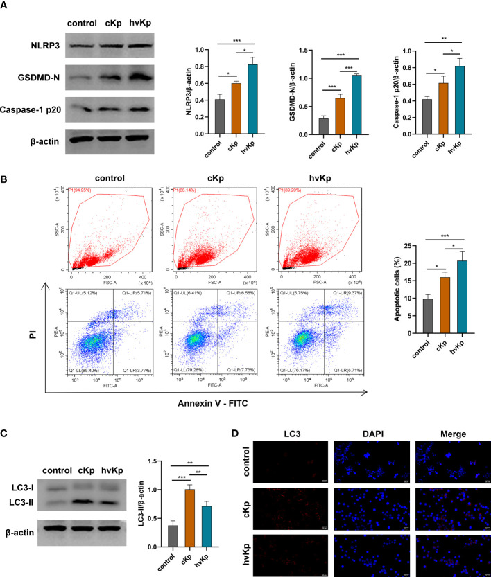
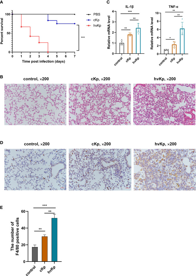
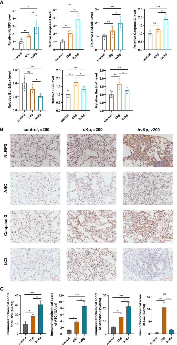
Klebsiella pneumoniae can cause widespread infections and is an important factor of hospital- and community-acquired pneumonia. The emergence of hypervirulent K. pneumoniae poses a serious clinical therapeutic challenge and is associated with a high mortality. The goal of this work was to investigate the influence of K. pneumoniae infection on host cells, particularly pyroptosis, apoptosis, and autophagy in the context of host-pathogen interactions to better understand the pathogenic mechanism of K. pneumoniae. Two clinical K. pneumoniae isolates, one classical K. pneumoniae isolate and one hypervirulent K. pneumoniae isolate, were used to infect RAW264.7 cells to establish an in vitro infection model. We first examined the phagocytosis of macrophages infected with K. pneumoniae. Lactate dehydrogenase (LDH) release test, and calcein-AM/PI double staining was conducted to determine the viability of macrophages. The inflammatory response was evaluated by measuring the pro-inflammatory cytokines and reactive oxygen species (ROS) production. The occurrence of pyroptosis, apoptosis, and autophagy was assessed by detecting the mRNA and protein levels of the corresponding biochemical markers. In addition, mouse pneumonia models were constructed by intratracheal instillation of K. pneumoniae for in vivo validation experiments. As for results, hypervirulent K. pneumoniae was much more resistant to macrophage-mediated phagocytosis but caused more severe cellular damage and lung tissues damage compared with classical K. pneumoniae. Moreover, we found increased expression of NLRP3, ASC, caspase-1, and GSDMD associated with pyroptosis in macrophages and lung tissues, and the levels were much higher following hypervirulent K. pneumoniae challenge. Both strains induced apoptosis in vitro and in vivo; the higher apoptosis proportion was observed in infection caused by hypervirulent K. pneumoniae. Furthermore, classical K. pneumoniae strongly triggered autophagy, while hypervirulent K. pneumoniae weakly activated this process. These findings provide novel insights into the pathogenesis of K. pneumoniae and may form the foundation for the future design of treatments for K. pneumoniae infection.
Keywords: Klebsiella pneumoniae; apoptosis; autophagy; inflammation; macrophage; pyroptosis.
Copyright © 2023 Wang, Bi, Xin, Zhang, Fu, Lan, Wang and Yan.
Conflict of interest statement
The authors declare that the research was conducted in the absence of any commercial or financial relationships that could be construed as a potential conflict of interest.
Figures
Similar articles
-
Resistance of hypervirulent Klebsiella pneumoniae to cathepsin B-mediated pyroptosis in murine macrophages.Front Immunol. 2023 Jun 29;14:1207121. doi: 10.3389/fimmu.2023.1207121. eCollection 2023. Front Immunol. 2023. PMID: 37457695 Free PMC article.
-
OmpA of Klebsiella pneumoniae ATCC 13883 induces pyroptosis in HEp-2 cells, leading to cell-cycle arrest and apoptosis.Microbes Infect. 2020 Oct;22(9):432-440. doi: 10.1016/j.micinf.2020.06.002. Epub 2020 Jun 19. Microbes Infect. 2020. PMID: 32569734
-
Enhanced invasion and survival of antibiotic- resistant Klebsiella pneumoniae pathotypes in host cells and strain-specific replication in blood.Front Cell Infect Microbiol. 2025 Feb 14;15:1522573. doi: 10.3389/fcimb.2025.1522573. eCollection 2025. Front Cell Infect Microbiol. 2025. PMID: 40028183 Free PMC article.
-
Antimicrobial Resistance of Hypervirulent Klebsiella pneumoniae: Epidemiology, Hypervirulence-Associated Determinants, and Resistance Mechanisms.Front Cell Infect Microbiol. 2017 Nov 21;7:483. doi: 10.3389/fcimb.2017.00483. eCollection 2017. Front Cell Infect Microbiol. 2017. PMID: 29209595 Free PMC article. Review.
-
Autophagy, cell death, and cytokines in K. pneumoniae infection: therapeutic perspectives.Emerg Microbes Infect. 2023 Dec;12(1):2140607. doi: 10.1080/22221751.2022.2140607. Emerg Microbes Infect. 2023. PMID: 36287114 Free PMC article. Review.
Cited by
-
Adaptive attenuation of virulence in hypervirulent carbapenem-resistant Klebsiella pneumoniae.mSystems. 2024 Jun 18;9(6):e0136323. doi: 10.1128/msystems.01363-23. Epub 2024 May 16. mSystems. 2024. PMID: 38752758 Free PMC article.
-
Multidrug-resistant hypervirulent Klebsiella pneumoniae: an evolving superbug.Future Microbiol. 2025 Apr;20(6):499-511. doi: 10.1080/17460913.2025.2482478. Epub 2025 Mar 26. Future Microbiol. 2025. PMID: 40135944 Review.
-
Itaconate inhibits SYK through alkylation and suppresses inflammation against hvKP induced intestinal dysbiosis.Cell Mol Life Sci. 2023 Oct 28;80(11):337. doi: 10.1007/s00018-023-04971-w. Cell Mol Life Sci. 2023. PMID: 37897551 Free PMC article.
-
Benzaldehyde stimulates autophagy via the sonic hedgehog signaling pathway in mouse brain astrocytes after treatment with Angiostrongylus cantonensis excretory-secretory products.Int J Parasitol Drugs Drug Resist. 2024 Dec;26:100560. doi: 10.1016/j.ijpddr.2024.100560. Epub 2024 Aug 12. Int J Parasitol Drugs Drug Resist. 2024. PMID: 39146602 Free PMC article.
-
ROS induced pyroptosis in inflammatory disease and cancer.Front Immunol. 2024 Jul 1;15:1378990. doi: 10.3389/fimmu.2024.1378990. eCollection 2024. Front Immunol. 2024. PMID: 39011036 Free PMC article. Review.
References
Publication types
MeSH terms
LinkOut - more resources
Full Text Sources
Molecular Biology Databases
Research Materials
Miscellaneous