. 2023 Jun 16;12(6):1845-1858.
doi: 10.1021/acssynbio.3c00231. Epub 2023 May 24.

Design and Selection of Heterodimerizing Helical Hairpins for Synthetic Biology

Affiliations

Design and Selection of Heterodimerizing Helical Hairpins for Synthetic Biology

Abigail J Smith et al. ACS Synth Biol. .

Abstract

Synthetic biology applications would benefit from protein modules of reduced complexity that function orthogonally to cellular components. As many subcellular processes depend on peptide-protein or protein-protein interactions, de novo designed polypeptides that can bring together other proteins controllably are particularly useful. Thanks to established sequence-to-structure relationships, helical bundles provide good starting points for such designs. Typically, however, such designs are tested in vitro and function in cells is not guaranteed. Here, we describe the design, characterization, and application of de novo helical hairpins that heterodimerize to form 4-helix bundles in cells. Starting from a rationally designed homodimer, we construct a library of helical hairpins and identify complementary pairs using bimolecular fluorescence complementation in E. coli. We characterize some of the pairs using biophysics and X-ray crystallography to confirm heterodimeric 4-helix bundles. Finally, we demonstrate the function of an exemplar pair in regulating transcription in both E. coli and mammalian cells.

Keywords: coiled coil; in-cell library screening; protein−protein interactions; rational peptide design; synthetic biology.


Conflict of interest statement

The authors declare no competing financial interest.

Figures
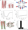
Figure 1
Figure 1
A de novo designed CC hairpin that dimerizes to form a 4HB. (A) Model of the designed homodimer CC-HP1.0 in a syn topology. This was built by grafting the loop LSKEPS from PDB entry 2fv2 (residues 204–209) onto the structure of apCC-Tet (PDB ID: 6q5s), and then truncating each helix by one heptad. (B) Helical-wheel representation of the syn model of CC-HP1.0. (C) CD spectra at 5 °C before (solid line) and after (dashed line) heating to 95 °C, and (D) thermal-response curves of the CD signal at 222 nm (heating, solid line; cooling, dashed line) for peptide CC-HP1.0. Conditions: 10 μM peptide, PBS, pH 7.4. (E) Structural overlay of the X-ray crystal structure of CC-HP1.0 dimer (green; PDB ID, 8bcs; Table S1) and the in silico designed syn model (pink); RMSDall-atom = 1.035 Å. Left: Backbone overlay of the entire structure. Right: Backbone and side-chain overlay for one heptad repeat. Note: Experimental structure reveals that CC-HP1.0 crystallized in the anti conformation (left panel, green cartoon). (F) Bar chart showing the reconstitution of split YFP activity by CC-HP1.0 for combinations of nYFP and cYFP fusions as indicated. YFP fluorescence was normalized to the OD600 of the bacterial cell culture, averaged from three different cultures, and shown with standard deviations.
Figure 2
Figure 2
Screening the CC-HP1.0-lib library for heterodimeric hairpins. (A) Cartoon showing the process of library sorting. E. coli cells harboring interacting variants of CC-HP1.0 reconstitute split-YFP fluorescence and were separated from cells expressing noninteracting variants by FACS. (B) Representative FACS plots showing E. coli cells expressing nYFP and cYFP in the absence of the hairpins (left), fused to CC-HP1.0 (middle), or fused to the degenerate codon library CC-HP1.0-lib (right). Side scatter (SSC) is plotted against YFP fluorescence. A large YFP+ gate was defined as containing cells in which split YFP fluorescence was reconstituted, and the two gates selecting the brightest cells (YFP1 and YFP2), used for sorting potential heterodimers, are outlined in red.
Figure 3
Figure 3
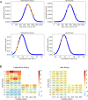
Statistical analysis of interacting hairpins selected from the library. (A) Calculated normal distributions of the summed side-chain volumes and hydrophobicities at the varied a and e positions for all the possible library variants (blue solid lines). Corresponding values for the sequences of the selected putative heterodimers and the dark library are overlaid on these curves and shown in orange. The values for CC-HP1.0 are plotted in cyan and indicated with an arrow. The values for the heterodimers, dark library, and CC-HP1.0 are shown plotted on the normal-distribution curve to ease comparisons only; however, only the x-axis values are relevant for these points. (B) Heatmap showing the z-score of the standard error of proportion of the observed selected amino acids compared to the expected numbers of those amino acids. The amino acids are ordered in increasing hydrophobicity from bottom to top. The a sites (positions 10, 17, 36, and 43) are shown left, and the e sites (7, 14, 33, and 40) right. A value of 0 indicates the amino acid is found at the same frequency as that expected by chance; >0 indicates favored residues (red scale); and <0 disfavored residues (blue scale).
Figure 4
Figure 4
Identifying hairpins from the library screen that form obligate heterodimers. (A) A heatmap showing split-YFP reconstitution between different hairpins fused to nYFP (α) and cYFP (β). The numbering scheme simply refers to pairs isolated from the library screen, with the coselected pairs highlighted by black boxes. Each hairpin was tested against 9 other hairpins. The scale is for YFP fluorescence normalized to the OD600 of the bacterial cell culture. Controls are 1, no CCs; and 2, CC-HP1.0. (B) Bar chart of normalized YFP fluorescence for the four selected heterodimer candidates. Each hairpin was fused to nYFP and cYFP in the combinations shown. Bars are colored black to show heterodimer interactions between the α and β hairpins, and white to show homodimerization of two α or two β hairpins. YFP fluorescence was normalized to the OD600 of the bacterial cell culture, averaged from three different cultures, and shown with standard deviations.
Figure 5
Figure 5
In vitro biophysical characterization of hairpin pair 26. (A) CD spectra at 5 °C of the peptides 26α (yellow) and 26β (blue) alone and when mixed (26α/26β, red). (B) Thermal unfolding following the CD signal at 222 nm. Same coloring as in panel A. Second derivatives of melting curves returned melting temperatures of 55 °C for 26α, 48 °C for 26β, and 60 °C for the mixture 26α/26β. (C) Sedimentation-velocity AUC data colored as in panel A. Fits returned molecular weights corresponding to dimers for each peptide. (D) Native mass spectrum (black line) for the 26α/26β mixture showing the region corresponding to the 5 H+ state of the dimers. Simulations of the 5 H+ protonated state for the homodimer 26α (yellow), the homodimer 26β (blue), and the heterodimer 26α/26β (red) are overlaid. The peaks at 2128.8 m/z and at 2144.2 m/z correspond to the TFA adduct (+ 113 Da) of the homodimer 26α and the heterodimer 26α/26β, respectively. Conditions for A–C: 50 μM peptide, PBS, 500 μM TCEP, pH 7.4. Conditions for D: 50 μM peptide, ammonium acetate buffer, 500 μM TCEP, pH 7.0.
Figure 6
Figure 6
X-ray crystal structure of the heterodimer 26α/26β (PDB ID, 8bct, Table S1). (A) Left: Cartoon with hairpin 26α depicted in yellow and 26β in blue. Right: Cross section through one heptad repeat showing the knobs-into-holes packing. (B) View into the core with modifications at a positions in cyan and at e in red. (C) Chainbow representation with each chain colored from blue (N terminus) to red (C terminus) and with charged side chains shown as sticks. (D) Fluorescence-quenching assay for the 26α/26β pair. 4CF refers to 4-cyano-l-phenylalanine fluorophore (bottom panels, yellow star), and MSE to l-selenomethionine fluorescence quencher (bottom panels, gray triangle); and “n” and “c” indicate the fluorophore is near the N and C termini, respectively. Color code as in panel A. Conditions: 50 μM concentration of each peptide in phosphate buffer, pH 7.4.
Figure 7
Figure 7
Heterodimeric 4HBs are orthogonal and portable into other subcellular contexts. (A) Heatmap showing YFP activity to examine the orthogonality of the different hairpin heterodimers. Each hairpin was fused to nYFP or cYFP and cells were transformed in the combinations indicated. Transformations were carried out in 96-well plates and YFP fluorescence was normalized to the OD600 of the bacterial cell culture. Each square is the average of at least two independent transformations. Controls: 1, no CCs; and 2, CC-HP1.0. (B) Cartoon and bar chart showing repression of a GFP gene expressed from the lacUV5 promoter in E. coli. Hairpins were fused to LacI*, a dimerization mutant of the LacI repressor also lacking the tetramerization domain. Fusion with CC-HP1.0 or 26α/26β increased repression of GFP. GFP fluorescence was normalized to the OD600 of the bacterial cell culture, averaged from three different cultures, and shown with standard deviations. (C) Cartoons and bar chart showing the activation of transcription in human HEK293 cells elicited by the dimerization of 26α/26β. The DNA-binding (DBD) and activation domains (AD) of a split transcription factor are brought together by the PPI, which initiates transcription of a luciferase reporter gene. As a positive control the iDimerize DmrA and DmrC proteins dimerize in the presence of A/C heterodimerizer (*), and also activate transcription of luciferase. Luminescence is shown as relative light units (RLU) and was the average of 6 independent experiments with standard deviations shown.

References

    1. Korendovych I. V.; DeGrado W. F. De novo protein design, a retrospective. Q. Rev. Biophys. 2020, 53, e3.10.1017/S0033583519000131. - DOI - PMC - PubMed
    1. Pan X.; Kortemme T. Recent advances in de novo protein design: Principles, methods, and applications. J. Biol. Chem. 2021, 296, 100558.10.1016/j.jbc.2021.100558. - DOI - PMC - PubMed
    1. Woolfson D. N. A Brief History of De Novo Protein Design: Minimal, Rational, and Computational. J. Mol. Biol. 2021, 433 (20), 167160.10.1016/j.jmb.2021.167160. - DOI - PubMed
    1. Nooren I. M.; Thornton J. M. Diversity of protein-protein interactions. EMBO J. 2003, 22 (14), 3486–92. 10.1093/emboj/cdg359. - DOI - PMC - PubMed
    1. Kuriyan J.; Eisenberg D. The origin of protein interactions and allostery in colocalization. Nature 2007, 450 (7172), 983–90. 10.1038/nature06524. - DOI - PubMed

Publication types