

Cerebral small vessel disease pathology in COVID-19 patients: A systematic review
- PMID: 37224885
- PMCID: PMC10202464
- DOI: 10.1016/j.arr.2023.101962
Cerebral small vessel disease pathology in COVID-19 patients: A systematic review
Abstract
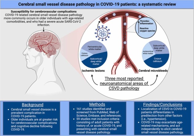
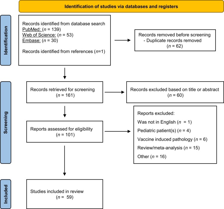
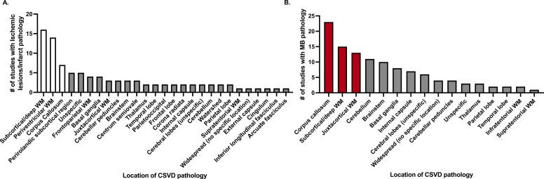
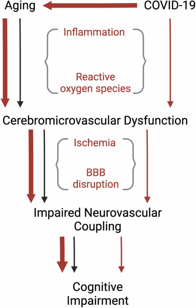
Cerebral small vessel disease (CSVD) is the leading cause of vascular cognitive impairment and is associated with COVID-19. However, contributing factors that often accompany CSVD pathology in COVID-19 patients may influence the incidence of cerebrovascular complications. Thus, a mechanism linking COVID-19 and CSVD has yet to be uncovered and differentiated from age-related comorbidities (i.e., hypertension), and medical interventions during acute infection. We aimed to evaluate CSVD in acute and recovered COVID-19 patients and to differentiate COVID-19-related cerebrovascular pathology from the above-mentioned contributing factors by assessing the localization of microbleeds and ischemic lesions/infarctions in the cerebrum, cerebellum, and brainstem. A systematic search was performed in December 2022 on PubMed, Web of Science, and Embase using a pre-established search criterion related to history of, or active COVID-19 with CSVD pathology in adults. From a pool of 161 studies, 59 met eligibility criteria and were included. Microbleeds and ischemic lesions had a strong predilection for the corpus callosum and subcortical/deep white matter in COVID-19 patients, suggesting a distinct CSVD pathology. These findings have important implications for clinical practice and biomedical research as COVID-19 may independently, and through exacerbation of age-related mechanisms, contribute to increased incidence of CSVD.
Keywords: Aging; COVID-19; Cerebral small vessel disease; Cognitive impairment.
Copyright © 2023 Elsevier B.V. All rights reserved.
Conflict of interest statement
Declaration of Competing Interest None.
Figures
References
-
- Al-Hakeim H.K., Al-Rubaye H.T., Al-Hadrawi D.S., Almulla A.F., Maes M. Long-COVID post-viral chronic fatigue and affective symptoms are associated with oxidative damage, lowered antioxidant defenses and inflammation: a proof of concept and mechanism study. Mol. Psychiatry. 2022 doi: 10.1038/s41380-022-01836-9. - DOI - PMC - PubMed
Publication types
MeSH terms
Grants and funding
- I01 CX000340/CX/CSRD VA/United States
- I01 CX002578/CX/CSRD VA/United States
- R03 AG070479/AG/NIA NIH HHS/United States
- T32 AG052363/AG/NIA NIH HHS/United States
- R01 AG068295/AG/NIA NIH HHS/United States
- R01 AG075834/AG/NIA NIH HHS/United States
- P30 CA225520/CA/NCI NIH HHS/United States
- I01 BX005592/BX/BLRD VA/United States
- 966924/AHA/American Heart Association-American Stroke Association/United States
- K01 AG073614/AG/NIA NIH HHS/United States
- R01 NS100782/NS/NINDS NIH HHS/United States
- RF1 AG072295/AG/NIA NIH HHS/United States
- U54 GM104938/GM/NIGMS NIH HHS/United States
- R21 AG075639/AG/NIA NIH HHS/United States
- R01 CA255840/CA/NCI NIH HHS/United States
LinkOut - more resources
Full Text Sources
Medical
Miscellaneous
