. 2023 Aug 17;74(15):4736-4750.
doi: 10.1093/jxb/erad188.

Combination of in vivo proximity labeling and co-immunoprecipitation identifies the host target network of a tumor-inducing effector in the fungal maize pathogen Ustilago maydis

Affiliations

Combination of in vivo proximity labeling and co-immunoprecipitation identifies the host target network of a tumor-inducing effector in the fungal maize pathogen Ustilago maydis

Wei Shi et al. J Exp Bot. .

Abstract

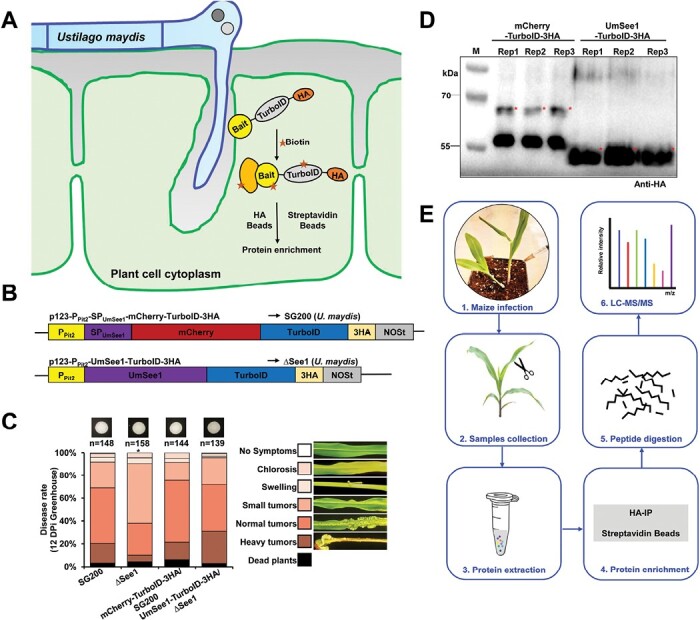
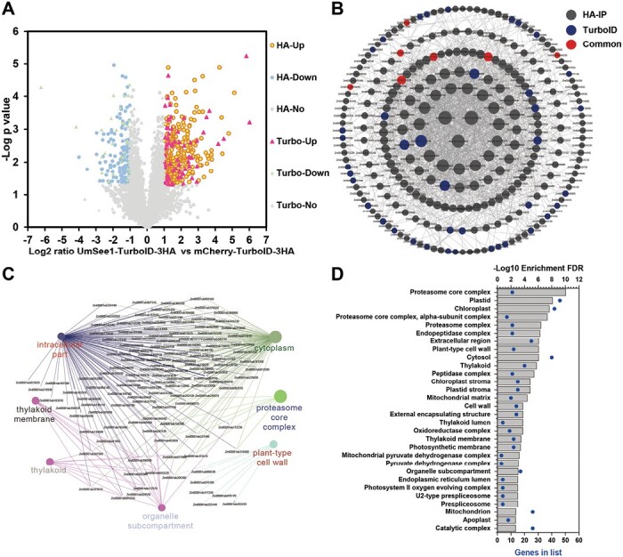
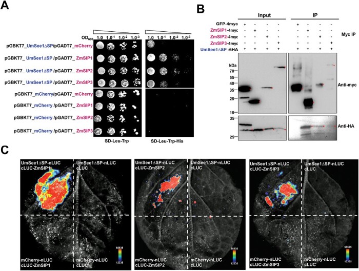
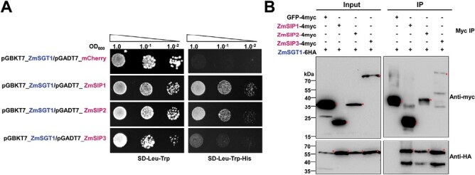
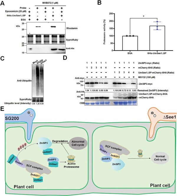
Plant pathogens secrete effectors, which target host proteins to facilitate infection. The Ustilago maydis effector UmSee1 is required for tumor formation in the leaf during infection of maize. UmSee1 interacts with maize SGT1 (suppressor of G2 allele of skp1) and blocks its phosphorylation in vivo. In the absence of UmSee1, U. maydis cannot trigger tumor formation in the bundle sheath. However, it remains unclear which host processes are manipulated by UmSee1 and the UmSee1-SGT1 interaction to cause the observed phenotype. Proximity-dependent protein labeling involving the turbo biotin ligase tag (TurboID) for proximal labeling of proteins is a powerful tool for identifying the protein interactome. We have generated transgenic U. maydis that secretes biotin ligase-fused See1 effector (UmSee1-TurboID-3HA) directly into maize cells. This approach, in combination with conventional co-immunoprecipitation, allowed the identification of additional UmSee1 interactors in maize cells. Collectively, our data identified three ubiquitin-proteasome pathway-related proteins (ZmSIP1, ZmSIP2, and ZmSIP3) that either interact with or are close to UmSee1 during host infection of maize with U. maydis. ZmSIP3 represents a cell cycle regulator whose degradation appears to be promoted in the presence of UmSee1. Our data provide a possible explanation of the requirement for UmSee1 in tumor formation during U. maydis-Zea mays interaction.

Keywords: Ustilago maydis; Fungal effectors; TurboID; maize; protein interactome; ubiquitin–proteasome.


Conflict of interest statement

The authors declare no conflicts of interest.

Figures

Fig. 1.
Fig. 1.
Experimental setup for TurboID-based biotin labeling in the Ustilago maydis–maize pathosystem. (A) Principle to isolate further UmSee1-interacting proteins and proteins in close proximity to UmSee1 in maize leaves via pull-down and TurboID-based proximity labeling (PL). (B) Schematic representation of the constructs used for HA-IP and Turbo-based PL. (C) Quantification of infection symptoms on EGB maize seedlings infected with U. maydis strains as indicated in (B) at 12 dpi. SG200, wild-type U. maydis; mCherry-TurboID-HA/SG200, mCherry–TurboID-3HA expressed in wild-type U. maydis; ΔSee1, See1 deletion mutant; UmSee1-TurboID-3HA/∆See1, UmSee1–TurboID-3HA expressed in the See1 deletion mutant. (D) Detection of mCherry–TurboID-HA and UmSee1–TurboID-HA in maize leaves upon delivery by U. maydis. For anti-HA immunoprecipitation, leaves were harvested at 3 dpi. Three independent replicates (Rep1–3) are shown. M, protein ladder. The asterisks represent target proteins. The expected sizes of mCherry–TurboID-3HA and UmSee1–TurboID-3HA proteins are 68.0 kDa and 54.5 kDa, respectively. (E) Overview of the workflow used to identify putative UmSee1 targets by Co-IP and TurboID-based biotin labeling in the U. maydis–maize pathosystem.
Fig. 2.
Fig. 2.
Identification of UmSee1-interacting proteins by Co-IP and TurboID-based biotin labeling. (A) Volcano plot analysis of identified proteins from HA-IP and TurboID-based biotin labeling by LC-MS/MS. Proteins significantly enriched in UmSee1 samples are shown in the top right corner (red and yellow dots). HA-up (yellow dots), significantly enriched proteins in UmSee1 compared with mCherry samples in the HA pull-down; HA-down (blue dots), significantly enriched proteins in mCherry compared with UmSee1 samples in the HA pull-down; HA-No (green dots), no significantly enriched proteins between UmSee1 and mCherry samples in the HA pull-down dataset; Turbo-up (red triangle), significantly enriched proteins in UmSee1 compared with mCherry samples in the TurboID-based proximity labeling; Turbo-down (purple triangles), significantly enriched proteins in mCherry compared with UmSee1 samples in the TurboID-based proximity labeling; Turbo-No (green triangles), no significantly enriched proteins between UmSee1 and mCherry samples in the TurboID-based proximity labeling dataset (P-value <0.05 and log2 fold change >1 set as significantly enriched). (B) PPI analysis of the proteins identified in UmSee1 samples using the STRING database. Proteins significantly enriched in the HA-IP and TurboID-based PL of UmSee1 samples were submitted to the STRING database, and the protein interaction network was modified and analyzed using the Cytoscape software (version 3.9.1). Edges represent protein–protein associations, including known interactions and predicted interactions (gene neighborhood, gene fusions, gene co-occurrence); the proteins identified by the different methods are marked with different colors, and the size of the shape indicates the degree of interaction. Proteins are represented by their gene ID. (C) Cluster analysis of the PPI network of the UmSee1-specific dataset by CluePedia (version 1.5.9) and ClueGo (version 2.5.9). (D) GO cellular component enrichment of the UmSee1-specific dataset using ShinyGO v0.66.
Fig. 3.
Fig. 3.
Three identified proteins enriched in theUmSee1 dataset interact with UmSee1 in one-to-one interaction assays. (A) Y2H assay of UmSee1 and three putative UmSee1 interaction partners (ZmSIPs). The yeast strain AH109 was co-transformed with mCherry or UmSee1ΔSP (UmSee1 without signal peptide) and the constructs of ZmSIPs (ZmSIP1, ZmSIP2, and ZmSIP3). A drop-out assay of a dilution series of the double transformants was carried out in the presence (SD-Trp-Leu) and absence (SD-Trp-Leu-His) of histidine. Yeast growth in the absence of histidine suggests the interaction of proteins. Pictures of three independent experiments were taken 5 d after plating. (B) Co-IP assays of UmSee1 and the three ZmSIPs. N. benthamiana leaves were transiently transformed with the constructs encoding UmSee1∆SP-6HA and ZmSIPs-4myc or GFP–4Myc. Leaves were harvested 3 d post-Agrobacterium-mediated transformation for protein extraction. Anti-myc immunoprecipitation (IP) was performed and total extracts and IP proteins were detected by western blot analysis using anti-HA and anti-myc. The expected sizes of GFP–4myc, ZmSIP1-4myc, ZmSIP2-4myc, ZmSIP3-4myc, and UmSee1∆SP-6HA proteins are 31.9, 20.5, 32.2, 86.1, and 26.4 kDa, respectively. (C) Split-luciferase assay of UmSee1 and the three ZmSIPs. N. benthamiana plants were transiently transformed with constructs of UmSee1∆SP–nLUC or mCherry–nLUC and cLUC–ZmSIPs (ZmSIP1, ZmSIP2, and ZmSIP3) or cLUC as indicated. Leaves were harvested 2 d post-Agrobacterium-mediated transformation and treated with 1 mM d-luciferin for 10 min in the dark. Shown are representative pictures of luminescence signals from three independent biological replicates. Images were detected by the CCD imaging system (ChemiDoc, Bio-Rad).
Fig. 4.
Fig. 4.
Three identified UmSee1 interactors are associated with the ZmSGT1-interacting protein complex. (A) Y2H assays of SGT1 and the three ZmSIPs. The yeast strain AH109 was co-transformed with ZmSGT1 and the three ZmSIPs, ZmSIP1, ZmSIP2, and ZmSIP3, or mCherry as a negative control. A drop-out assay of dilution series of the double transformants was carriedout in the presence (SD-Trp-Leu) and absence (SD-Trp-Leu-His) of histidine. Yeast growth in the absence of histidine suggests the interaction of proteins. Shown are representative pictures of three independent biological experiments taken 5 d after drop-out plating. (B) Co-IP assays of ZmSGT1 and the ZmSIPs. N. benthamiana leaves were transiently transformed with the ZmSGT1-6HA and ZmSIPs (ZmSIP1, ZmSIP2, and ZmSIP3)-4myc constructs. Leaves were harvested 3 d post-Agrobacterium-mediated transformation for protein extraction. Anti-myc immunoprecipitation (IP) was performed and total extracts and IP proteins were detected by western blot analysis using anti-HA and anti-myc. The expected sizes of GFP–4myc, ZmSIP1-4myc, ZmSIP2-4myc, ZmSIP3-4myc, and ZmSGT1-6HA proteins are 31.9, 20.5, 32.2, 86.1, and 53.6 kDa, respectively.
Fig. 5.
Fig. 5.
UmSee1 enhances proteasome activity and promotes ZmSIP3 degradation. (A) Activity-based protein profiling (ABPP) of the effect of recombinant UmSee1 on proteasome activity. Total proteins extracted from maize leaves were mixed with the probe MVB072. Samples were pre-incubated with BSA or recombinant UmSee1_His and with or without the proteasome inhibitor epoxomicin. Samples were analyzed by gel electrophoresis and monitored using a Rhodamine filter. SyproRuby staining served as loading control. Purified 6His-UmSee1∆SP was detected by western blot using an anti-His antibody. (B) Relative proteasome activity quantification of (A). (C) Maize ubiquitin activity upon infection with U. maydis strains SG200 and SG200ΔSee1. Leaves were harvested 3 dpi and total protein extracts were analyzed by anti-ubiquitin western blot. SyproRuby staining serves as the loading control. (D) In vitro protein degradation analysis of ZmSIP3. ZmSIP3-4myc, mCherry-6HA, and UmSee1∆SP–mCherry-6HA were separately expressed in N. benthamiana leaves. Total ZmSIP3-4myc protein lacking protease inhibitor was mixed in different proportions with mCherry-6HA or UmSee1∆SP–mCherry-6HA protein extracts in the presence of either 100 µM MG132 or DMSO (control), and incubated at 28 °C for 45 min before the reactions were stopped by boiling samples at 95 °C in 2× SDS loading buffer for 10 min. Samples were analyzed by western blot using anti-myc and anti-HA as indicated. Coomassie brilliant blue (CBB) staining served as loading control. (E) Hypothetical model for the function of UmSee1. U. maydis secretes UmSee1 protein which binds to ZmSGT1 and ZmSIPs (ZmSIP1, ZmSIP2, and ZmSIP3) to modify the SCF complex and enhance proteasome activity, affecting the degradation of cell cycle-related proteins, including ZmSIP3.

Similar articles

Cited by

References

    1. Bashir T, Dorrello NV, Amador V, Guardavaccaro D, Pagano M.. 2004. Control of the SCFSkp2–Cks1 ubiquitin ligase by the APC/CCdh1 ubiquitin ligase. Nature 428, 190–193. - PubMed
    1. Bindea G, Galon J, Mlecnik B.. 2013. CluePedia Cytoscape plugin: pathway insights using integrated experimental and in silico data. Bioinformatics 29, 661–663. - PMC - PubMed
    1. Bindea G, Mlecnik B, Hackl H, Charoentong P, Tosolini M, Kirilovsky A, Fridman WH, Pages F, Trajanoski Z, Galon J.. 2009. ClueGO: a Cytoscape plug-in to decipher functionally grouped gene ontology and pathway annotation networks. Bioinformatics 25, 1091–1093. - PMC - PubMed
    1. Bindics J, Khan M, Uhse S, et al. . 2022. Many ways to TOPLESS—manipulation of plant auxin signalling by a cluster of fungal effectors. New Phytologist 236, 1455–1470. - PubMed
    1. Branon TC, Bosch JA, Sanchez AD, Udeshi ND, Svinkina T, Carr SA, Feldman JL, Perrimon N, Ting AY.. 2018. Efficient proximity labeling in living cells and organisms with TurboID. Nature Biotechnology 36, 880–887. - PMC - PubMed

Publication types

Supplementary concepts