. 2023 May 24;14(1):3002.
doi: 10.1038/s41467-023-38684-2.

A ZFYVE21-Rubicon-RNF34 signaling complex promotes endosome-associated inflammasome activity in endothelial cells

Affiliations

A ZFYVE21-Rubicon-RNF34 signaling complex promotes endosome-associated inflammasome activity in endothelial cells

Xue Li et al. Nat Commun. .

Erratum in

Abstract

Internalization of complement membrane attack complexes (MACs) assembles NLRP3 inflammasomes in endothelial cells (EC) and promotes IL-β-mediated tissue inflammation. Informed by proteomics analyses of FACS-sorted inflammasomes, we identify a protein complex modulating inflammasome activity on endosomes. ZFVYE21, a Rab5 effector, partners with Rubicon and RNF34, forming a "ZRR" complex that is stabilized in a Rab5- and ZFYVE21-dependent manner on early endosomes. There, Rubicon competitively disrupts inhibitory associations between caspase-1 and its pseudosubstrate, Flightless I (FliI), while RNF34 ubiquitinylates and degradatively removes FliI from the signaling endosome. The concerted actions of the ZRR complex increase pools of endosome-associated caspase-1 available for activation. The ZRR complex is assembled in human tissues, its associated signaling responses occur in three mouse models in vivo, and the ZRR complex promotes inflammation in a skin model of chronic rejection. The ZRR signaling complex reflects a potential therapeutic target for attenuating inflammasome-mediated tissue injury.


Conflict of interest statement

The authors declare no competing interests.

Figures

Fig. 1
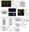
Fig. 1. Rubicon and RNF34 Modulate Endosome-Associated Inflammasome Activity.
IFN-γ-pretreated HUVECs were exposed to PRA at the indicated times prior to performing Western blotting (a, g, j) and Rab5 pulldowns (b, e, h). HUVEC stably transduced with Rab5 WT-GFP were treated with PRA for 30 min and subjected to proximity ligation assays (PLAs, c). HUVECs were co-transduced with Rubicon-GFP or RNF34-GFP along with Rab5-RFP constructs, treated with PRA for 30 min and analyzed by confocal I.F. (d, f, i). Two-tailed Student’s t-test (c, f, i) was used for statistical comparisons where p < 0.05 is considered statistically significant. Experiments repeated 2 times (c, f, i), 3 times (b, d, e, g, h, j), and 5 times (a) with different HUVEC donors. All scale bars, 200 μm. p-values are indicated in the figures.
Fig. 2
Fig. 2. ZFYVE21 sequesters Rubicon and RNF34 on Rab5 endosomes to enhance their stability.
HUVECs stably transduced with Rab5 WT or Rab5 DN were treated with PRA for the indicated times prior to I.F. analyses (a), co-IPs (b) and Western blotting (c). HUVECs were co-transduced with ZFYVE21-RFP, Rubicon-BNP, and RNF34-GFP constructs prior to I.F. analysis (d). HUVECs co-transduced with Rubicon and Rab5 or RNF34 and Rab5 constructs were analyzed following control or ZFYVE21 siRNA transfection (e). HUVECs were transfected with ZFYVE21 siRNA (f) or pre-treated vehicle or PIKIII (5 nM) prior to PRA treatment for 30 min prior to Western blot analysis (g). HUVECs were exposed to PRA in the presence or absence of cycloheximide (CHX, h, 10 μg/mL), MG132 (i, 25 μM), or leupeptin (j, 50 μM) for the indicated times prior to analysis by Western blotting. For Fig. 2h, p values involve comparisons of the respective timepoint between treatment with PRA and PRA plus cycloheximide where * indicates p < 0.05. For Fig. 2i, j, p-values involve comparisons of respective timepoint to timepoint zero where * indicates p < 0.05. HUVECs were transduced with Rab5 DN, pre-treated with MG132 and analyzed at the times indicated (k). Two-tailed Student’s t test (a, e) and one-way ANOVA (Fig. 2h-j) followed by Tukey’s pairwise comparison were used for statistical comparisons where p < 0.05 is considered statistically significant. Experiments repeated 3 times. All scale bars, 200 μm. Experiments repeated 3 times (ak). Data are presented as mean values+/-SD. p-values are indicated in the figures.
Fig. 3
Fig. 3. ZFYVE21, Rubicon, and RNF34 form heterotrimeric complexes.
PRA-treated HUVECs were co-transduced with Rubicon-GFP and RNF34-RFP constructs and analyzed by confocal I.F. a HUVECs stably transduced with Rubicon-GFP were transiently transfected with increasing concentrations of RNF34-His plasmid prior to GFP co-IPs and Western blotting (b). HUVECs stably transduced with ZFYVE21-GFP were treated with PRA for 30 min (c). Rubicon (1μg) and RNF34 (1 μg) were co-incubated at 1 h at room temperature (lanes 1-3) or overnight at 4 °C (lanes 4-6) prior to resolution via SDS-PAGE (d). Purified ZFYVE21-GFP (1 μg) was used as a probe using recombinant human Rubicon (1 μg), RNF34 (1 μg), or a 1:1 mixture of Rubicon and RNF34 as bait (e). Rubicon-GST (1 μg), RNF34-His (1 μg), or a 1:1 mixture of Rubicon and RNF34 pre-incubated for 4 h at room temperature were added to ZFYVE21-GFP-conjugated agarose beads. Protein complexes were eluted and analyzed via Western blotting under non-denaturing conditions (f). Plate-bound ZFYVE21, Rubicon, or RNF34 were incubated with varying concentrations of prey proteins as indicated in ELISAs (g). GFP-tagged constructs were overexpressed in HeLa cells following by GFP co-IPs (h, i). Rab5 DN-mCherry was stably co-transduced with ZFYVE21-GFP (j) or Rubicon-GFP (k) in HUVECs and treated with MG132 as indicated prior to GFP co-IPs. p values involve relative comparisons with no prey protein added using one-way ANOVA followed by Tukey’s pairwise comparison where * indicates p < 0.05. Experiments repeated 2 times (a, b, df, j, k), 3 times (c, j, k) or 4 times (gi) with different HUVEC donors. All scale bars, 200 μm. Data are presented as mean values+/-SD. p-values are indicated in the figures.
Fig. 4
Fig. 4. FliI constrains Rab5-associated inflammasome activity.
HUVECs were exposed to PRA at the indicated times and whole cell lysates (a) or Rab5 pulldowns (b) were analyzed by Western blot. HUVECs were treated for PRA for 30 min and assessed in confocal I.F. (c) and PLA (d) assays. HUVECs were transiently transfected with FliI siRNA (e) or a FliI overexpression construct (f) prior to treatment with PRA for 30 min in Western blot studies. HUVECs were transfected with control or FliI siRNA and treated for 4 h prior to analyses of culture supernatants (g). Two-tailed Student’s t-test was used for statistical comparisons (c, d) where p < 0.05 is considered statistically significant. Experiments repeated 3 times (ag) with different HUVEC donors. All scale bars, 200 μm. p-values are indicated in the figures.
Fig. 5
Fig. 5. Rubicon and RNF34 cooperatively reduce the stability of Rab5-associated fliI.
HUVECs were exposed to PRA for the indicated times prior to performing pulldowns for FliI (a) and caspase-1 (b). HeLa cells were transiently transfected with various GFP-tagged overexpression vectors prior to GFP co-IPs (c). HUVECs were transiently transfected with increasing plasmid concentrations encoding AA 625-760 of Rubicon fused with GFP, (d) or full-length FliI (e) followed by pulldowns for caspase-1. HUVECs stably transduced with Ubiquitin WT-HA (Ub-HA) were exposed to PRA for the indicated times followed by pulldowns for FliI (f). Ub-HA HUVECs were transfected with control or RNF34 siRNA as indicated and treated with PRA for 30 min (g). HUVECs were transfected with siRNA as indicated followed by Rab5 co-IPs following PRA treatment for 30 min (h). In ubiquitin ligase assays, recombinant human FliI (0.5 μg) was incubated as substrate with activated Ub, the indicated E2 ligase(s), and native RNF34 protein (0.5 μg, i) or RNF34 truncation mutants prior to Western blotting (j). Experiments repeated 3 times with different HUVEC donors.
Fig. 6
Fig. 6. ZRR complexes form in patient tissues.
Cortical renal tissue from control patients were placed in organ culture and treated with PRA for 4 h prior to ZFYVE21 co-IP and Western blot analysis (a, n = 3). Cortical renal tissues from healthy control (HC) or antibody-mediated rejection (ABMR) were analyzed by co-IP (n = 6 per group, b). ABMR tissues were analyzed by dual immune-electron microscopy for ZFYVE21 (arrowheads, n = 3, c) and Rubicon (arrows). FFPE biopsies from patients with CABMR (n = 6, d), RA (n = 4, e), or SLE (n = 5, f) were stained and analyzed as indicated. Transcriptomic data from renal tissues from renal transplant patients with CABMR (n = 110), synovial tissues from patients with RA (n = 23), and renal tissues from patients with SLE nephritis (n = 21) were obtained from the Gene Expression Omnibus. One-way ANOVA with Tukey’s post-hoc comparison (a), Two-tailed Student’s t test (b, df), and Pearson’s correlations (g, h) were used for statistical comparisons where p < 0.05 is considered statistically significant. All scale bars, 400 μm. Data are presented as mean values+/-SD. p-values are indicated in the figures. All scale bars, 200 μm.
Fig. 7
Fig. 7. ZRR complex-associated signaling occurs in vivo.
Human artery xenografts implanted in immunodeficient SCID/beige hosts were exposed to PRA for 24 h (200 μL i.v. tail vein injection per mouse) and analyzed in proximity ligation assays (a, n = 3 per group). Sera from SCID/beige hosts bearing PRA-treated human artery xenografts were analyzed by Western blot (b, n = 3 per group). HUVECs stably transduced with Rab5 WT mCherry or Rab5 DN mCherry (c), Rubicon shRNA (d) or RNF34 shRNA (e) were embedded in collagen gel matrices, implanted in SCID/beige mice for 2–4 weeks, and microvessels were exposed to PRA for 24 h (200 μL i.v. tail vein injection per mouse) prior to staining and analysis by I.F (n = 3 per group). Sera from SCID/beige hosts implanted with collagen gel matrices were analyzed by Western blot (f, n = 3 per group). C57/Bl6 or Rubicon-/-mice were injected with 750 μg MOPC or anti-H-2b Abs (g), and kidneys were harvested for Rab5 co-IPs 24 h later (n = 3 per group, h). Figure 7g was created with BioRender.com. Two-tailed Student’s t-test was used for statistical comparisons (a, ce) where p < 0.05 is considered statistically significant. Experiments repeated 3 times. All scale bars, 400 μm. p-values are indicated in the figures.
Fig. 8
Fig. 8. ZRR complexes regulate complement-induced vascular injury in vivo.
HUVECs stably transduced with an NF-κB luciferase reporter were treated with PRA and luciferase activity were assessed following treatment with PRA for 6 h (a). Quantitative epifluorescence analysis of nuclear translocation of the P65 NF-κB subunit in HUVECs transfected with siRNA and treated with PRA for 4 h (b). qRT-PCR of siRNA-transfected HUVECs exposed to PRA for 4 h (c). EC:T cell cocultures were performed using HUVECs transfected with various siRNA as indicated, and CFSE-labeled T cells were harvested and analyzed by FACS after 10–14 days in coculture (d). Male WT or Rubicon-/-mice received 200 μL tail vein injection of MOPC or anti-H-2b Ab (750 μg) and 24 h later, male skin grafts were harvested and analyzed by I.F. implanted onto female SCID/bg hosts that had passively received 1 × 106 female WT C57/Bl6 splenocytes. Three weeks post-implantation, skin grafts were analyzed (e). Following MOPC or anti-H-2b Ab injection i.v. (750 μg), kidney tissues from age-matched WT or Rubicon-/- mice were harvested 24 h later and analyzed for glomerular VCAM-1 staining on CD31+ECs prior to host implantation (f, n = 8 mice per group). Three weeks post-implantation, male skin grafts treated with MOPC or anti-H-2b Ab as above were analyzed by I.F. (g, n = 8 mice per group) and H&E sections (h, n = 8 mice per group). Schema of the proposed mechanism for ZRR complexes (i). For Fig. 8a–d, experiments repeated 3 times with different HUVEC donors. For Fig. 8a–d, f–h two-way ANOVA followed by Tukey’s pairwise comparison was used for statistical analysis where p < 0.05 is considered statistically significant. Figures 8e and 8i were created with BioRender.com. All scale bars, 400 μm. p-values are indicated in the figures.

References

    1. Liu L, et al. Endothelial Cell-Derived Interleukin-18 Released During Ischemia Reperfusion Injury Selectively Expands T Peripheral Helper Cells to Promote Alloantibody Production. Circulation. 2020;141:464–478. doi: 10.1161/CIRCULATIONAHA.119.042501. - DOI - PMC - PubMed
    1. Xie CB, et al. Complement Membrane Attack Complexes Assemble NLRP3 Inflammasomes Triggering IL-1 Activation of IFN-γ-Primed Human Endothelium. Circ. Res. 2019;124:1747–1759. doi: 10.1161/CIRCRESAHA.119.314845. - DOI - PMC - PubMed
    1. Xie CB, et al. Complement-activated interferon-γ-primed human endothelium transpresents interleukin-15 to CD8+ T cells. J. Clin. Invest. 2020;130:3437–3452. doi: 10.1172/JCI135060. - DOI - PMC - PubMed
    1. Migita K, et al. Serum amyloid A triggers the mosodium urate -mediated mature interleukin-1β production from human synovial fibroblasts. Arthritis Res Ther. 2012;14:R119. doi: 10.1186/ar3849. - DOI - PMC - PubMed
    1. Lei-Leston AC, Murphy AG, Maloy KJ. Epithelial Cell Inflammasomes in Intestinal Immunity and Inflammation. Front Immunol. 2017;8:1168. doi: 10.3389/fimmu.2017.01168. - DOI - PMC - PubMed

Publication types