. 2023 May 24;13(1):8382.
doi: 10.1038/s41598-023-34934-x.

Topographical influence of electrospun basement membrane mimics on formation of cellular monolayer

Affiliations

Topographical influence of electrospun basement membrane mimics on formation of cellular monolayer

Puja Jain et al. Sci Rep. .

Abstract

Functional unit of many organs like lung, kidney, intestine, and eye have their endothelial and epithelial monolayers physically separated by a specialized extracellular matrix called the basement membrane. The intricate and complex topography of this matrix influences cell function, behavior and overall homeostasis. In vitro barrier function replication of such organs requires mimicking of these native features on an artificial scaffold system. Apart from chemical and mechanical features, the choice of nano-scale topography of the artificial scaffold is integral, however its influence on monolayer barrier formation is unclear. Though studies have reported improved single cell adhesion and proliferation in presence of pores or pitted topology, corresponding influence on confluent monolayer formation is not well reported. In this work, basement membrane mimic with secondary topographical cues is developed and its influence on single cells and their monolayers is investigated. We show that single cells cultured on fibers with secondary cues form stronger focal adhesions and undergo increased proliferation. Counterintuitively, absence of secondary cues promoted stronger cell-cell interaction in endothelial monolayers and promoted formation of integral tight barriers in alveolar epithelial monolayers. Overall, this work highlights the importance of choice of scaffold topology to develop basement barrier function in in vitro models.


Conflict of interest statement

The authors declare no competing interests.

Figures
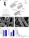
Figure 1
Figure 1
(A) Schematic representation of the mechanism of pore formation on fiber surface during the electrospinning process. The secondary nano-structure on the microfiber is obtained through a process of phase separation when an optimum ratio of good and bad solvent is used during solution preparation. (B) represents homogenous non-woven electron micrograph of smooth surface fibers and (C) porous surface fibers. The graphs represent fabrication of non-woven meshes with (D) similar fiber diameter, and (E) mean mesh pore area of both fiber types, (F) represents pore area distribution of the secondary surface pores present on the fiber surface shown in c; n = 5.
Figure 2
Figure 2
Cell proliferation was analyzed using XTT assay for (A) H441 epithelial cells and (E) HUVEC endothelial cells on smooth and porous fiber surfaces on day 3 and 9, where proliferation of cells was higher on porous surface fibers on day 3 but no difference was observed for longer culture period day 9; n = 3. Single cell area was investigated using ImageJ where (B) H441 cells that did not show significant difference but (F) HUVEC displayed a larger cell area on porous surface fibers; n = 115. Confocal images where (C), (D) H441 cells and (G), (H) HUVEC are immunostained to visualize F-actin filaments (green), and nucleus (blue). H441 cells appear round on both fiber meshes, however, HUVEC appear elongated and occupy larger area on the porous surface fiber meshes; n = 115. Confocal images were also taken to visualize HUVEC on (I) smooth surface fiber meshes and (J) porous surface fiber meshes for focal adhesion vinculin (red) and nucleus (blue). The dotted boxes in (I), (J) represent mature vinculin on the porous compared to weak intensity points by cells on smooth fiber surfaces. (K) Using ImageJ, the fluorescence intensity of vinculin was quantified. Higher intensity of vinculin was displayed by cells on the porous surface fibers; n = 25. The line in the middle of the box denotes the median and the whiskers denote the minimum and maximum values. Scale bar: 20 µm.
Figure 3:
Figure 3:
10-day culture of HUVEC endothelial monolayers on (A) smooth and (B) porous fiber surfaces were immune-stained to visualize F-actin filaments (green), cell–cell adhesion junction VE-cadherin (red) and nucleus (blue). Area occupied by individual cells in the monolayer was lower on smooth compared to that on porous surface fibers as seen in graph (C), where the line in the middle of the box denotes the median and the whiskers denotes the minimum and maximum values. The organization of F-actin filaments is majorly on the periphery of cells on the smooth fibers compared to the porous surface fibers where the actin filaments are more spread and depict stress fiber formation. Scale bar: 20 µm. Endothelial to mesenchymal transition is absent as cells did not stain positive for the mesenchymal marker αSMA on both (D) smooth and (E) porous surface fibers, (F) The actin profile was quantified for single cells along their long axis. n = 30. Scale bar: 50 µm.
Figure 4
Figure 4
Integrity and spread of H441 epithelial cells was analyzed with increasing culture period to mark the time point at which tight junctions are formed as well as confluency is achieved. (AH) H441 cells were immunostained for cell–cell tight junction ZO-1 (red), vinculin (green) and nucleus (blue). H441 cells appear to form (A) non-confluent monolayers on smooth surface fibers as seen in the cross-section view compared to (E) non-confluent multilayered formation on porous surface fibers. The initial tight junctions are formed already on (BD) day 2 cultures of smooth fiber surfaces and tend to increase with time where cells reach confluent barrier after day 5, (FH) but initial tight junction formation is only observed later on day 4 and attain confluent barriers after day 5 on porous surface fibers. This trend is supported by (I) cell spread area where H441 cells form multilayered island groups on porous surface fibers and monolayer islands on smooth surface fibers. The area increases with time as cells cover and spread more to form integral monolayers by day 6; (J) the barrier properties were analyzed quantitatively by periodic measurements of TEER as well where cells initially display higher values from day 2 onwards on smooth surface fibers. The values reach a plateau phase with culture time when cells reach confluency in both the cases. n = 3. Scale bar: 100 µm.
Figure 5
Figure 5
Monolayer H441 cells were analyzed for presence of characteristic properties of alveolar type II cells including lamellar bodies and microvilli formation. Day-10 H441 cells were stained to visualize lamellar bodies using Nile red on (A) smooth and (B) porous surface fibers, Scale bar: 50 µm, and quantitatively analyzed using ImageJ (C) where a significant increase was found on smooth surface fibers; n = 25. Closer view of the lamellar bodies is shown on (D) smooth and (E) porous surface, Scale bar: 20 µm, (F) This surfactant storing organelle was also quantified to have a diameter of nearly 1 µm in both the cases; n = 535. Microvilli, an important feature involved in increasing cell surface area was also well developed as seen on electron micrographs of (G) smooth than on (H) porous surface fibers. For box graphs (C and F), the line in the middle of the box denotes the median and the whiskers denotes the minimum and maximum values. Scale bar: 10 µm.

References

    1. Marastoni S, Ligresti G, Lorenzon E, Colombatti A, Mongiat M. Extracellular matrix: A matter of life and death. Connect. Tissue Res. 2008;49:203–206. doi: 10.1080/03008200802143190. - DOI - PubMed
    1. Kruegel J, Miosge N. Basement membrane components are key players in specialized extracellular matrices. Cell. Mol. Life Sci. 2010;67:2879–2895. doi: 10.1007/s00018-010-0367-x. - DOI - PMC - PubMed
    1. Morrissey MA, Sherwood DR. An active role for basement membrane assembly and modification in tissue sculpting. J. Cell Sci. 2015;128:1661–1668. - PMC - PubMed
    1. Farquhar MG. The glomerular basement membrane: Not gone, just forgotten. J. Clin. Investig. 2006;116:2090–2093. doi: 10.1172/JCI29488. - DOI - PMC - PubMed
    1. Vukicevic S, et al. Identification of multiple active growth factors in basement membrane Matrigel suggests caution in interpretation of cellular activity related to extracellular matrix components. Exp. Cell Res. 1992;202:1–8. doi: 10.1016/0014-4827(92)90397-Q. - DOI - PubMed

Publication types