. 2023 Dec;37(18):3734-3742.
doi: 10.1038/s41433-023-02581-1. Epub 2023 May 24.

Deep phenotypic characterization of the retinal dystrophy in patients with RNU4ATAC-associated Roifman syndrome

Affiliations

Deep phenotypic characterization of the retinal dystrophy in patients with RNU4ATAC-associated Roifman syndrome

Brian G Ballios et al. Eye (Lond). 2023 Dec.

Abstract

Purpose: To characterize the retinal phenotype in RNU4ATAC-associated Roifman syndrome.

Methods: Ten patients (including 8 males) with molecularly confirmed Roifman syndrome underwent detailed ophthalmologic evaluation including fundus imaging, fundus autofluorescence (FAF) imaging, spectral-domain optical coherence tomography (SD-OCT), and electroretinography (ERG). Six patients had follow-up eye exams. All patients also underwent comprehensive examination for features of extra-retinal Roifman syndrome.

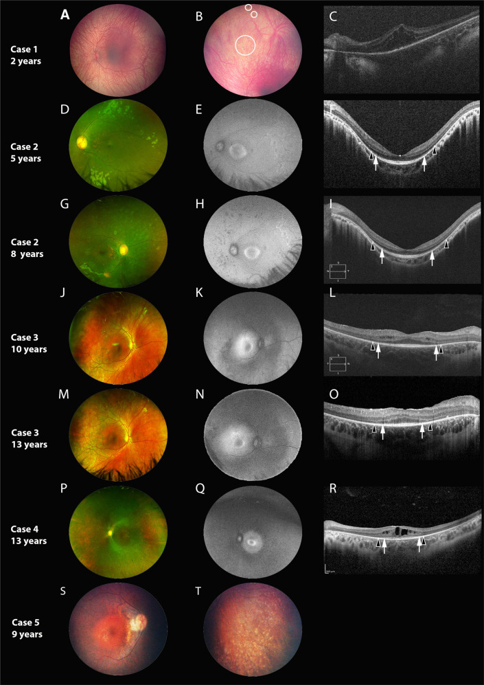
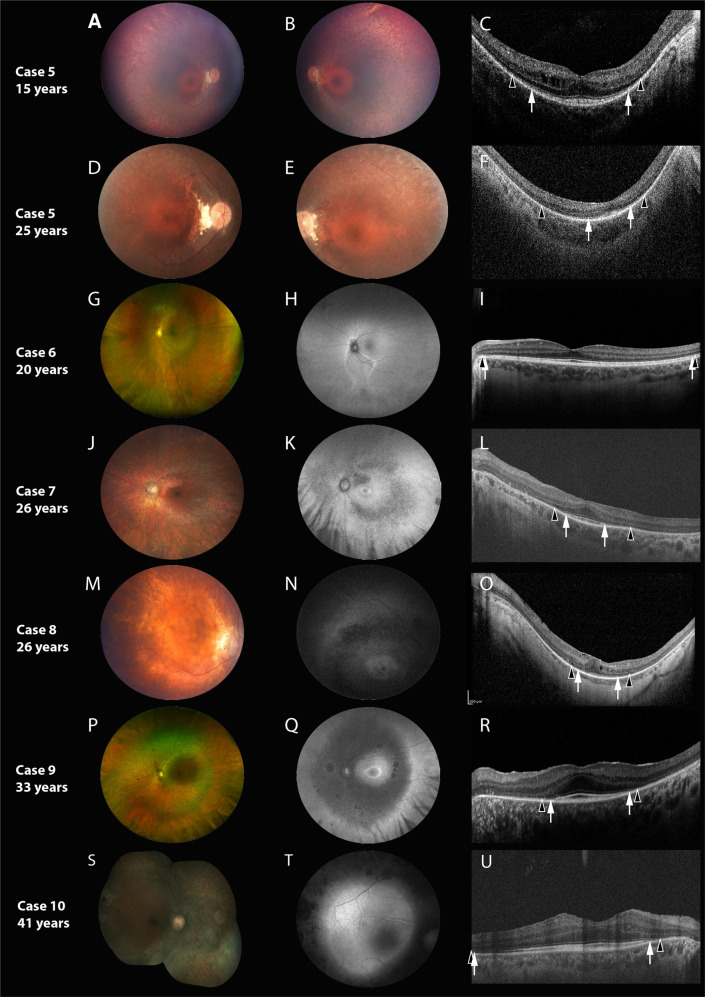
Results: All patients had biallelic RNU4ATAC variants. Nyctalopia was common (7/10). Visual acuity at presentation ranged from 20/20 to 20/200 (Age Range: 5-41 years). Retinal exam revealed features of generalized retinopathy with mid-peripheral pigment epithelial changes. A para or peri-foveal ring of hyper-autofluorescence was the commonest FAF abnormality noted (6/8). The SD-OCT demonstrated relative preservation of the foveal ellipsoid zone in six cases; associated features included cystoid changes (5/10) and posterior staphyloma (3/10). The ERG was abnormal in all patients; nine showed generalized rod-cone dystrophy, whilst one patient with sectoral retinal involvement only had isolated rod dystrophy (20 years old). On follow-up examination (Mean duration: 8.16 years), progressive loss of visual acuity (2/6), mid-peripheral retinal atrophy (3/6) or shortening of ellipsoid zone width (1/6) were observed.

Conclusion: This study has characterized the retinal phenotype in RNU4ATAC-associated Roifman syndrome. Retinal involvement is universal, early-onset, and overall, the retinal and FAF features are consistent with rod-cone degeneration that is slowly progressive over time. The sub-foveal retinal ultrastructure is relatively preserved in majority of patients. Phenotypic variability independent of age exists, and more study of allelic- and sex-based determinants of disease severity are necessary.


Conflict of interest statement

The authors declare no competing interests.

Figures

Fig. 1
Fig. 1. Retinal imaging of Cases 1–5.
Each horizontal panel represents a single patient visit with fundus photo, autofluorescence (FAF, if available) and optical coherence tomography (OCT, if available). A–C represent Case 1, (D–I) represent Case 2, (J–O) represent Case 3, (P–R) represent Case 4 and (S, T) represent Case 5. Cases are organized in terms of increasing age at characterization, with repeat imaging for Cases 2 and 3 provided at follow-up. Black arrowheads demarcate the preserved horizontal extent of the external limiting membrane (ELM); white arrows demarcate the extent of the ellipsoid zone (EZ). The fundus is characterized by retinal pigment epithelial and pigmentary changes along the arcades and mid-periphery, with associated arteriolar attenuation, without significant disc pallor. A dull foveal reflex is also characteristic. In the youngest patient (A) the fundus is blonde with prominent choroidal vasculature and no vessel attenuation, with pigmentary changes only visible in the far periphery (B, circles). A para-foveal (Q) or peri foveal (E, H, K, N) hyper autofluorescent ring was commonly observed. The ring demonstrated a reduction in diameter, becoming more distinct and closer to the foveal centre with increasing age. The peripheral areas of hypofluorescence on FAF corresponded well with the retinal areas of retinal atrophy. Findings on macular OCT are characterized by relatively preserved foveal outer nuclear layer, with most cases showing preservation of outer retinal lamination (EZ and ELM) in the central 2 mm (F, I, L, O, R). Around the margins of EZ loss, ELM could be identified. Three cases showed cystoid/schitic macular changes (C, L, R).
Fig. 2
Fig. 2. Detailed retinal characteristics of Cases 5-10.
Each horizontal panel represents a single patient visit with fundus photo, autofluorescence (FAF, if available) and optical coherence tomography (OCT). A–F Represent Case 5, (G–I) represent Case 6, (J–L) represent Case 7, (M–O) represent Case 8, (P–R) represent Case 9 and (S–U) represent Case 10. Cases are organized in terms of increasing age at characterization, with repeat imaging for Cases 5 provided at follow-up. Black arrowheads demarcate the preserved horizontal extent of the external limiting membrane (ELM); white arrows demarcate the extent of the ellipsoid zone (EZ). The fundus is characterized by retinal pigment epithelial and pigmentary changes along the arcades and mid-periphery, with associated arteriolar attenuation, without significant disc pallor. Bilateral disc drusen was observed in Case 5 (D, E). A parafoveal (K, N) or peri-foveal (Q) hyper autofluorescent ring was commonly observed. The peripheral areas of hypofluorescence corresponded well with the retinal areas of retinal atrophy. Cases 6 had normal macular autofluorescence (H) or whereas, Case 10 had generalized macular hyper autofluorescence, respectively (T). Widefield imaging in Case 6 shows (G) inferior sectoral RPE and pigmentary mottling; FAF (H) shows a corresponding area of sectoral hypo-autofluorescence, with a hyper-autofluorescent border. Two cases showed relative preservation of outer retinal lamination (EZ and ELM) across most of temporal macular line scan (I, U); whereas Case 5 at the last visit (F) and Case 8 (O) showed mild disruption of the sub-foveal EZ. Two cases showed mild cystoid macular changes (C, O).

References

    1. Roifman CM. Antibody deficiency, growth retardation, spondyloepiphyseal dysplasia and retinal dystrophy: a novel syndrome. Clin Genet. 1999;55:103–9. doi: 10.1034/j.1399-0004.1999.550206.x. - DOI - PubMed
    1. Merico D, Roifman M, Braunschweig U, Yuen RK, Alexandrova R, Bates A, et al. Compound heterozygous mutations in the noncoding RNU4ATAC cause Roifman Syndrome by disrupting minor intron splicing. Nat Commun. 2015;6:8718. doi: 10.1038/ncomms9718. - DOI - PMC - PubMed
    1. Dinur Schejter Y, Ovadia A, Alexandrova R, Thiruvahindrapuram B, Pereira SL, Manson DE, et al. A homozygous mutation in the stem II domain of RNU4ATAC causes typical Roifman syndrome. NPJ Genom Med. 2017;2:23. doi: 10.1038/s41525-017-0024-5. - DOI - PMC - PubMed
    1. He H, Liyanarachchi S, Akagi K, Nagy R, Li J, Dietrich RC, et al. Mutations in U4atac snRNA, a component of the minor spliceosome, in the developmental disorder MOPD I. Science. 2011;332:238–40. doi: 10.1126/science.1200587. - DOI - PMC - PubMed
    1. Abdel-Salam GM, Miyake N, Eid MM, Abdel-Hamid MS, Hassan NA, Eid OM, et al. A homozygous mutation in RNU4ATAC as a cause of microcephalic osteodysplastic primordial dwarfism type I (MOPD I) with associated pigmentary disorder. Am J Med Genet A. 2011;155A:2885–96. doi: 10.1002/ajmg.a.34299. - DOI - PubMed

Supplementary concepts

LinkOut - more resources