. 2023 Oct;44(10):2048-2064.
doi: 10.1038/s41401-023-01103-9. Epub 2023 May 25.

β-arrestin2 deficiency ameliorates S-100-induced autoimmune hepatitis in mice by inhibiting infiltration of monocyte-derived macrophage and attenuating hepatocyte apoptosis

Affiliations

β-arrestin2 deficiency ameliorates S-100-induced autoimmune hepatitis in mice by inhibiting infiltration of monocyte-derived macrophage and attenuating hepatocyte apoptosis

Ting-Ting Chen et al. Acta Pharmacol Sin. 2023 Oct.

Abstract

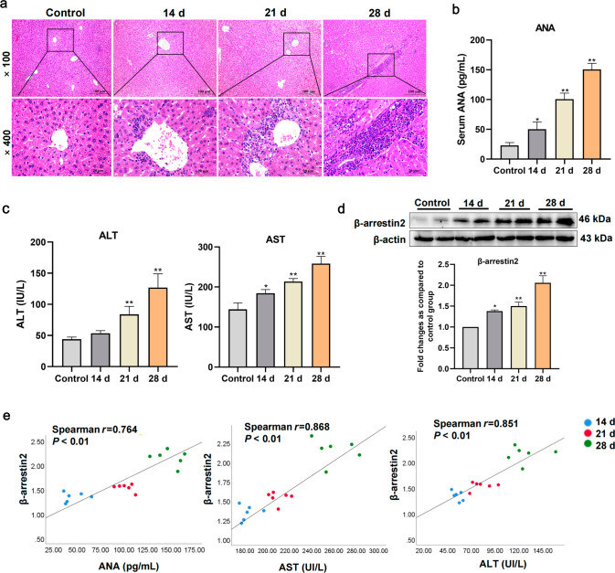
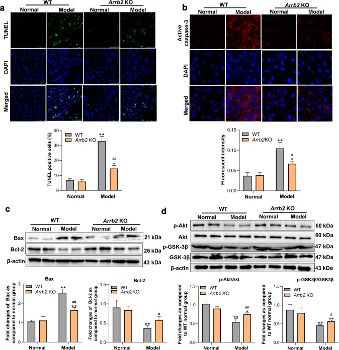
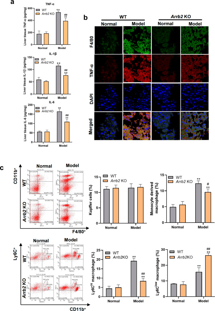
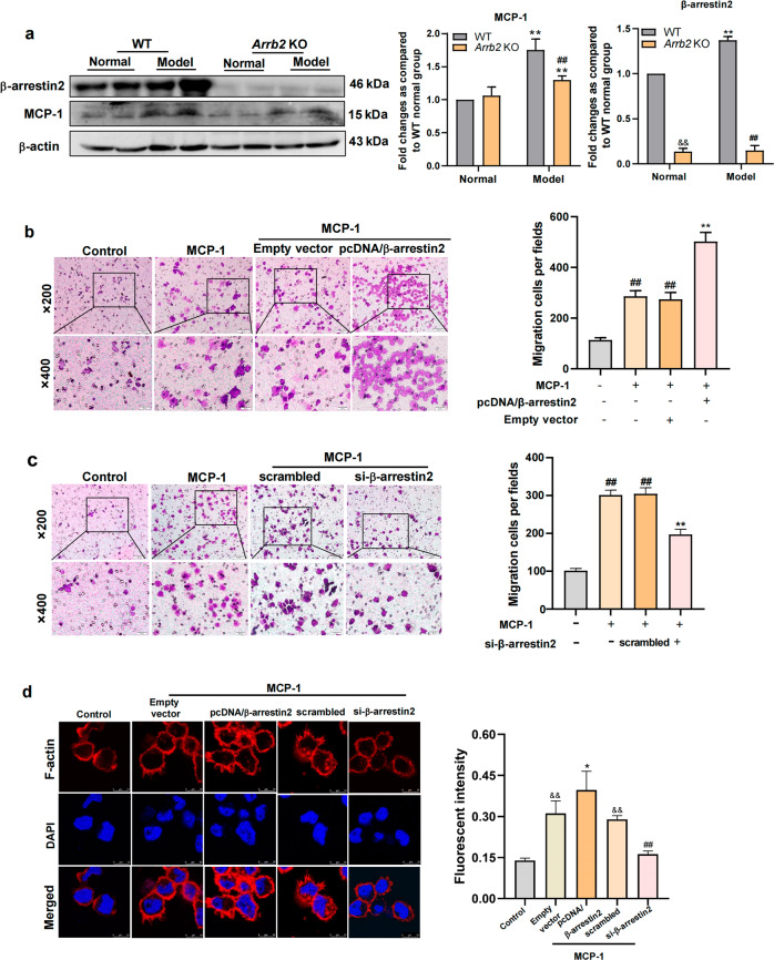
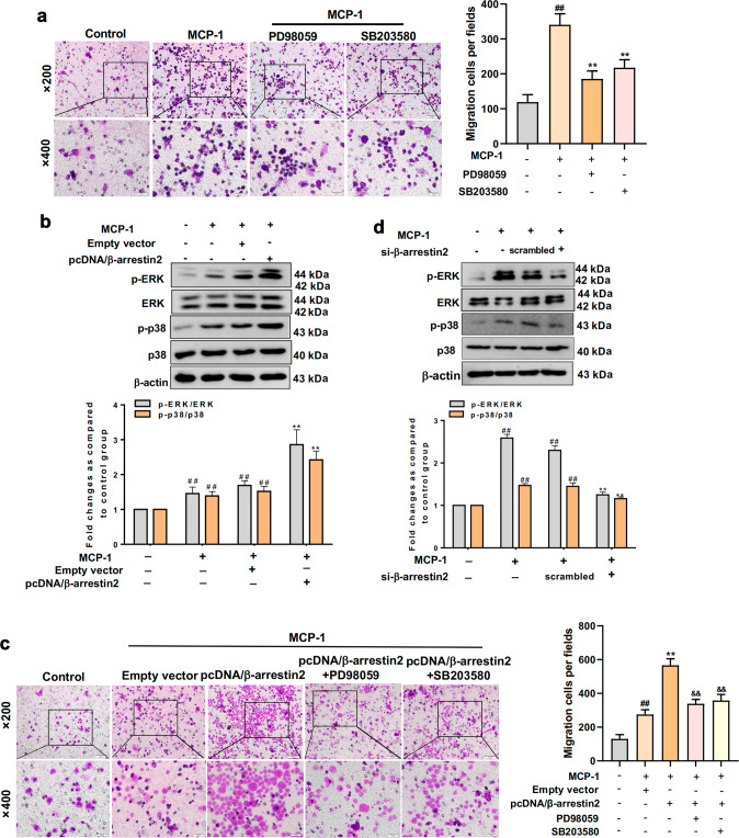
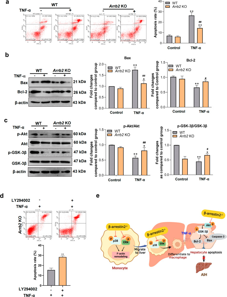
Autoimmune hepatitis (AIH) is a progressive hepatitis syndrome characterized by high transaminase levels, interface hepatitis, hypergammaglobulinemia, and the presence of autoantibodies. Misdiagnosis or delayed treatment of AIH can lead to cirrhosis or liver failure, which poses a major risk to human health. β-Arrestin2, a key scaffold protein for intracellular signaling pathways, has been found to be involved in many autoimmune diseases such as Sjogren's syndrome and rheumatoid arthritis. However, whether β-arrestin2 plays a role in AIH remains unknown. In the present study, S-100-induced AIH was established in both wild-type mice and β-arrestin2 knockout (Arrb2 KO) mice, and the experiments identified that liver β-arrestin2 expression was gradually increased, and positively correlated to serum ANA, ALT and AST levels during AIH progression. Furthermore, β-arrestin2 deficiency ameliorated hepatic pathological damage, decreased serum autoantibody and inflammatory cytokine levels. β-arrestin2 deficiency also inhibited hepatocyte apoptosis and prevented the infiltration of monocyte-derived macrophages into the damaged liver. In vitro experiments revealed that β-arrestin2 knockdown suppressed the migration and differentiation of THP-1 cells, whereas β-arrestin2 overexpression promoted the migration of THP-1 cells, which was regulated by the activation of the ERK and p38 MAPK pathways. In addition, β-arrestin2 deficiency attenuated TNF-α-induced primary hepatocyte apoptosis by activating the Akt/GSK-3β pathway. These results suggest that β-arrestin2 deficiency ameliorates AIH by inhibiting the migration and differentiation of monocytes, decreasing the infiltration of monocyte-derived macrophages into the liver, thereby reducing inflammatory cytokines-induced hepatocytes apoptosis. Therefore, β-arrestin2 may act as an effective therapeutic target for AIH.

Keywords: MCP-1; TNF-α; autoimmune hepatitis; hepatocyte; macrophage; monocyte; β-arrestin2.


Conflict of interest statement

The authors declare no competing interests.

Figures

Fig. 1
Fig. 1. Expression of β-arrestin2 increased in liver during autoimmune hepatitis (AIH) development.
a Representative hematoxylin-eosin staining images of liver tissue in control mice and S-100-treated mice at 14, 21, and 28 days (×100 and ×400 magnifications). b Detection of antinuclear antibody (ANA) levels in serum of mice treated with S-100 at different times. c Detection of serum alanine aminotransferase (ALT) and aspartate aminotransferase (AST) activities in S-100-treated mice at different times. d β-arrestin2 expression in liver tissue of S-100-induced AIH mice at different times was detected using Western blotting. The value of the control group was 1. Representative pictures of the bands are shown. e The correlation analyses of β-arrestin2 and serum ANA, ALT, and AST levels during AIH progression. n = 6 mice per group. *P < 0.05, **P < 0.01 compared with the control group.
Fig. 2
Fig. 2. β-arrestin2 deficiency ameliorates AIH in mice.
a Representative liver tissue hematoxylin-eosin staining images from wild-type (WT) and Arrb2 KO mice treated with S-100 (×100 and ×400 magnifications). b Detection of serum levels of ANA in mice treated with S-100. c Detection of serum alanine aminotransferase and aspartate aminotransferase activities in mice treated with S-100. d The percentages of CD3+CD4+ T and CD3+CD8+ T cells in the liver were analyzed. e The percentages of Treg (CD4+CD25+Foxp3+) and Th17 (CD4+IL-17+) cells in the liver were analyzed. f The percentages of Th1 (CD4+IFN-γ+) and Th2 (CD4+IL-4+) cells in the liver were analyzed. n = 6 mice per group. *P < 0.05, **P < 0.01 compared with the normal mice; #P < 0.05, ##P < 0.01 compared with the WT model mice.
Fig. 3
Fig. 3. β-arrestin2 deficiency reduces hepatocyte apoptosis in AIH.
a Livers were removed from mice. Apoptotic cells were determined using the terminal deoxynucleotidyl transferase biotin-dUTP nick end labeling staining assay. Representative images in different groups using a fluorescence microscope. b The immunofluorescence staining of active caspase-3 in liver tissue was performed using a fluorescence microscope. c Western blotting of Bax and Bcl-2 expression, apoptosis-related proteins, from the liver tissue of WT and Arrb2 KO mice treated with S-100. d Liver tissue protein extracts were subjected to immunoblotting for Akt, p-Akt, GSK-3β, and p-GSK-3β. The value of the normal group was visualized as 1. n = 6 mice per group. *P < 0.05, **P < 0.01 compared with the normal group; #P < 0.05, ##P < 0.01 compared with the WT model group.
Fig. 4
Fig. 4. β-arrestin2 deficiency inhibits inflammation and infiltration of monocyte-derived macrophages in AIH.
a Enzyme-linked immunosorbent assay was used to detect the effect of β-arrestin2 deficiency on the production of TNF-α, IL-6, and IL-1β in the liver of AIH mice. b Immunofluorescence staining of fixed liver tissue using the murine macrophage-specific monoclonal F4/80 antibodies (green) and TNF-α (red). The nuclei were examined using 4′,6-diamidino-2-phenylindole (blue). c CD45, F4/80, CD11b, and Ly6C were used as markers to analyze the changes in three different hepatic macrophage populations in AIH using flow cytometry. A representative staining image is shown. n = 6 mice per group. *P < 0.05, **P < 0.01 compared with the normal mice; #P < 0.05, ##P < 0.01 compared with the WT model mice.
Fig. 5
Fig. 5. β-arrestin2 plays a crucial role in THP-1 monocyte migration.
a Western blotting of β-arrestin2 and monocyte chemoattractant protein 1 (MCP-1) from liver tissue in AIH. The value of the normal group was visualized as 1. Representative pictures of the bands are shown. **P < 0.01 compared with normal mice; ##P < 0.01 compared with WT model mice. &&P < 0.01 compared with WT normal mice. b Overexpression of β-arrestin2 increased the numbers of migrated THP-1 cells induced by MCP-1. ##P < 0.01 compared with control group; **P < 0.01 compared with empty vector group. c β-arrestin2 knockdown decreased the number of migrated THP-1 cells induced by MCP-1. ##P < 0.01 compared with control group; **P < 0.01 compared with the scrambled group. d Immunocytochemical staining for F-actin remodeling using TRITC phalloidin (red). The nuclei were examined by 4′,6-diamidino-2-phenylindole (blue). &&P < 0.01 compared with the control group; *P < 0.05 compared with the empty vector group; ##P < 0.01 compared with the scrambled group.
Fig. 6
Fig. 6. β-arrestin2 regulates the migration of THP-1 monocytes via ERK and p38 MAPK signaling pathways.
a Effects of PD98059 and SB203580 on the migration of THP-1 cells induced by MCP-1 detected using the Transwell assay. ##P < 0.01 compared with control group, **P < 0.01 compared with the MCP-1 group. b β-arrestin2 overexpression promoted the activation of ERK and p38 MAPK pathways in THP-1 cells. ##P < 0.01 compared with control group. **P < 0.01 compared with empty vector group. c Effects of β-arrestin2 overexpressed plasmid on the migration abilities of THP-1 cells treated with PD98059 or SB203580. ##P < 0.01 compared with control group; **P < 0.01 compared with empty vector group; &&P < 0.01 compared with the β-arrestin2 overexpression group. d Effects of β-arrestin2 siRNA on the activation of ERK and p38 MAPK pathways in MCP-1-stimulated THP-1 cells. ##P < 0.01 compared with control group; **P < 0.01 compared with the scrambled group.
Fig. 7
Fig. 7. Silencing of β-arrestin2 inhibits differentiation of THP-1 cells to macrophages and reduces hepatocyte apoptosis induced by TNF-α secreted from macrophages.
a After treatment with phorbol-12-myristate-13-acetate (PMA) for 12–48 h, β-arrestin2 expression in THP-1 cells was analyzed using Western blotting. **P < 0.01 compared with the control group. b After transfection with β-arrestin2 siRNA, macrophage surface marker CD68 (red) expression was analyzed using immunofluorescence. The nuclei were examined by 4′,6-diamidino-2-phenylindole (blue). c The expressions of CD68 and CD36 in THP-1 cells treated with PMA were detected using flow cytometry after interfering with β-arrestin2 siRNA. ##P < 0.01 compared with control group; **P < 0.01 compared with scrambled group. d Immunofluorescence was used to observe the effects of PD98059 or SB203580 on CD68 expression in THP-1 cells stimulated by PMA. ##P < 0.01 compared with control group; **P < 0.01 compared with PMA group. e Western blotting revealed the expression of p-ERK and p-p38 in THP-1 cells stimulated with PMA after transfection with β-arrestin2 siRNA. ##P < 0.01 compared with the control group; **P < 0.01 compared with the scrambled group. f Effects of β-arrestin2 on TNF-α secretion by monocyte-derived macrophages detected using enzyme-linked immunosorbent assay. &&P < 0.01 compared with the control group; **P < 0.01compared with the empty vector group; ##P < 0.01 compared with the scrambled group. g The apoptosis rates of L02 cells cultured with conditioned media (CM). &&P < 0.01 compared with the control-CM group; **P < 0.01 compared with the empty vector-CM group; ##P < 0.01 compared with the scrambled-CM group. $$P < 0.01 compared with the pcDNA-β-arrestin2-CM group.
Fig. 8
Fig. 8. Deletion of β-arrestin2 reduces apoptosis of primary hepatocytes by activating the Akt/GSK-3β pathway.
a Effects of β-arrestin2 deficiency on apoptosis of TNF-α-induced primary hepatocytes were detected using flow cytometry. b Western blotting revealed the expression of Bax and Bcl-2 in TNF-α stimulated primary hepatocytes. c The protein levels of Akt, GSK-3β, p-Akt, and p-GSK-3β in hepatocytes after TNF-α stimulation were assessed using Western blotting. *P < 0.05, **P < 0.01 compared with the control group, #P < 0.05, ##P < 0.01 compared with the TNF-α group. d Effect of LY294002 (Akt inhibitor) on apoptosis of hepatocytes from Arrb2 KO mice. **P < 0.01 compared with the TNF-α group. e The schematic diagram of the mechanism underlying the regulation of monocyte migration and differentiation by β-arrestin2, further mediating hepatocyte apoptosis in autoimmune hepatitis.

Similar articles

Cited by

References

    1. Beer A, Dienes HP. Autoimmune hepatitis-is histology conclusive? Ann Transl Med. 2021;9:733. doi: 10.21037/atm-20-5084. - DOI - PMC - PubMed
    1. Katsumi T, Ueno Y. Epidemiology and surveillance of autoimmune hepatitis in Asia. Liver Int. 2022;42:2015–22. doi: 10.1111/liv.15155. - DOI - PubMed
    1. Baeck C, Tacke F. Balance of inflammatory pathways and interplay of immune cells in the liver during homeostasis and injury. EXCLI J. 2014;13:67–81. - PMC - PubMed
    1. Longhi MS, Mitry RR, Samyn M, Scalori A, Hussain MJ, Quaglia A, et al. Vigorous activation of monocytes in juvenile autoimmune liver disease escapes the control of regulatory T-cells. Hepatology. 2009;50:130–42. doi: 10.1002/hep.22914. - DOI - PubMed
    1. Bartneck M, Fech V, Ehling J, Govaere O, Warzecha KT, Hittatiya K, et al. Histidine-rich glycoprotein promotes macrophage activation and inflammation in chronic liver disease. Hepatology. 2016;63:1310–24. doi: 10.1002/hep.28418. - DOI - PubMed