Review
. 2023 Sep;24(9):607-632.
doi: 10.1038/s41580-023-00606-x. Epub 2023 May 24.

Exercise metabolism and adaptation in skeletal muscle

Affiliations
Review

Exercise metabolism and adaptation in skeletal muscle

Jonathon A B Smith et al. Nat Rev Mol Cell Biol. 2023 Sep.

Abstract

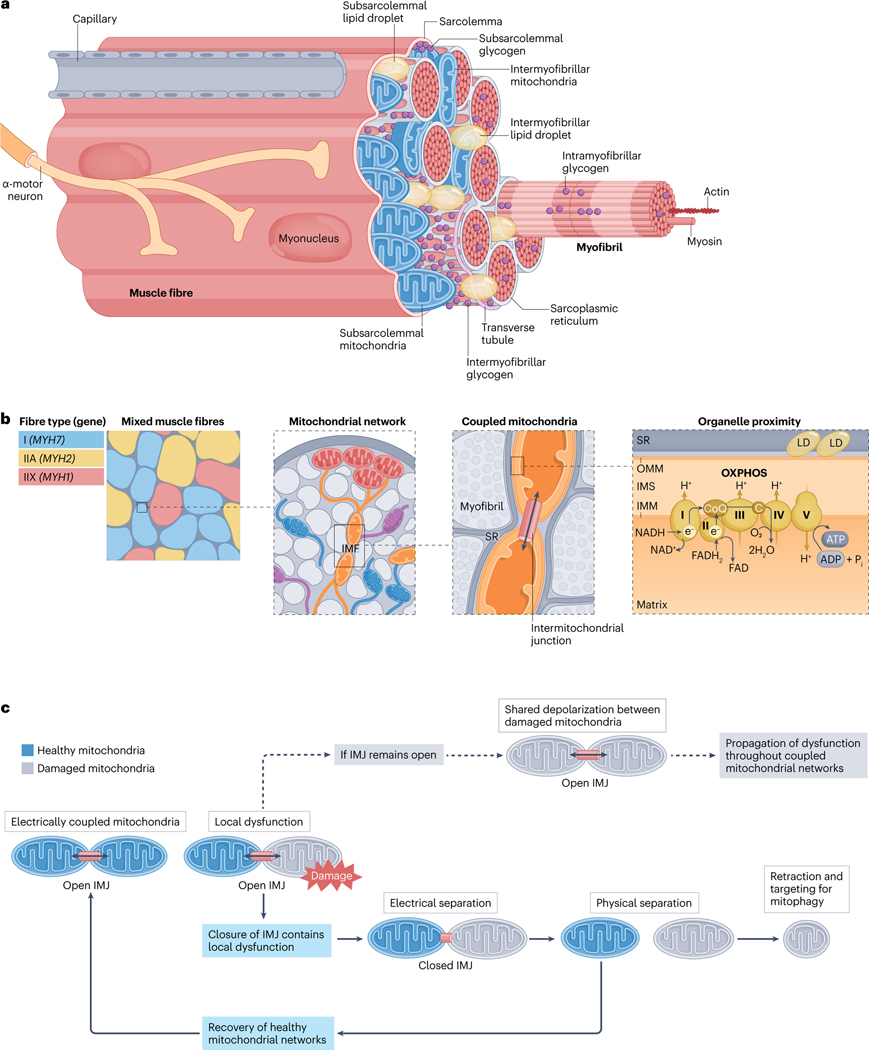
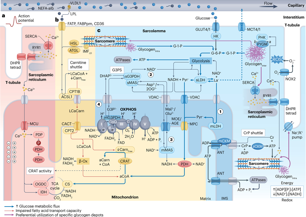
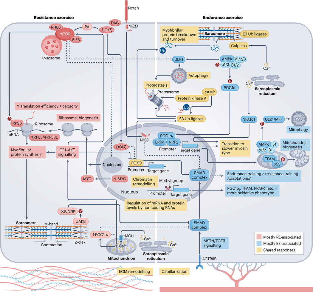
Viewing metabolism through the lens of exercise biology has proven an accessible and practical strategy to gain new insights into local and systemic metabolic regulation. Recent methodological developments have advanced understanding of the central role of skeletal muscle in many exercise-associated health benefits and have uncovered the molecular underpinnings driving adaptive responses to training regimens. In this Review, we provide a contemporary view of the metabolic flexibility and functional plasticity of skeletal muscle in response to exercise. First, we provide background on the macrostructure and ultrastructure of skeletal muscle fibres, highlighting the current understanding of sarcomeric networks and mitochondrial subpopulations. Next, we discuss acute exercise skeletal muscle metabolism and the signalling, transcriptional and epigenetic regulation of adaptations to exercise training. We address knowledge gaps throughout and propose future directions for the field. This Review contextualizes recent research of skeletal muscle exercise metabolism, framing further advances and translation into practice.


Conflict of interest statement

Competing interests

The authors declare no competing interests.

Figures

Fig. 1 |
Fig. 1 |. Skeletal muscle fibre ultrastructure.
a, Location of mitochondrial subpopulations and energy stores in muscle fibres. Skeletal muscle is composed of layers of connective tissue and fascicles (also known as muscle bundles). Fascicles contain organized arrangements of individual syncytial muscle fibres, each covered by an endomysium, or basal lamina, which is anchored to the fibre membrane (also known as the sarcolemma). Muscle stem cells, termed ‘satellite cells’, reside within this sarcolemma-basal lamina ‘niche’ (Supplementary Fig. 3). Specialized components, such as sodium/potassium pumps (Na+/K+-ATPase), triads (consisting of transverse tubule and sarcoplasmic reticulum (SR)) and proteins of the myofibrils (long arrangements of connected sarcomeres) enable fibre contraction through the process of excitation–contraction coupling and sliding filament theory (Supplementary Fig. 2). Free ATP in muscle is limited,, and fibres possess additional energy depots to maintain contractile activity, including creatine phosphate, glycogen and intramyocellular lipids (Box 3). Glycogen granules are nonuniformly distributed between intramyofibrillar, intermyofibrillar and subsarcolemmal pools,,. Alternatively, intramyocellular lipids are stored in lipid droplets (LDs) found predominantly at central (intermyofibrillar) but also peripheral (subsarcolemmal) regions within healthy muscle fibres,. During submaximal and longer-duration high-intensity interval exercise most ATP in muscle is regenerated by mitochondrial oxidative phosphorylation (OXPHOS) (see the section ‘Acute exercise muscle metabolism’) (Fig. 2). b, Spatial distribution of the mitochondrial reticulum within muscle fibres. Human muscle comprises three main fibre types,,,,, type I (marked by MYH7 expression), type IIA (with MYH2 expression) and type IIX (expressing MYH1). Differences in mitochondrial protein content, and mitochondrial network configuration, between fibre types directly impacts muscle metabolism and function. Muscle mitochondria form an interconnected reticulum that enables swift and efficient distribution of potential energy from subsarcolemmal (also known as peripheral) mitochondria to intermyofibrillar mitochondria (IMF), deep within the fibre. The positioning of mitochondria in the intermyofibrillar space influences the structure of adjacent sarcomeres, resulting in variable cross-sectional areas and myofilament spacing at different regions across the sarcomere length. The branching morphology of IMF also accommodates functional interactions with nearby cellular components, such as the sarcoplasmic reticulum and intermyofibrillar lipid droplets,,. In oxidative mouse muscle, ~20% of all IMF are connected to lipid droplets, which may facilitate efficient ATP production and distribution. c, Adjacent mitochondria form networks and share energy potential through the intermitochondrial junction (IMJ). Analogous to circuit breakers, intermitochondrial junctions split the reticulum into smaller subnetworks, permitting swift separation of defective mitochondria before their removal through mitophagy. In this way, intermitochondrial junctions provide a dynamic layer of quality control, rapidly rewiring the mitochondrial reticulum through healthy network components to sustain muscle function. C, cytochrome c; CoQ, coenzyme Q; IMM, inner mitochondrial membrane; IMS, intermembrane space; OMM, outer mitochondrial membrane.
Fig. 2 |
Fig. 2 |. Skeletal muscle metabolism during higher-intensity exercise.
a, Exercise-onset metabolic inertia (red area). Acetyl-carnitine (aCarn) abundance and the acetyl coenzyme A (aCoA)-producing capacities of carnitine acetyltransferase (CRAT), and pyruvate dehydrogenase (PDH) appear rate-limiting for oxidative adenosine triphosphate (ATP) regeneration at the onset of moderate-high intensity exercise. Contraction-induced calcium (Ca2+) transients promote mitochondrial Ca2+ uptake into the matrix space through the inner-membrane mitochondrial calcium uniporter (MCU) complex,. Increased matrix Ca2+ can upregulate PDH through activation of its phosphatase (PDP), and can upregulate isocitrate dehydrogenase (IDH) and 2-oxoglutarate dehydrogenase (OGDC),, directly to fine-tune oxidative metabolism via stimulation of the tricarboxylic acid (TCA) cycle. Ca2+ kinetics probably precede an allosteric rise in the [ADP][Pi] to [ATP] and [creatine][Pi] to [creatine phosphate] ratios in part because ADP and creatine (Cr) are buffered by the adenylate kinase (not shown) and creatine phosphate (CrP) shuttle reactions. Thus, synchrony between mechanisms of substrate provision, Ca2+-feedforward and metabolite feedback regulation might underlie acute metabolic inertia. This could be particularly prominent in type II fibres, which have lower CRAT and MCU abundance and slower mitochondrial Ca2+ import rates. Furthermore, metabolic inertia is more pronounced in metabolically compromised and older untrained adults, related to the lower CRAT activity and acetyl-carnitine content of muscle in these individuals. b, Carbohydrates outcompete non-esterified fatty acids (NEFAs) for oxidation at higher intensities (yellow area). Muscle glucose uptake and carbohydrate utilization,, increases with exercise intensity. At workloads above maximum fat oxidation (>60–65% of maximal oxygen consumption (V.O2max)) flux of pyruvate to acetyl-CoA progressively exceeds rates of TCA cycle entry at citrate synthase (CS), leading to depletion of the muscle free-carnitine (Carnfree) pool through CRAT-dependent acetylation to acetyl-carnitine. After higher-intensity submaximal exercise, acetylation of the free-carnitine pool is greatest in type I fibres. Insufficient free-carnitine availability would inhibit NEFA mitochondrial import at the first step of the carnitine shuttle — that is, carnitine palmitoyl transferase 1B (CPT1B) conjugation of carnitine to long-chain acyl-CoA (LCaCoA). Reduced fat oxidation is associated with diminished free-carnitine levels at ~70% of V.O2max (ref. 72), whereas medium-chain NEFA metabolism bypasses carnitine shuttling and is maintained at higher submaximal workloads. Therefore, free-carnitine levels appear rate-limiting for long-chain NEFA utilization at increasing exercise intensities. c, Lactate and pyruvate oxidation and NADH shuttles (blue area). Downstream of glycolysis, pyruvate (Pyr) and/or lactate (La) pass through voltage-dependent anion channels (VDAC), where lactate is converted to pyruvate by mitochondrial lactate dehydrogenase (mLDH) in the intermembrane space (step 1). Pyruvate then enters the mitochondrial matrix through the mitochondrial pyruvate carrier (MPC),. The glycerol-3-phosphate (G3P2−) shuttle (G3PS) and malate (Mal2−)/aspartate (Asp2−) shuttle enables mitochondrial oxidation of lactate and pyruvate through compartmentalized redox shuttling. G3PS and MAS recycle extra-matrix nicotinamide adenine dinucleotide (NAD+) (step 2) and transport reducing power from glycolysis to the mitochondrial matrix. This occurs through reactions associated with Mal2− and Asp2− delivery into the matrix space, (step 3) and G3P2− donation of electrons directly to coenzyme Q (CoQ) of the electron transport chain (step 4). As such, saturation of these shuttles increases lactate accumulation and upregulates the lactate-favouring LDHA isoform in vitro. See Box 3 for discussion of CrP, intermyofibrillar and intramyofibrillar glycogen (glycogeninter and glycogenintra, respectively) and intermyofibrillar lipid droplet (IMFLD) stores, and section ‘Acute exercise muscle metabolism’ for details of their metabolism during acute exercise. β-Ox, β-oxidation; ACSL1, acyl-CoA synthetase long-chain family member 1; AGE, aspartate/glutamate exchanger; ANT, adenine nucleotide translocator; ATGL, adipose triacylglyceride lipase; ba denotes posttranslational modification of PYGM from its less-active b form to the constitutively active a form by PHK; C, cytochrome c; CACT, carnitine acylcarnitine translocase; mCKM mitochondrial creatine kinase muscle; sCKM, sarcoplasmic CKM; DHAP2−, dihydroxyacetone phosphate; DHPR tetrad, four dihydropyridine receptors associated with one ryanodine receptor 1 (RYR1); FABPpm, fatty acid binding protein plasma membrane; FAD, flavin AD; FADH2, reduced FAD; FATP, FA transporter protein; GLUT4/1; glucose transporters 4 and 1; G-1-P, glucose-1-phosphate; G-6-P, glucose-6-phosphate; HK, hexokinase; HSL, hormone-sensitive lipase; IMS, intermembrane space; LCaCarn, long-chain acyl carnitine; LPL, lipoprotein lipase; sLDH, sarcoplasmic LDH; mMAS, mitochondrial MAS; MCT4/1, monocarboxylate transporters 4 and 1; mG3PDH, mitochondrial glycerol-3-phosphate dehydrogenase; MOE, malate/2-oxoglutarate exchanger; NADH, reduced NAD; NEFA-alb, albumin-bound NEFA; NOX2, NAD phosphate oxidase 2; O2 superoxide; 2OG2−, 2-oxoglutarate; OXPHOS, oxidative phosphorylation and associated respiratory complexes; PHK, muscle phosphorylase kinase; PYGM, glycogen phosphorylase muscle-associated; ROS, reactive oxygen species; SERCA, sarcoplasmic/endoplasmic reticulum Ca2+-ATPase; sG3PDH, sarcoplasmic glycerol-3-phosphate dehydrogenase; sMAS, sarcoplasmic MAS; T-tubule, transverse tubule; VLDL1, very low density lipoprotein 1.
Fig. 3 |
Fig. 3 |. Molecular responses to acute exercise and exercise training.
Exercise-induced alterations in circulating molecules, and the intramuscular milieu,, together with mechanical tension, initiates a temporal series of biochemical and molecular events that lead to muscle adaptation. Activation of signalling cascades promote substantial posttranslational modification of the muscle proteome, and DNA accessibility,,. Collectively, this drives transcription factor-dependent changes in gene expression,, alongside microRNAs and long-non-coding RNAs that are thought to ‘fine-tune’ the molecular responses to exercise. Endurance exercise (EE) and resistance exercise (RE) are often considered divergent stimuli, primarily driving oxidative versus hypertrophic muscle adaptations, respectively. However, common processes among exercise modalities can result in shared enrichment of signalling cascades and transcriptional networks in the post-exercise period. For example, coordinated proteolysis is detected following acute exercise, irrespective of exercise type. This may require cAMP–protein kinase A (PKA), and ensures protein quality control and physiological muscle remodelling. 5′-AMP-activated protein kinase (AMPK) activity and the expression of total PGC1A mRNA are also increased after a single bout of either endurance or resistance exercise. AMPK phosphorylation activates peroxisome proliferator-activated receptor-γ coactivator 1α isoform 1 (PGC1α1), and both AMPK and PGC1α1 potentiate angiogenic factors, and mitochondrial bioenergetics,,, in muscle. After endurance exercise, a distinct pool of mitochondrial AMPK (composed of α1, α2, β2 and γ1 isoforms) regulates mitophagy and promoter hypomethylation facilitates the transcription of peroxisome proliferator-activated receptor-δ (PPARδ) and mitochondrial transcription factor A (TFAM). Whether resistance exercise elicits these same effects is unclear. Despite similarities, there are more distinct than overlapping post-exercise responses between modalities,. Rapamycin-sensitive substrates of mammalian target of rapamycin (mTOR) complex 1 (mTORC1) are phosphorylated to a greater extent after resistance exercise. Mechanical overload initiates translocation of the mTOR–lysosomal complex and diacylglycerol (DAG) kinase-ζ (DGKζ) to the sarcolemma. Here, mTOR colocalization with RAS homologue enriched in brain (RHEB) and eukaryotic initiation factor 3 (EIF3) and phosphatidic acid (PA) produced by DGKζ might coalesce to fully stimulate mTORC1 (ref. 175) and the translation of contraction-associated mRNAs. Acute attenuation of UNC-51-like autophagy activating kinase 1 (ULK1) autophagic signalling after resistance exercise and nuclear DGKζ-mediated suppression of forkhead box protein O (FOXO)-dependent proteasomal degradation could also support muscle mass by moderating the breakdown of myofibrillar proteins. At the sarcomere, contraction recruits ZAKβ to the Z-disc where it acts through JUN N-terminal kinase 1 (JNK1) and JNK2 (refs. 179,181), potentially alongside Notch, to inhibit myostatin (MSTN)/transforming growth factor-β (TGFβ) signalling. This represents one of many intracellular changes permitting resistance training-induced hypertrophy over endurance-like adaptations. The upregulation of MYC with resistance exercise stimulates ribosomal biogenesis,, and the formation of a specialized pool of ribosomes with a high ratio of ribosomal protein large 3 (RPL3) to RPL3-like (RPL3L) may favour protein synthesis over translational fidelity. MYC expression is mTOR-independent but MYC cooperation with mTOR is necessary to successfully increase ribosomal content, possibly requiring an mTOR-driven reorganization of nucleoli to aid rRNA transcription. Divergence between endurance and resistance exercise at the transcriptomic level is magnified after a period of training. Endurance training increases electron transport chain complex expression, mitochondrial content and muscle oxidative capacity. Conversely, growth-related pathways, ribosomal abundance and muscle mass are augmented more by resistance training. This could be mediated in part by PGC1α isoforms. Nuclear localization of PGC1α1 and Ser15 phosphorylation of p53 are greater in resting muscle after high-intensity interval training, which might help to preserve mitochondrial content and function. By contrast, PGC1α1 protein is unchanged after resistance training, whereas the PGC1α isoform 4 (PGC1α4) protein is preferentially enriched. PGC1α4 stimulates muscle hypertrophy and is associated with enhanced Igf1 expression, insulin-like growth factor 1 (IGF1)–serine/threonine protein kinase (AKT) and mTORC1 signalling and downregulation of Mstn mRNA in mouse muscle. However, unlike PGC1α1 (ref. 280), PGC1α4 does not coactivate oestrogen-related receptor-α (ERRα) and has no effect on oxidative phosphorylation enzymes,. Appreciable overlap in the initial stages of exercise training probably underlies the degree of shared adaptation between endurance and resistance exercise (see the sections ‘Skeletal muscle responses to acute exercise’ and ‘Skeletal muscle adaptations to long-term exercise’). Depending on individual predisposition (Box 1), dedicated training of a certain exercise modality could amplify discrete differences in the adaptive response, resulting in distinct muscle adaptations and the development of specific phenotypes over time. Evidence showing that combined endurance and resistance training can blunt muscle hypertrophy in humans is scarce, but concurrent training could impede gains in explosive strength. Still, a combined exercise regime may offer dual benefits for most individuals. ACTRIIB, activin receptor type 2B; ECM, extracellular matrix; MFF, mitochondrial fission factor; MCU, mitochondrial calcium uniporter; NFATc1, nuclear factor of activated T cells, cytoplasmic 1; NICD, Notch intracellular domain; NRF2, nuclear factor erythroid 2-related factor 2; p38, p38 mitogen-activated protein kinase; RPS6, ribosomal protein S6; SMAD, mothers against decapentaplegic homologue; Ub, ubiquitin; ZAKβ, MAP3K20 isoform-β.

References

    1. Bilet L. et al. One-leg inactivity induces a reduction in mitochondrial oxidative capacity, intramyocellular lipid accumulation and reduced insulin signalling upon lipid infusion: a human study with unilateral limb suspension. Diabetologia 63, 1211–1222 (2020). - PMC - PubMed
    1. Bergouignan A. et al. Effect of contrasted levels of habitual physical activity on metabolic flexibility. J. Appl. Physiol 114, 371–379 (2013). - PubMed
    1. Kivimaki M. et al. Physical inactivity, cardiometabolic disease, and risk of dementia: an individual-participant meta-analysis. Brit. Med. J 365, l1495 (2019). - PMC - PubMed
    1. Zhang D. et al. Leisure-time physical activity and incident metabolic syndrome: a systematic review and dose-response meta-analysis of cohort studies. Metabolism 75, 36–44 (2017). - PubMed
    1. Millard LAC, Tilling K, Gaunt TR, Carslake D. & Lawlor DA Association of physical activity intensity and bout length with mortality: an observational study of 79,503 UK Biobank participants. PLoS Med. 18, e1003757 (2021). - PMC - PubMed

Publication types

MeSH terms