. 2023 Jun;618(7965):543-549.
doi: 10.1038/s41586-023-06100-w. Epub 2023 May 24.

A median fin derived from the lateral plate mesoderm and the origin of paired fins

Affiliations

A median fin derived from the lateral plate mesoderm and the origin of paired fins

Keh-Weei Tzung et al. Nature. 2023 Jun.

Abstract

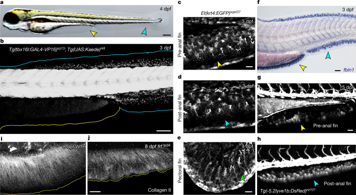
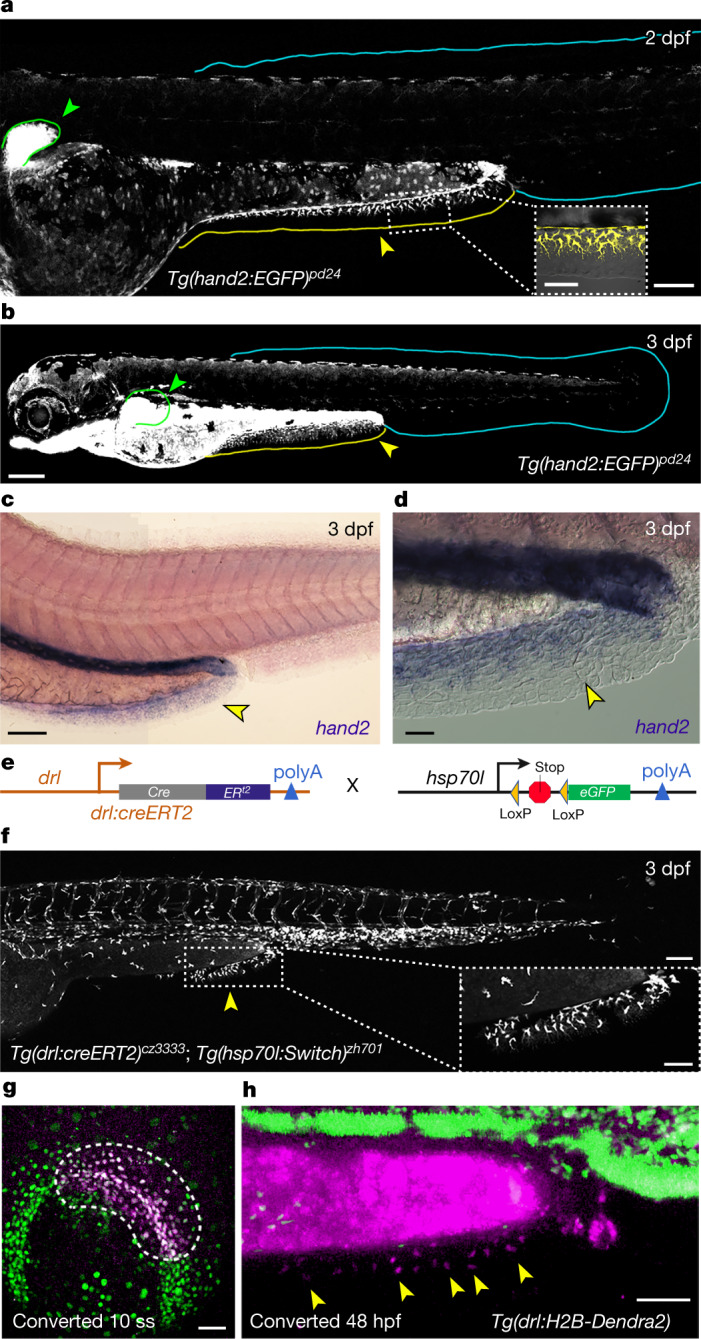
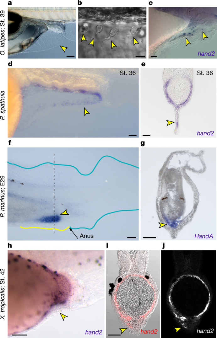
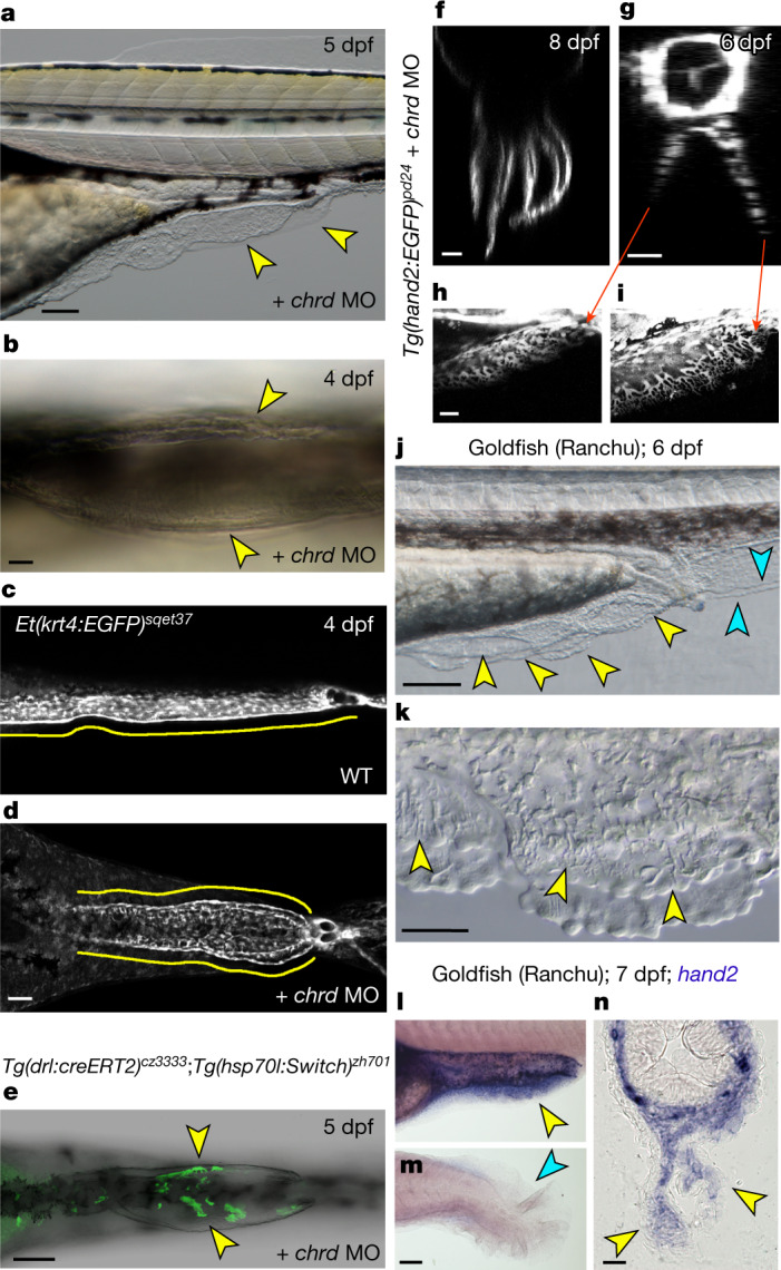
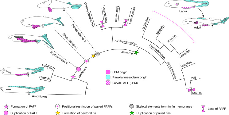
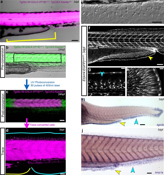
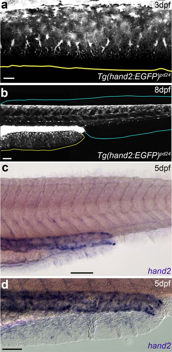
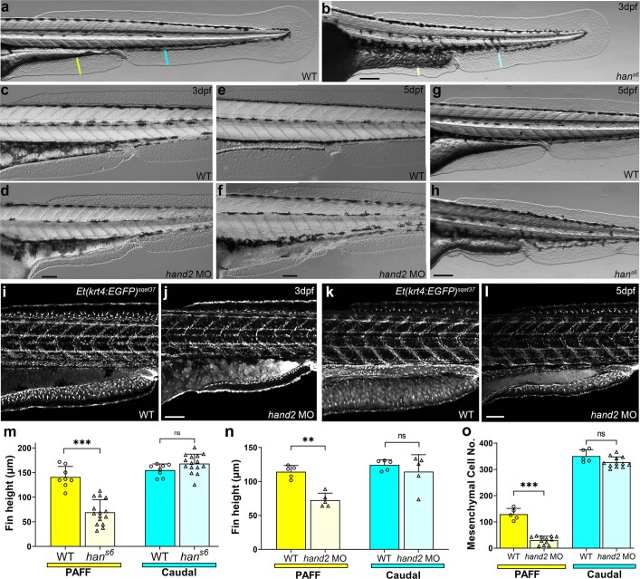
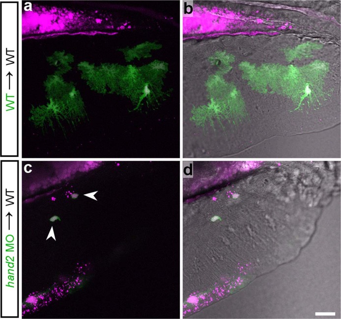
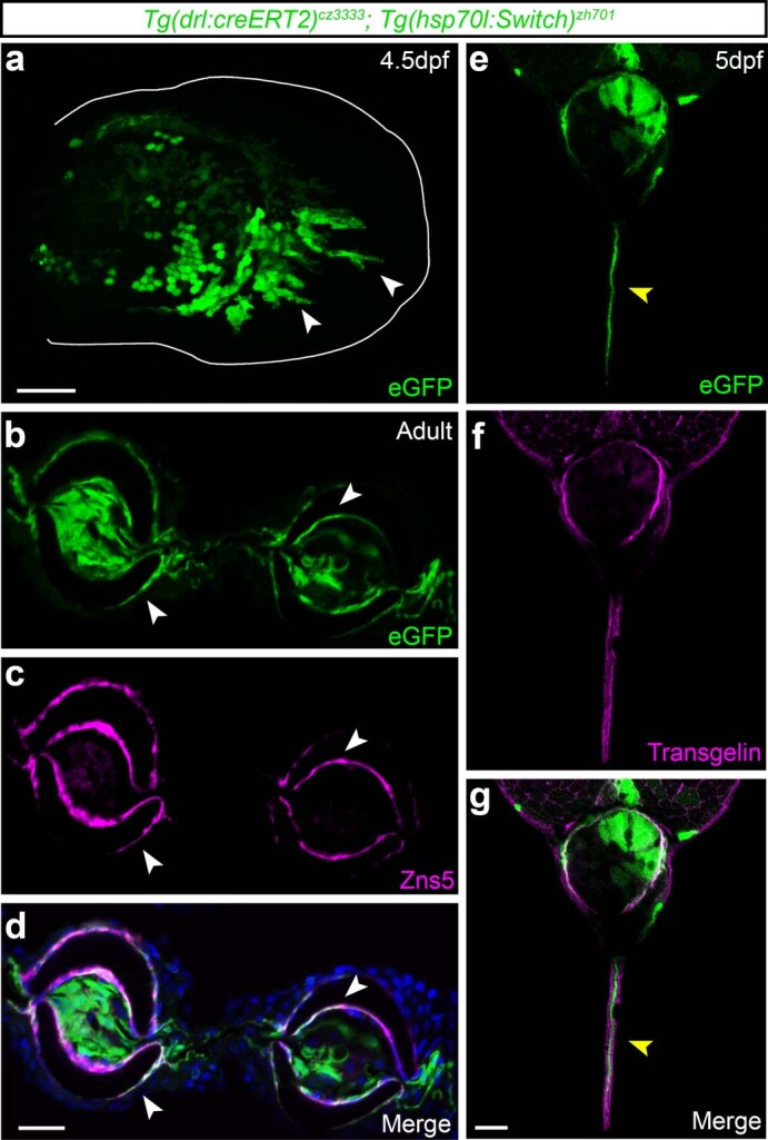
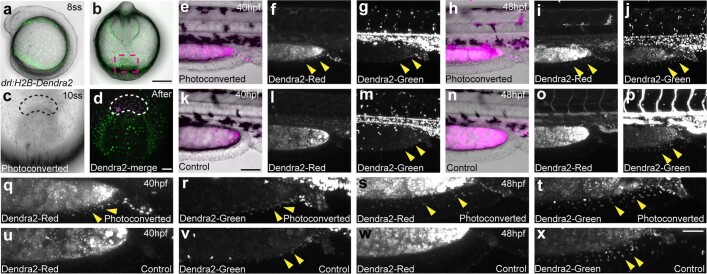
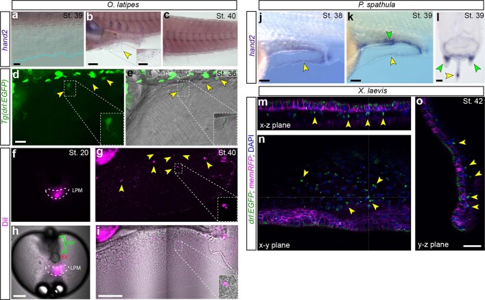
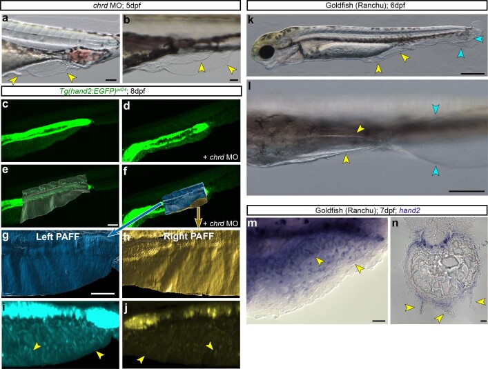
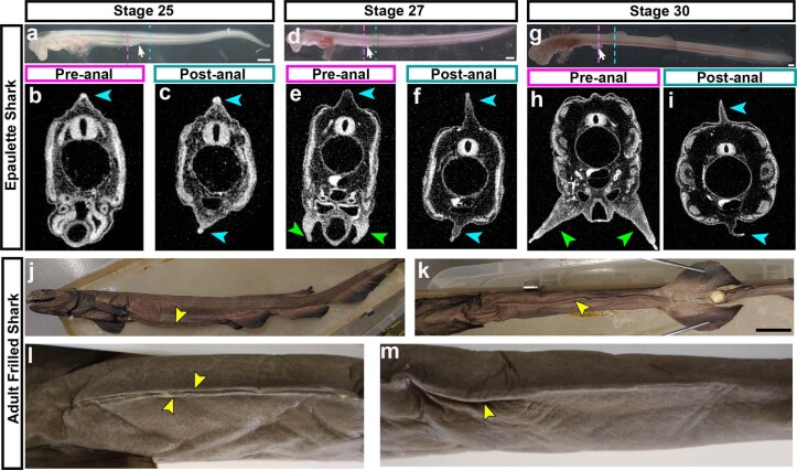
The development of paired appendages was a key innovation during evolution and facilitated the aquatic to terrestrial transition of vertebrates. Largely derived from the lateral plate mesoderm (LPM), one hypothesis for the evolution of paired fins invokes derivation from unpaired median fins via a pair of lateral fin folds located between pectoral and pelvic fin territories1. Whilst unpaired and paired fins exhibit similar structural and molecular characteristics, no definitive evidence exists for paired lateral fin folds in larvae or adults of any extant or extinct species. As unpaired fin core components are regarded as exclusively derived from paraxial mesoderm, any transition presumes both co-option of a fin developmental programme to the LPM and bilateral duplication2. Here, we identify that the larval zebrafish unpaired pre-anal fin fold (PAFF) is derived from the LPM and thus may represent a developmental intermediate between median and paired fins. We trace the contribution of LPM to the PAFF in both cyclostomes and gnathostomes, supporting the notion that this is an ancient trait of vertebrates. Finally, we observe that the PAFF can be bifurcated by increasing bone morphogenetic protein signalling, generating LPM-derived paired fin folds. Our work provides evidence that lateral fin folds may have existed as embryonic anlage for elaboration to paired fins.


Conflict of interest statement

The authors declare no competing interests.

Figures

Fig. 1
Fig. 1. A non-PM-derived median fin fold.
a, Larval 4 dpf zebrafish possess a median PAFF (yellow arrowhead) in addition to the caudal median fin fold (cyan arrowhead). b, Confocal image of a 3 dpf Tg(tbx16l:GAL4-VP16); Tg(UAS:Kaede) embryo with PM labelled by Kaede showing PM-derived mesenchyme in the caudal median fin fold (cyan outline) but not the PAFF (yellow outline). ce, Confocal images of pre-anal (c), ventral caudal (d) and pectoral (e) fins of the ET37 Enhancer Trap transgenic line indicating that PAFF contains morphologically comparable mesenchyme (indicated by arrowheads) to other larval fin folds. f, In situ hybridization of the fin mesenchyme marker fbln1 in both PAFF (yellow arrowhead) and caudal fin fold (cyan arrowhead) at 3 dpf. g,h, DsRed expression in mesenchyme of both pre-anal (g) and caudal (h) fin folds of the 5 dpf Tg(-5.2lyve1b:DsRed) transgenic line. i,j, Immunostaining for collagen II in 8 dpf frf mutants (j) shows loss of fibril organization compared with WT (i). Scale bars, 200 µm (a); 100 µm (b); 20 µm (c,e); 50 µm (f,g,j).
Fig. 2
Fig. 2. The PAFF is an LPM-derived median fin fold.
a,b, Confocal images of Tg(hand2:EGFP) embryos at 2 dpf (a) and 3 dpf (b) showing eGFP labelling of the mesenchyme of the pectoral (green outline and arrowheads) and PAFFs (yellow outline and arrowheads, and magnified in inset) but not the caudal fin fold (cyan outline). c,d, In situ hybridization of hand2 at 3 dpf shows fin expression of hand2 only in the PAFF (yellow arrowheads (c), and higher magnification with Nomarski optics indicates expression in the mesenchyme (d). e, Schematic of the LPM lineage tracing transgenes. f, Lineage tracing of LPM using transgenics depicted in (e) following 4-OHT treatment and heat shock before imaging shows that PAFF mesenchyme is derived from the LPM (yellow arrowhead and magnified in inset). g,h, Ventral (g) and lateral (h) confocal images of the drl:H2B-Dendra2 transgenic line at the 10-somite stage (10 ss) (g) and 48 hpf (h) following ultra-violet laser photoconversion in the region of the LPM outlined in (g). h, Photoconverted PAFF mesenchyme is indicated by yellow arrowheads. Scale bars, 100 µm (a,c,f); 50 µm (a (inset),f (inset),g,h); 200 µm (b); 20 µm (d).
Fig. 3
Fig. 3. PAFF mesenchyme expression of hand2 is conserved across vertebrates.
a,b, Nomarski images of 9 dpf medaka showing PAFF (a) with dispersed mesenchymal cells (yellow arrowheads) (b). c, In situ hybridization of medaka at stage 39 showing hand2 expression in pre-anal fin mesenchyme (yellow arrowheads). d,e, In situ hybridization of hand2 in stage 36 paddlefish embryos shown laterally (d) or in transverse section (e). hand2-positive PAFF fin mesenchyme is indicated by yellow arrowheads. f,g, HandA in situ hybridization of stage E29 lamprey (P. marinus) embryos shown laterally (f) or in transverse section (g). Lamprey show strong expression of HandA in a fin anterior to the anus (yellow arrowhead in f) corresponding to cells in the interior of the fin (g). The section location of (g) is indicated by the dashed line in (f). hj, Chromogenic (h) or fluorescent (i,j) in situ hybridization of hand2 in stage 42 X. tropicalis embryos shown laterally (h) or in transverse section (i,j). Fluorescent image (j) is overlayed on the Nomarski image (i). The small PAFF in Xenopus contains sparse hand2-positive fin mesenchyme (yellow arrowheads). St., stage. Scale bars, 100 µm (a,d,h); 10 µm (b); 20 µm (c); 50 µm (eg,i).
Fig. 4
Fig. 4. Duplication of the PAFF into paired fin folds.
a,b, Lateral (a) and ventral (b) Nomarski images of PAFFs of 5 dpf (a) and 4 dpf (b) embryos injected with MO targeting chordin, resulting in duplication of PAFF (yellow arrowheads). c,d, Confocal micrographs, imaged ventrally, of PAFFs in 4 dpf ET37 transgenic larvae uninjected (c) or injected with a chrd MO (d). PAFFs are indicated with yellow lines. d, Note the duplicated anal openings following chrd MO injection. e, Ventral confocal image of chrd MO duplicated PAFFs of the transgenic line shown above. Hydroxytamoxifen treatment at the 12-somite stage and heat shock before imaging show that mesenchyme of duplicated PAFFs is derived from the LPM (yellow arrowheads). fi, Light-sheet (f) and confocal (gi) images of Tg(hand2:EGFP) larvae at 8 dpf (f) and 6 dpf (gi). Orthogonal display through the xz plane (f,g) shows that multiple PAFFs can form and that duplicated PAFFs contain eGFP-positive mesenchyme (gi). Lateral views of left (h) and right (i) duplicated PAFFs of sample in g are given. j,k, Lateral low- (j) and high-power (k) Nomarski images of the PAFF of the Ranchu goldfish at 6 dpf. The multiple PAFFs (j) and individual mesenchyme cells (k) are indicated by yellow arrowheads. Duplicated caudal fin folds are indicated by cyan arrowheads (j). ln, Lateral (l,m) and transverse (n) views of pre-anal (l,n) and caudal (m) fin folds of 7 dpf Ranchu larvae stained by in situ hybridization for hand2, where hand2-positive PAFF mesenchyme is indicated by yellow arrowheads. Absence of hand2 in the caudal fin fold is indicated by the cyan arrowhead (m). Scale bars, 100 µm (a,e,m); 20 µm (b,fh,n); 50 µm (d,k); 200 µm (j).
Fig. 5
Fig. 5. Hypothesis of the elaboration of the PAFF to paired fins.
Simplified evolutionary scenario of vertebrates showing the presence of a PAFF and subsequent modifications leading to paired fins. Dashed lines and dagger symbols indicate extinct lineages, and solid lines indicate extant lineages. PM-derived fins and fin folds are in cyan, while LPM-derived fins are in pink. Larval PAFF is hatched. Black arrows indicate the position of the anus.
Extended Data Fig. 1
Extended Data Fig. 1. A non-paraxial mesoderm derived median fin fold.
a, Confocal image of 3 dpf Tg(tbx16l:GAL4-VP16); Tg(UAS:Kaede) embryo with paraxial mesoderm labelled by photoconverted Kaede (magenta). Unlabelled PAFF underscored by yellow bracket. c-d, Expression of Kaede in the dorsal caudal fin fold of 3 dpf Tg(tbx16l:GAL4-VP16); Tg(UAS:Kaede) is not due to localised de novo expression from the tbx16l promoter. Confocal images of 24 hpf trunks of both prior to (b) and after (c) UV photoconversion. Unconverted Kaede is in the green channel overlaid with converted Kaede in magenta. Region of paraxial mesoderm conversion and Nomarski image given in (b). At 3 dpf, converted cells can be seen in the adjacent fin fold dorsally indicating Kaede is reporting lineage (d). e, Nomarski image of PAFF at 3 dpf showing presence of mesenchymal cells. fh, Confocal images of the pre-anal fin fold (f), dorsal (g) and caudal (h) regions of the caudal fin fold of the ET37 Enhancer Trap transgenic line at 3 dpf, indicating PAFF contains numerous mesenchymal cells which are morphologically comparable to mesenchyme of other larval fin folds. ij, In situ hybridisation of fin mesenchyme markers itgb3b (i; 3 dpf) and bmp1a (j; 5 dpf) in both PAFFs (yellow) and caudal fin folds (cyan arrowhead). Scale Bars: 100 µm (a,f,i), 50 µm (c,d,e,j), 20 µm (g,h).
Extended Data Fig. 2
Extended Data Fig. 2. Expression of hand2 in the pre-anal fin fold.
ab, High (a) and low (b) magnification confocal images of Tg(hand2:EGFP) zebrafish embryos at 3 dpf (a) and 8 dpf (b) showing eGFP labelling of mesenchyme of the PAFF (yellow outline, a,b), but not the caudal fin fold (cyan outline, b). cd, In situ hybridisation of hand2 at 5 dpf showing expression of hand2 only in the PAFF (c). Higher magnification with Nomarski optics indicates expression in mesenchyme (d). Scale Bars: 100 µm (b,c), 50 µm (d), 20 µm (a).
Extended Data Fig. 3
Extended Data Fig. 3. Loss of Hand2 leads to reduction of pre-anal fin folds but not caudal fin folds.
al, Lateral Nomarski images of uninjected WT (a,c,e,g), hans6 mutants (b,h) and hand2 morphants (d,f) at 3 dpf (ad) or 5 dpf (eh). il, Confocal images of 3 dpf (i,j) and 5 dpf (k,l) ET37 Enhancer Trap transgenic larvae, uninjected (i,k) or injected with a hand2 morpholino (j,l). m,n Quantification of 3 dpf fin height of PAFFs (yellow) and caudal fin folds (cyan) in uninjected WT, hand2 mutants (hans6; m) and hand2 morphants (n). Loss of Hand2 leads to significant reduction of PAFFs but not caudal fin folds. n = 8 (WT), n = 15 (hans6), n = 5 (hand2 MO). Location of measurements of PAFF and caudal fin shown in (a) and (b) by yellow and cyan lines respectively. ***: p = 0.000008; **: p = 0.008; ns: p = 0.12 (m); ns: p > 0.9999 (n). o, Quantification of mesenchymal cell numbers in pre-anal (yellow bars) and ventral caudal (cyan bars) fin folds at 3 dpf. There is a significant reduction of PAFF mesenchyme in hand2 morphants compared to WT, but ventral caudal fin mesenchymal cell numbers are unaffected. n = 5 (WT), n = 12 (hand2 MO). ***: p = 0.000323; ns: p = 0.06. mo: Data are presented as mean values with error bars representing standard deviations. In all cases n refers to biologically independent embryos. Two-sided Mann-Whitney test. Scale Bars: 100 µm (d,f,j,l), 200 µm (b,h). Source data
Extended Data Fig. 4
Extended Data Fig. 4. Hand2 acts cell autonomously in PAFF mesenchyme.
ad, Lateral confocal images of PAFF of 3 dpf embryos. Cells were transplanted into WT hosts from ET37 embryos that were injected with H2B-mCherry mRNA (a,b) or co-injected with H2B-mCherry mRNA and 500 µM hand2 MO (c,d). Fluorescent confocal images of mCherry (magenta) and eGFP (green) (a,c) are overlayed on Nomarski image (b,d). Arrowheads indicate transplanted cells from hand2 MO injected donors (c). Scale Bar: 20 µm (d).
Extended Data Fig. 5
Extended Data Fig. 5. Lineage tracing of the LPM into paired fins and the PAFF.
ag, Permanently labelled LPM cells can be seen within pectoral and pelvic fins and pre-anal fin fold (arrowheads). Confocal images of the pectoral fin at 4.5 dpf (a), adult pelvic fin (bd) and pre-anal fin fold at 5 dpf (eg) of Tg(drl:creERT2); Tg(hsp70l:Switch) transgenics following Hydroxytamoxifen treatment and heat-shock prior to imaging. Pelvic fins and PAFF are shown as transverse sections (bg) and were fluorescently immunostained for eGFP (b,e), Zns5 (c) or Transgelin (f). Merged images are shown in (d,g). LPM-derived mesenchyme cells of the pectoral and pre-anal fin folds are indicated by white and yellow arrowheads respectively (a, eg). LPM-derived Zns5-positive osteoblasts of the pelvic fins are indicated by white arrowheads (bd). Scale Bars: 20 µm (d,g), 50 µm (a).
Extended Data Fig. 6
Extended Data Fig. 6. Pre-anal fin fold mesenchyme originates from posterior-most drl-positive cells.
ad, Lateral (a) and ventral (bd) views of 8ss (ab) or 10ss (cd) drl:H2B-Dendra2 transgenic line either prior to (ab) or following (cd) photoconversion of posterior-most LPM (region outlined by dotted line) using UV laser illumination. ep, Lateral confocal images of the PAFF region at 40 hpf (eg, km) and 48 hpf (hj, np) of a photoconverted embryo (ej) and an unconverted control (kp). Panels show Dendra2-red (f,i,l,o), Dendra2-green (g,j,m,p) and merged (e,h,k,n) channels. PAFFs are indicated by yellow arrowheads, and are visible in green channel in both converted and controls, but only converted embryos show red labelled PAFFs (f,i; n = 4 photoconverted fish). Magnified views of panels (f,g,i,j,l,m,o,p) are given in panels (qx) respectively. Scale Bars: 200 µm (b), 50 µm (d,x), 100 µm (k).
Extended Data Fig. 7
Extended Data Fig. 7. LPM origin of PAFF mesenchyme is conserved in medaka, paddlefish and Xenopus.
ac, In situ hybridisation of medaka hand2 at Stage 39 (a) and Stage 40 (bc) showing exclusive expression in pre-anal fin mesenchyme (yellow arrowhead, b), but not in the caudal fin mesenchyme (cyan outline). Ventral caudal fin imaged in (a). de, Lateral confocal images of PAFF of Stage 36 transgenic Tg(-6.35drl:EGFP) medaka embryo. Fluorescent confocal images of eGFP (d) are overlayed on Nomarski image (e). Faint eGFP perdurance is seen in nascent PAFF mesenchymal cells (yellow arrowheads and inset). fi, Ventral (f,h) and lateral (g,i) confocal images of medaka embryo injected with DiI in the posterior LPM at Stage 20 (f,h) and traced to PAFF at Stage 40 (g,i). DiI within the mesenchyme indicated by yellow arrowheads, with higher magnification example shown in inset (g,i). DiI signal (f,g) is overlayed with Nomarski (h,i). Location of paraxial mesoderm (PM), Kupffer’s vesicle (KV) and region of posterior LPM are indicated in green, red, and white respectively in (h). jl, In situ hybridisation of hand2 in Stage 38 (j) and Stage 39 (kl) paddlefish embryos imaged laterally (j,k) or in transverse section (l). hand2-positive PAFF fin mesenchyme indicated by yellow arrowheads is located distally. Expression of hand2 is also seen in the nascent pelvic fins at Stage 39 (green arrowheads k,l). mo, Confocal images of a region of Xenopus laevis PAFF at NF Stage 42 injected with -6.35drl:EGFP. Lateral x-y view (n) is shown with orthogonal sections in the x-z plane (m) and y-z plane (o). Transient mosaic expression in the interstitial mesenchyme is highlighted with yellow arrowheads (mn). Scale Bars: 20 µm (a,d), 100 µm (b,i), 50 µm (b – inset, l,o), 200 µm (h,j,k).
Extended Data Fig. 8
Extended Data Fig. 8. Reduced Chordin leads to paired duplication of PAFF.
ab, Low (a) and high (b) power Nomarski images of duplicated PAFFs (yellow arrowheads) in 5 dpf larvae injected with chrd morpholino. cj, Confocal images of PAFFs from 8 dpf Tg(hand2:EGFP) either uninjected (c,e) or injected with chrd morpholino (d,f,gj). Confocal projections (c,d,i,j) and total surface renderings (e,f,g,h) highlight duplicated PAFFs (blue and yellow, f) compared to single PAFF in uninjected larvae (grey, e). Imaging of left (g) and right (h) duplicated PAFFs of chrd morphants indicated eGFP positive mesenchymal cells populated both fin folds (yellow arrowheads, i,j). kl, Lateral (k) and ventral (l) Nomarski images of the duplicated PAFFs of the Ranchu goldfish strain at 6 dpf. Duplicated PAFFs and ventral caudal fin folds indicated by yellow and cyan arrowheads, respectively. mn, Lateral (m) and transverse (n) views of 7 dpf Ranchu larvae stained by in situ hybridisation for hand2. Expression in individual mesenchymal cells of the PAFF indicated by yellow arrowheads (m). Occurrence of three PAFFs with core hand2 expression indicated by yellow arrowheads (n). Scale Bars: 100 µm (a,e,g), 50 µm (b), 500 µm (k), 200 µm (l), 20 µm (m,n).
Extended Data Fig. 9
Extended Data Fig. 9. Variable presence of a pre-anal fin in sharks.
ai, The Epaulette shark does not possess a PAFF. Whole mount (a,d,g) and virtual cross sections from microCT scans (bc, ef, hi) of the Epaulette shark Hemiscyllium ocellatum, at the developmental stages of 25 (ac), 27 (df), and 30 (gi). Virtual cross sections for each stage show the region either anterior (Pre-anal; b,e,h), or posterior (Post-anal; c,f,i) to the developing cloaca (white arrow, a,d,g). The dashed lines (a,d,g) indicate approximate pre-anal (magenta) and post-anal (cyan) microCT section locations (separate specimens). The developing pelvic and median fin folds are indicated by green and cyan arrowheads respectively (bc, ef, hi). jm: The tropeic folds found in a pre-anal position of the adult frilled shark, Chlamydoselachus anguineus, shown in lateral (j) and ventral (k) views (yellow arrowheads) (adult male specimen, ZRC 54430 - LKC Natural History Museum, Singapore). lm, Ventral view of another adult frilled shark exhibits partially paired tropeic folds (yellow arrowheads) in anterior half (l), which then merges to become singular (yellow arrowhead) in posterior half (m) (anterior: left, CSIRO H 7115-01, adult male 1310 mm TL, Tasmania, Australia). Scale Bars: 1 mm (a,d,g), 5 cm (k).

References

    1. Coates MI. The evolution of paired fins. Theory Biosci. 2003;122:266–287. doi: 10.1007/s12064-003-0057-4. - DOI
    1. Freitas R, Zhang G, Cohn MJ. Evidence that mechanisms of fin development evolved in the midline of early vertebrates. Nature. 2006;442:1033–1037. doi: 10.1038/nature04984. - DOI - PubMed
    1. Gegenbaur, C. Elements of Comparative Anatomy (Macmillan and Company, 1878).
    1. Balfour FM. On the development of the skeleton of the paired fins of Elasmobranchii, considered in relation to its bearings on the nature of the limbs of the Vertebrata. Proc. Zool. Soc. Lond. 1881;49:656–670. doi: 10.1111/j.1096-3642.1881.tb01323.x. - DOI
    1. Mivart SG. Notes on the fins of elasmobranchs, with considerations on the nature and homologues of vertebrate limbs. Trans. Zool. Soc. Lond. 1879;10:439–484. doi: 10.1111/j.1096-3642.1879.tb00460.x. - DOI

Publication types

MeSH terms

Substances