. 2023 Sep 1;31(5):526-535.
doi: 10.4062/biomolther.2023.057. Epub 2023 May 25.

Cremastranone-Derived Homoisoflavanes Suppress the Growth of Breast Cancer Cells via Cell Cycle Arrest and Caspase-Independent Cell Death

Affiliations

Cremastranone-Derived Homoisoflavanes Suppress the Growth of Breast Cancer Cells via Cell Cycle Arrest and Caspase-Independent Cell Death

Yeram Choi et al. Biomol Ther (Seoul). .

Abstract

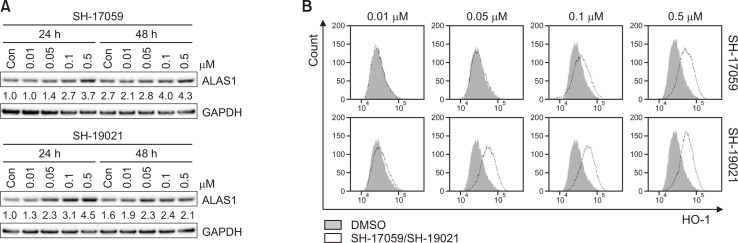
Breast cancer is the most common cancer and a frequent cause of cancer-related deaths among women wordlwide. As therapeutic strategies for breast cancer have limitations, novel chemotherapeutic reagents and treatment strategies are needed. In this study, we investigated the anti-cancer effect of synthetic homoisoflavane derivatives of cremastranone on breast cancer cells. Homoisoflavane derivatives, SH-17059 and SH-19021, reduced cell proliferation through G2/M cell cycle arrest and induced caspase-independent cell death. These compounds increased heme oxygenase-1 (HO-1) and 5-aminolevulinic acid synthase 1 (ALAS1), suggesting downregulation of heme. They also induced reactive oxygen species (ROS) generation and lipid peroxidation. Furthermore, they reduced expression of glutathione peroxidase 4 (GPX4). Therefore, we suggest that the SH-17059 and SH-19021 induced the caspase-independent cell death through the accumulation of iron from heme degradation, and the ferroptosis might be one of the potential candidates for caspase-independent cell death.

Keywords: Anti-cancer; Breast cancer; Caspase-independent cell death; Cell cycle arrest; Cremastranone; Homoisoflavane.


Figures

Fig. 1
Fig. 1
The cytotoxic effect of homoisoflavane derivatives. (A) Structure of cremastranone and synthetic homoisoflavane derivatives (Shin et al., 2022). (B-E) T47D (B, D) and ZR-75-1 (C, E) cells were treated with the indicated doses of SH-17059, SH-19017, SH-19021, SH-19026, SH-19027, and SHA-035 for the indicated periods. The cell viability of the T47D and ZR-75-1 cells was measured using the WST assay. Values are the means ± SD. *p<0.05, **p<0.01, ***p<0.005 vs DMSO control. These are representatives of three independent experiments.
Fig. 2
Fig. 2
The homoisoflavane derivatives reduce cell proliferation via cell cycle arrest at the G2/M phase. (A, B) T47D (A) and ZR-75-1 (B) cells were treated with SH-17059, SH-19021, SH-19027, and SHA-035 for the indicated periods, and then BrdU incorporation assay was performed. (C, E) The T47D cells were treated with the indicated doses of SH-17059 and SH-19021, and the cell lysates were subjected to western blot analysis. GAPDH was used as a loading control. (D) The T47D cells were obtained after treatment with the indicated dose of SH-17059 and SH-19021 for 48 h. The cell cycle was analyzed by flow cytometry after PI staining. The distribution of cells in the G0/G1, S, and G2/M phases of the cell cycle are presented. Values are the means ± SD. *p<0.05, **p<0.01, ***p<0.005 vs DMSO controls. These are representatives of three independent experiments.
Fig. 3
Fig. 3
SH-17059 and SH-19021 induce caspase-independent cell death. (A) The T47D cells were obtained after treatment with the indicated doses of SH-17059 and SH-19021 for 48 h. The percentages of apoptotic cells were detected by flow cytometry after annexin V-FITC and PI staining. (B) T47D cells were treated with 0.1 μM of SH-17059 or SH-19021 for 48 h. The caspase activity was measured using each colorimetric caspase substrate. (C) The T47D cells were untreated or pre-treated with Z-VAD-FMK for 1 h followed by treatment with the indicated doses of SH-17059 and SH-19021 for 72 h. The cell viability of the T47D cells was measured using the WST assay. Cell viability was normalized with each DMSO control. Values are the means ± SD. *p<0.05, **p<0.01, ***p<0.005 vs DMSO controls. These are representatives of three independent experiments.
Fig. 4
Fig. 4
SH-17059 and SH-19021 upregulate the expression levels of HO-1 and ALAS1. (A) The T47D cells were treated with the indicated doses of SH-17059 and SH-19021 for the indicated times. (A) The protein of ALAS1 was detected by western blot analysis. The relative expression of ALAS1 was indicated after normalization with the value of DMSO control at 24 h. (B) The T47D cells were treated with the indicated doses of SH-17059 and SH-19021 for 48 h. The HO-1 expression level was analyzed by flow cytometry. These are representatives of three independent experiments.
Fig. 5
Fig. 5
SH-17059 and SH-19021 induce the ROS generation, lipid peroxidation, and GPX4 reduction. (A, B) The T47D cells were treated with SH-17059 or SH-19021 at the indicated doses for 48 h. Intracellular ROS levels were detected by flow cytometry using H2DCF-DA (A). The levels of lipid peroxidation were analyzed using flow cytometry and BODIPY™ 581/591 C11 (B). (C) The T47D cells were treated with the indicated doses of SH-17059 and SH-19021 for 24 h or 48 h, and the cell lysates were subjected to western blot analysis. The relative expression of GPX4 was indicated after normalization with the value of each DMSO control at 24 h. These are representatives of three independent experiments.

Similar articles

Cited by

References

    1. Basavarajappa H. D., Lee B., Lee H., Sulaiman R. S., An H., Magaña C., Shadmand M., Vayl A., Rajashekhar G., Kim E. Y., Suh Y. G., Lee K., Seo S. Y., Corson T. W. Synthesis and biological evaluation of novel homoisoflavonoids for retinal neovascularization. J. Med. Chem. 2015;58:5015–5027. doi: 10.1021/acs.jmedchem.5b00449. - DOI - PMC - PubMed
    1. Basavarajappa H. D., Sulaiman R. S., Qi X., Shetty T., Sheik Pran Babu S., Sishtla K. L., Lee B., Quigley J., Alkhairy S., Briggs C. M., Gupta K., Tang B., Shadmand M., Grant M. B., Boulton M. E., Seo S. Y., Corson T. W. Ferrochelatase is a therapeutic target for ocular neovascularization. EMBO Mol. Med. 2017;9:786–801. doi: 10.15252/emmm.201606561.3e824506a6224c1eb5ab5fcfef835898 - DOI - PMC - PubMed
    1. Chiabrando D., Vinchi F., Fiorito V., Mercurio S., Tolosano E. Heme in pathophysiology: a matter of scavenging, metabolism and trafficking across cell membranes. Front. Pharmacol. 2014;5:61. doi: 10.3389/fphar.2014.00061.dc6732984bd14740a3fc850a397cdfda - DOI - PMC - PubMed
    1. Chiang S. K., Chen S. E., Chang L. C. A dual role of heme oxygenase-1 in cancer cells. Int. J. Mol. Sci. 2018;20:39. doi: 10.3390/ijms20010039.abbf585dfafc4e95bf9bb01ac85969b7 - DOI - PMC - PubMed
    1. Cragg G. M., Pezzuto J. M. Natural products as a vital source for the discovery of cancer chemotherapeutic and chemopreventive agents. Med. Princ. Pract. 2016;25 Suppl 2:41–59. doi: 10.1159/000443404. - DOI - PMC - PubMed