. 2023 Jul;13(7):e3075.
doi: 10.1002/brb3.3075. Epub 2023 May 24.

Effects of chronic sleep restriction on the neuro-phenotypes of Ctnnd2 knockout mice

Affiliations

Effects of chronic sleep restriction on the neuro-phenotypes of Ctnnd2 knockout mice

Man Xu et al. Brain Behav. 2023 Jul.

Abstract

Introduction: Sleep abnormalities are highly correlated with neurodevelopmental disorders, such as intellectual disability, attention deficit hyperactivity disorder, and autism spectrum disorders (ASD). The severity of behavioral abnormalities is correlated with the presence of sleep abnormalities. Based on previous research, we investigated that Ctnnd2 gene deletion in mice lead to ASD-like behaviors and cognitive defects. Given the importance of sleep in individuals with ASD, this study aimed to determine the effects of chronic sleep restriction (SR) on wild-type (WT) mice and on Ctnnd2 deletion-induced, neurologically related phenotypes in mice.

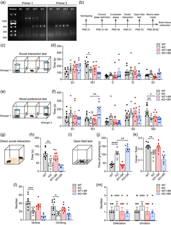
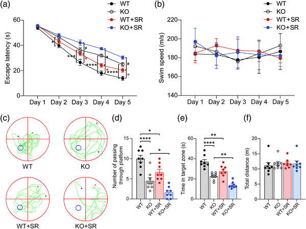
Method: WT and Ctnnd2 knockout (KO) mice were both subjected to manual SR (5 h per day) for 21 consecutively days separately, then we compared neurologically related phenotypes of WT mice, WT mice subjected to SR, KO mice, and KO mice subjected to SR using a three-chamber assay, direct social interaction test, open-field test, Morris water maze, Golgi staining, and Western blotting.

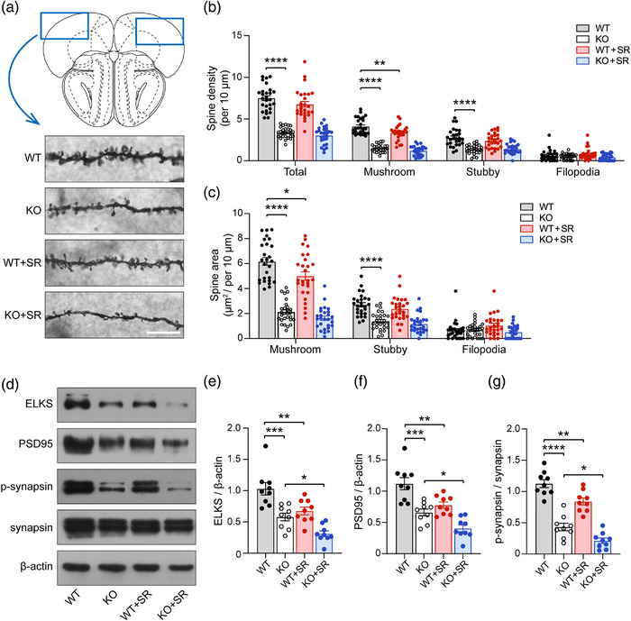
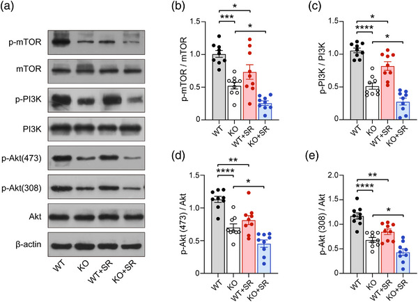
Results: The effects of SR on WT and KO mice were different. After SR, social ability and cognition were impaired in both WT and KO mice. Repetitive behaviors were increased, and exploration abilities were decreased in KO mice but not in WT mice. Moreover, SR reduced the density and area of mushroom-type dendritic spines in WT rather than KO mice. Finally, the PI3K/Akt-mTOR pathway was found to be involved in the effects induced by SR-impaired phenotypes in WT and KO mice.

Conclusion: Overall, results of the present study may have implications for the role of disrupted sleep in patients with CTNND2 gene-related autism and the evolution of neurodevelopmental disorders.

Keywords: Ctnnd2; autism; cognition; dendritic spines; sleep restriction; synapse.


Conflict of interest statement

The authors declare that there are no conflict of interests.

Figures

FIGURE 1
FIGURE 1
The results of autism‐like behaviors, anxiety, and exploration behaviors of four groups mice: (a) genotyping results of wild type (WT), heterozygote (HET), and knockout (KO) mice; (b) timelines of experiments; (c–f) three‐chamber assay of four groups mice. Schematic presentation (c) and statistical analysis (d) of social interaction test. Schematic presentation (e) and statistical analysis (f) of novelty preference test. (g and h) Schematic presentation (g) and statistical analysis of time in direct social interaction (h). (i–m) Open‐field test of four groups mice. Schematic presentation (i) and statistical analysis of time of grooming (j), ratio of center/total grids (k), frequency of vertical and climbing (l), and number of excretions (m). PND, postnatal; S1, time of test mouse staying in stranger 1 mouse chamber without interaction with stranger 1; IS1, time of test mouse interaction with Stranger 1; C, time of test mouse staying center chamber; O, time of test mouse staying in object chamber, IO, time of test mouse interaction with object; S2, time of test mouse staying in stranger 2 mouse chamber withour interaction with stranger 2; IS2, time of test mouse interaction with Stranger 2; SR, sleep restriction. All groups: n = 8. Data are the means ± SEM. *p < .05, **p < .01, ***p < .001, and ****p < .0001.
FIGURE 2
FIGURE 2
The spatial learning and memory ability of four groups mice: (a) escape latency time to find the submerged platform from day 1 to 5 of learning phase; (b) average swim speed from day 1 to 5; (c) representative swimming traces were shown for probe test of four groups mice; (d–f) the number of platform crossings (d), the time in the target zone (e), and the total distances (f) during the probe trial on day six were analyzed to evaluate the mice's spatial memory. WT, wild type; KO, knockout; SR, sleep restriction. All groups: n = 8. *p < .05, **p < .01, ***p < .001, ****p < .0001, and # p < .05.
FIGURE 3
FIGURE 3
The development of dendritic spines and synapses of four groups mice: (a) the density and area of dendritic spines in prefrontal cortex (PFC) was assessed by Golgi stanning. Scale bar = 10 μm; (b) the density of total, mushroom, stubby, and filopodia; (c) the spine area of mushroom, stubby, and filopodia; (d) the schematic diagram of expression levels in ELKS, PSD95, and p‐synapsin, measured by Western blot; (e–g) the semiquantitative analysis of the expression of ELKS (e), PSD95 (f), and p‐synapsin (g). WT, wild type; KO, knockout; SR, sleep restriction. All groups: n = 3 (three times repeated). Data are the means ± SEM. *p < .05, **p < .01, ***p < .001, ****p < .0001.
FIGURE 4
FIGURE 4
The expression of PI3K/Akt‐mTOR signal pathway in four groups mice: (a) the schematic diagram of expression levels in p‐mTOR, mTOR, p‐PIK3, PI3K, p‐Akt(473), p‐Aky(308), and Akt, measured by Western blot; (b–e) the semiquantitative analysis of the expression of p‐mTOR (b), p‐PI3K (c), p‐Akt(473) (d), and p‐Akt(308) (e). WT, wild type; KO, knockout; SR, sleep restriction. All groups: n = 3 (three times repeated). Data are the means ± SEM. *p < .05, **p < .01, ***p < .001, ****p < .0001.

Similar articles

Cited by

References

    1. Amaral, D. G. , Schumann, C. M. , & Nordahl, C. W. (2008). Neuroanatomy of autism. Trends in Neuroscience (Tins), 31(3), 137–145. 10.1016/j.tins.2007.12.005 - DOI - PubMed
    1. Areal, C. C. , Warby, S. C. , & Mongrain, V. (2017). Sleep loss and structural plasticity. Current Opinion in Neurobiology, 44, 1–7. 10.1016/j.conb.2016.12.010 - DOI - PubMed
    1. Arikkath, J. , Peng, I. F. , Ng, Y. G. , Israely, I. , Liu, X. , Ullian, E. M. , & Reichardt, L. F. (2009). Delta‐catenin regulates spine and synapse morphogenesis and function in hippocampal neurons during development. Journal of Neuroscience, 29(17), 5435–5442. 10.1523/JNEUROSCI.0835-09.2009 - DOI - PMC - PubMed
    1. Bagni, C. , & Zukin, R. S. (2019). A synaptic perspective of fragile X syndrome and autism spectrum disorders. Neuron, 101(6), 1070–1088. 10.1016/j.neuron.2019.02.041 - DOI - PMC - PubMed
    1. Bandyopadhyay, A. , & Sigua, N. L. (2019). What is sleep deprivation? American Journal of Respiratory and Critical Care Medicine, 199(6), P11–P12. 10.1164/rccm.1996P11 - DOI - PubMed

Publication types

Substances