. 2023 Dec;64 Suppl 3(Suppl 3):S3-S12.
doi: 10.1111/epi.17664. Epub 2023 Jun 5.

Sleep, oscillations, and epilepsy

Affiliations

Sleep, oscillations, and epilepsy

Christophe Bernard et al. Epilepsia. 2023 Dec.

Abstract

Sleep and wake are defined through physiological and behavioral criteria and can be typically separated into non-rapid eye movement (NREM) sleep stages N1, N2, and N3, rapid eye movement (REM) sleep, and wake. Sleep and wake states are not homogenous in time. Their properties vary during the night and day cycle. Given that brain activity changes as a function of NREM, REM, and wake during the night and day cycle, are seizures more likely to occur during NREM, REM, or wake at a specific time? More generally, what is the relationship between sleep-wake cycles and epilepsy? We will review specific examples from clinical data and results from experimental models, focusing on the diversity and heterogeneity of these relationships. We will use a top-down approach, starting with the general architecture of sleep, followed by oscillatory activities, and ending with ionic correlates selected for illustrative purposes, with respect to seizures and interictal spikes. The picture that emerges is that of complexity; sleep disruption and pathological epileptic activities emerge from reorganized circuits. That different circuit alterations can occur across patients and models may explain why sleep alterations and the timing of seizures during the sleep-wake cycle are patient-specific.

Keywords: REM; interictal; non-REM; ripples; spindle.


Conflict of interest statement

None of the authors has any conflict of interest to disclose.

Figures

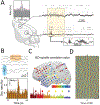
Figure 1:
Figure 1:
A. Impact of the type of arousal. Top example: no-slow wave arousal (predominant fast frequencies). A decrease in spiking is observed during the arousal in the fusiform cortex (light grey dotted arrow). Bottom example: slow wave arousal (mixed low and fast frequencies). An increase in spiking is observed during the arousal in the fusiform cortex (black dotted arrow). EMG = electromyogram; EOG = electro-oculogram. B. Real-time fluctuation before and during arousals. During the pre-arousal time window (ATW), spectral ratio and spike index of mesiotemporal channels are highly correlated, whereas for neocortical channels no time shift is observed. During the ATW, the spectral ratio variation remains significantly correlated and delayed as compared to the spike index of mesiotemporal channels, whereas a positive time shift is observed for neocortical channels. EMG = electromyogram; EOG = electro-oculogram, SI = spike index. Both figures are from Peter-Derex et al., Ann Neurol 2020.
Figure 2:
Figure 2:
Physiological and pathological coupling of brain oscillations. A. LFP traces and raster of neural spiking activity from the hippocampus and the medial prefrontal cortex (mPFC), along with a diagram of the rat brain indicating the recording sites. The highlighted box illustrates temporal coupling of hippocampal ripples and cortical spindles, quantified using a cross-correlogram (top). B. Raw traces of detected IEDs (shaded blue box) and coupled cortical spindles (shaded orange box) from a patient with temporal lobe epilepsy. Scale bar = 1 s, 200 μV (upper). Sample cross-correlogram demonstrating significant IED-spindle coupling in a patient with epilepsy. IED occurrence times served as reference (time = 0, vertical dashed line) and horizontal dashed lines represent 95% confidence intervals (lower). C. Anatomical distribution of electrode locations demonstrating spindles highly coupled with IEDs (warmer colors) from a sample patient with epilepsy. IED-spindle cross-correlograms with a range of significant correlations corresponding to electrode colors (upper panel; 10-s duration). D. Stacked trials of delta phase in a cortical electrode centered on IED occurrence (blue, –pi;red, pi). Scale bars, 25 trials, 500 ms; arrows, time of IED; n = 477 IED trials from one sample patient with epilepsy.

References

    1. Langdon-Down MRB, W. TIME OF DAY IN RELATION TO CONVULSIONS IN EPILEPSY. The Lancet 1929;213:1029–32.
    1. Karoly PJ, Rao VR, Gregg NM, et al. Cycles in epilepsy. Nat Rev Neurol 2021;17:267–84. - PubMed
    1. Karoly PJ, Freestone DR, Boston R, et al. Interictal spikes and epileptic seizures: their relationship and underlying rhythmicity. Brain 2016;139:1066–78. - PubMed
    1. Baud MO, Kleen JK, Mirro EA, et al. Multi-day rhythms modulate seizure risk in epilepsy. Nat Commun 2018;9:88. - PMC - PubMed
    1. Baud MO, Ghestem A, Benoliel JJ, et al. Endogenous multidien rhythm of epilepsy in rats. Exp Neurol 2019;315:82–7. - PubMed

Publication types