. 2024 Jan;115(1):e23539.
doi: 10.1002/bip.23539. Epub 2023 May 25.

The basal and major pilins in the Corynebacterium diphtheriae SpaA pilus adopt similar structures that competitively react with the pilin polymerase

Affiliations

The basal and major pilins in the Corynebacterium diphtheriae SpaA pilus adopt similar structures that competitively react with the pilin polymerase

Christopher K Sue et al. Biopolymers. 2024 Jan.

Abstract

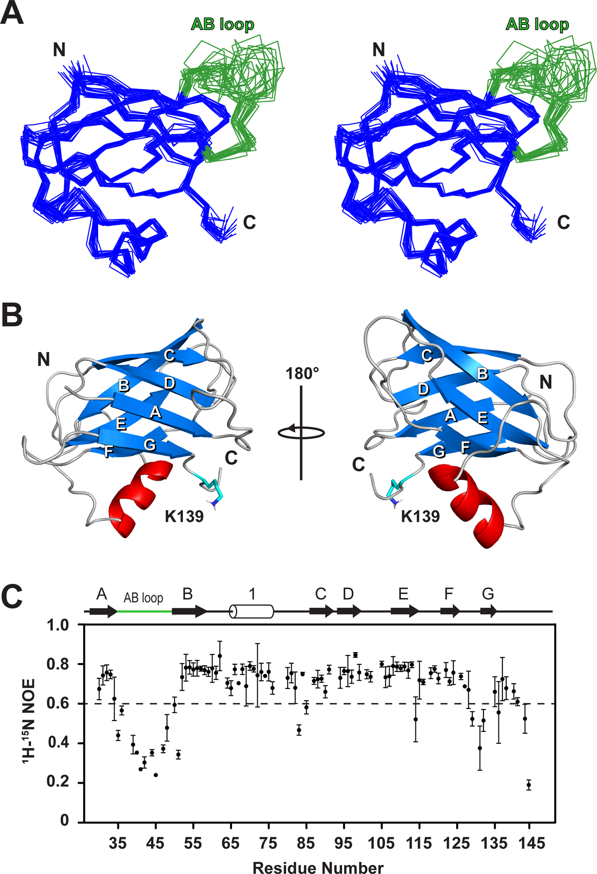
Many species of pathogenic gram-positive bacteria display covalently crosslinked protein polymers (called pili or fimbriae) that mediate microbial adhesion to host tissues. These structures are assembled by pilus-specific sortase enzymes that join the pilin components together via lysine-isopeptide bonds. The archetypal SpaA pilus from Corynebacterium diphtheriae is built by the Cd SrtA pilus-specific sortase, which crosslinks lysine residues within the SpaA and SpaB pilins to build the shaft and base of the pilus, respectively. Here, we show that Cd SrtA crosslinks SpaB to SpaA via a K139(SpaB)-T494(SpaA) lysine-isopeptide bond. Despite sharing only limited sequence homology, an NMR structure of SpaB reveals striking similarities with the N-terminal domain of SpaA (N SpaA) that is also crosslinked by Cd SrtA. In particular, both pilins contain similarly positioned reactive lysine residues and adjacent disordered AB loops that are predicted to be involved in the recently proposed "latch" mechanism of isopeptide bond formation. Competition experiments using an inactive SpaB variant and additional NMR studies suggest that SpaB terminates SpaA polymerization by outcompeting N SpaA for access to a shared thioester enzyme-substrate reaction intermediate.

Keywords: NMR; kinetics; pilus; sortase; virulence factors.


Figures

Figure 1.
Figure 1.. The lysine-isopeptide bond forming reactions catalyzed by CdSrtA.
(A) Schematic of SpaA pilus showing the location of the SpaA, SpaB, and SpaC pilins. The CSpaA-NSpaA and SpaB-CSpaA isopeptide crosslinks that are installed by CdSrtA are shown in expanded detail to the left and right, respectively. (B) Schematic showing the elongation reaction. Shown is the major SpaA pilin that contains N-terminal (N, square), middle (M, circle) and C-terminal (C, triangle) domains. SpaA proteins are joined when a lysine residue (K) within the N-terminal domain (red) is joined to the LPLTG sorting signal (blue). (C) Schematic showing the termination reaction. The LPLTG sorting signal within the major SpaA pilin (red) is joined to the lysine residue within the SpaB pilin (blue). In both images a circle containing a dashed line indicates the lysine-isopeptide bond. The SpaA, SpaB and SpaC pilins are 525, 205, and 1872, amino acids in length, respectively. However, in the figure they are shown as being nearly equal in size.
Figure 2.
Figure 2.. CdSrtA selectively uses K139 within SpaB for crosslinking.
(A) In vitro reconstitution of polymerase catalyzed NSpaA-CSpaA crosslinking reaction. The reactions contained CdSrtAΔ, NSpaA, and CSpaA at concentrations of 100 µM, 300 µM, and 300 µM, respectively. Reactions were incubated at 25°C for 0, 24, and 48 hours (left, middle, and right of each panel). (B) In vitro reconstitution of the SpaB-CSpaA crosslinking reaction. Reactions were performed as in panel (A), except that NSpaA was replaced with wild-type SpaB (left, SpaBWT) or SpaB containing K53A (middle, +SpaBK53A) or K115A (right, +SpaBK115A) amino acid substitutions. (C) Reconstituted reactions containing CdSrtAΔ and CSpaA, and either wild-type SpaB (left, +SpaBWT) or the K139A variant (right, +SpaBK139A). (D) Mass spectrometry (LC-MS/MS) identification of the residues used to form the lysine isopeptide bond between SpaA and SpaB. The panel shows the fragmentation spectra of the linked peptide to SpaB K139 (sequence shown in insert, α is the peptide sequence from SpaB and β is the synthetic LPLTG peptide sequence). Detected fragment ions (M, a-, b-, and y-ions) are labeled accordingly and reported in Supplemental Table S1.
Figure 3.
Figure 3.. Kinetic measurements of SpaB crosslinking.
(A) Schematic showing the termination reaction catalyzed by CdSrtAΔ that joins the LPLTG peptide within the CWSS of SpaA to K139 within the SpaB basal pilin. (B) Overlaid HPLC traces showing reaction progress as a function of time. The reactions contained: 25 μM enzyme, 100 μM SpaB, and 1 mM LPLTG peptide. The amount of SpaB reactant and SpaB-x-LPLT product were quantified. (C) Linear fitting of the Lineweaver-Burk plot of reciprocal initial velocities versus reciprocal SpaB concentration. The data were acquired in triplicate. The insert shows the same data, but with the initial velocity versus substrate SpaB concentration plotted.
Figure 4.
Figure 4.. In vitro crosslinking competition between NSpaA and SpaB.
The panels show time courses of crosslinking reactions containing NSpaA and wild-type and mutant SpaB proteins. (A) A plot of the time course of pilin protein crosslinking by CdSrtAΔ with the LPLTG peptide. The fraction of NSpaA (blue) and SpaB (orange) converted to their crosslinked product is shown. In the reactions 25 μM CdSrtAΔ and 4 mM LPLTG peptide were incubated with either NSpaA or SpaB (200 μM) and the fraction of pilin protein that was crosslinked with the peptide was determined by HPLC. Data were simulated using Equation 1 (see methods). (B) Same as panel (A) showing the fraction of NSpaA converted to its crosslinked product in the absence (blue) or presence of 200 μM SpaB (orange). In the reactions the concentration of NSpaA was 200 μM. (C) Same as panel (B), except that instead of wild type SpaB, an unreactive SpaBK139A variant was employed. The mutant does not react with the enzyme, but nevertheless inhibits SpaA crosslinking. The error bars for the experimental data are the standard deviations of the measured values obtained from three experiments. The dashed lines are the predicted values obtained from simulations (described in the Methods section).
Figure 5.
Figure 5.. SpaB binds to the apo-form of the pilin-specific sortase with very weak affinity.
1H-15N HSQC spectra of SpaB (A) alone, (B) in the presence of unlabeled CdSrtA2M, and (C) an overlay of the spectra shown in panels (A) and (B). Spectra were recorded using 50 µM 15N-labeled SpaB and 350 µM unlabeled CdSrtA2M. No significant changes in SpaB’s spectrum occur upon adding the enzyme. This data indicates that the SpaB pilin does not interact with the apo-form of the enzyme with appreciable affinity (KD > ~2 mM).
Figure 6.
Figure 6.. NMR structure of SpaB.
(A) Stereoview showing the bundle of 20 lowest energy structures of SpaB. Residues that are structured in SpaB are colored blue, while residues in the disordered AB loop are colored green. Residues A27-D142 are shown. (B) Cartoon representation of the structure of SpaB with the strands of the β-sheet elements labeled A to G. The side chain of K139 that is crosslinked by the sortase enzyme is also shown. (C) Plot showing {1H}15N heteronuclear NOE (hetNOE) data for the backbone amide residues in SpaB. Flexible residues are located below the dashed line and have hetNOE values <0.6. The secondary structure is shown above the figure. The data was collected in triplicate and the error bars are the standard deviation of these measurements. The hetNOE data indicate that the AB loop is dynamic on the pico- to nano-second time scales.
Figure 7.
Figure 7.. Comparison of SpaB and NSpaA.
(A) Schematic representation of the secondary structure topologies of SpaB and NSpaA. The β-strands are designated A through G and the helices are numbered. The reactive lysine and sorting signals are shown. (B) Overlay of the backbone structures of SpaB (blue) and the NSpaA (red) (PDB: 3HR6). (C) Comparison of the structures of NSpaA and SpaB. The surface representation of NSpaA crosslinked to its sorting signal peptide is shown on the left (PDB: 7K7F). The structures of the apo-forms of NSpaA and SpaB are shown in the middle and right panels, respectively. Color code: AB loop (green), peptide (magenta), reactive lysine (cyan) and pilin motif residues (red).
Scheme 1.
Scheme 1.

Update of

References

    1. Kline KA; Dodson KW; Caparon MG; Hultgren SJ Trends Microbiol 2010, 18, 224–232. - PMC - PubMed
    1. Ramirez NA; Das A; Ton-That H Trends Microbiol 2020, 28, 999–1009. - PMC - PubMed
    1. Telford JL; Barocchi MA; Margarit I; Rappuoli R; Grandi G Nat Rev Microbiol 2006, 4, 509–519. - PubMed
    1. Hospenthal MK; Costa TRD; Waksman G Nat Rev Microbiol 2017, 15, 365–379. - PubMed
    1. Shanmugasundarasamy T; Karaiyagowder Govindarajan D; Kandaswamy K Cell Surf 2022, 8, 100077. - PMC - PubMed

MeSH terms

LinkOut - more resources