. 2023 May 25;13(1):28.
doi: 10.1186/s12348-023-00340-7.

Combined central retinal vein occlusion and cilioretinal artery occlusion as the initial presentation of frosted branch angiitis: a case report and literature review

Affiliations

Combined central retinal vein occlusion and cilioretinal artery occlusion as the initial presentation of frosted branch angiitis: a case report and literature review

Abdullah Albahlal et al. J Ophthalmic Inflamm Infect. .

Abstract

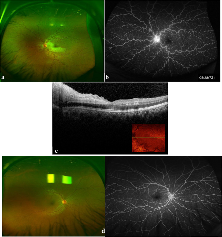
Purpose: To report a case of combined central retinal vein occlusion (CRVO) with cilioretinal artery occlusion (CLRAO) that heralded the development of frosted branch angiitis (FBA).

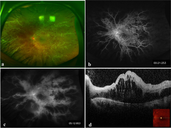
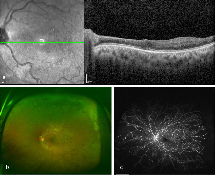
Case report: A 25-year-old healthy male presented with sudden painless visual loss in his left eye with a visual acuity (VA) of 20/300. Fundus exam and fluorescein angiography showed signs of combined CRVO and CLRAO. Without treatment, his vision gradually improved until it reached 20/30 within four months. Five months after initial presentation, he returned with severe visual loss (20/400) in the same eye and a clinical picture of severe occlusive periphlebitis resembling a frosted branch angiitis pattern associated with severe macular edema. This was promptly and successfully treated with systemic steroids and immunosuppressive medications.

Conclusion: CRVO in young population can have an unusual course and one should carefully rule out underlying uveitic etiologies in each visit. Clinical suspicion and close follow‑up are required for early detection and timely management of FBA.

Keywords: Behcet’s disease; Central retinal vein occlusion; Cilioretinal artery occlusion; Frosted branch angiitis; Uveitis.


Conflict of interest statement

The authors declare no competing interests.

Figures

Fig. 1
Fig. 1
Imaging at initial presentation. a Color fundus photograph of the left eye showing dilated tortuous veins, swollen optic disc, and ischemic retinal whitening at the distribution of cilioretinal artery, Note: the two white areas above the superior retinal arcades represent a reflection artefact from the fundus camera. b A fluorescein angiogram showing disc leakage, with no abnormal vascular leakage or capillary non-perfusion (note: consecutive angiography frames revealed an obvious delay in the venous filling). c SD-OCT image showing inner retinal hyperreflectivity and thickening involving the nasal macula in the left eye. d Color fundus photo and fluorescin angiogram image of the normal right eye
Fig. 2
Fig. 2
Imaging five months after the initial presentation. a The same eye shows increased disc edema with hyperemia, macular edema, and thick perivascular sheathing along the major vessels (predominantly venous), typical of frosted branch angiitis. b and c Early and late fluorescein angiograms revealed extensive retinal capillary non-perfusion and leakage from the optic nerve head and retinal veins. d SD-OCT shows severe macular edema
Fig. 3
Fig. 3
Imaging following treatment. a SD-OCT showed resolved macular edema following serial anti-VEGF injections and inner retinal atrophy from previous cilioretinal artery occlusion. At 12 months following immunosuppressive therapy, b fundus photo shows resolution of inflammatory signs, residual venous tortuosity and intraretinal hemorrhages and (c) late frame of fluorescein angiogram shows resolution of leakage from the optic nerve head and retinal veins and persistence of the retinal capillary non-perfusion

Similar articles

Cited by

References

    1. Kleiner RC. Frosted branch angiitis: clinical syndrome or clinical sign? Retina. 1997;17(5):370–371. doi: 10.1097/00006982-199709000-00002. - DOI - PubMed
    1. Walker S, Iguchi A, Jones NP. Frosted branch angiitis: a review. Eye. 2004;18(5):527–533. doi: 10.1038/sj.eye.6700712. - DOI - PubMed
    1. Agarwal A, Rübsam A, ZurBonsen L, Pichi F, Neri P, Pleyer U. A Comprehensive Update on Retinal Vasculitis: Etiologies, Manifestations and Treatments. J Clin Med. 2022;11(9):2525. doi: 10.3390/jcm11092525. - DOI - PMC - PubMed
    1. International Team for the Revision of the International Criteria for Behçet's Disease (ITR-ICBD) The International Criteria for Behçet's Disease (ICBD): a collaborative study of 27 countries on the sensitivity and specificity of the new criteria. J Eur Acad Dermatol Venereol. 2014;28(3):338–347. doi: 10.1111/jdv.12107. - DOI - PubMed
    1. Kleiner RC, Kaplan HJ, Shakin JL, Yannuzzi LA, Crosswell HH, Jr, McLean WC., Jr Acute frosted retinal periphlebitis. Am J Ophthalmol. 1988;106(1):27–34. doi: 10.1016/S0002-9394(14)76383-4. - DOI - PubMed

LinkOut - more resources