Review
. 2023 Aug;16(8):358-370.
doi: 10.1007/s12178-023-09845-z. Epub 2023 May 25.

Management of Glenoid Bone Loss in Primary Reverse Total Shoulder Arthroplasty

Affiliations
Review

Management of Glenoid Bone Loss in Primary Reverse Total Shoulder Arthroplasty

Nabil Mehta et al. Curr Rev Musculoskelet Med. 2023 Aug.

Abstract

Purpose of review: Glenoid bone loss presents distinct challenges in reverse total shoulder arthroplasty (rTSA) which, if unaddressed, can cause complications including poor outcomes and early implant failure. The purpose of this review is to discuss the etiology, evaluation, and management strategies of glenoid bone loss in primary rTSA.

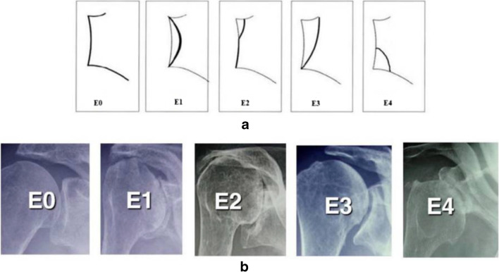
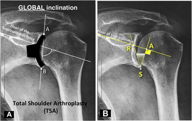
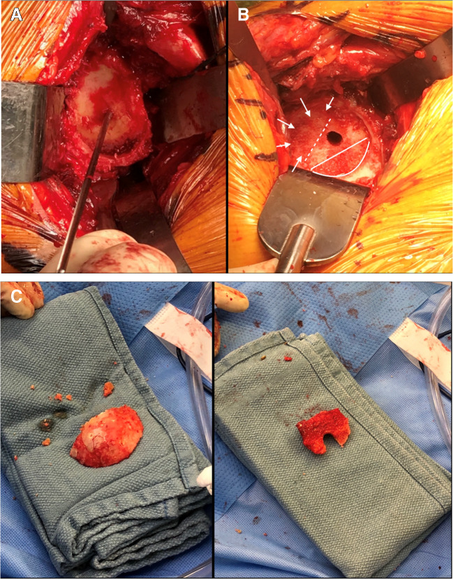
Recent findings: Three-dimensional computed tomography (3D CT) imaging and preoperative planning software have revolutionized the understanding of complex glenoid deformity and wear patterns from bone loss. With this knowledge, a detailed preoperative plan can be created and implemented for a more optimal management strategy. When appropriately indicated, deformity correction techniques with biologic or metal augmentation are successful in addressing the glenoid bone deficiency, creating optimal implant position, and thus providing stable baseplate fixation and improving outcomes. Thorough evaluation and characterization of the degree of glenoid deformity with 3D CT imaging is necessary prior to treatment with rTSA. Eccentric reaming, bone grafting, and augmented glenoid components have shown promising results in correcting glenoid deformity due to bone loss, but long-term outcomes are currently unknown.

Keywords: Glenoid augments; Glenoid bone grafting; Glenoid bone loss; Reverse total shoulder arthroplasty.


Conflict of interest statement

Dr. Nabil Mehta declares that he has no conflict of interest. Dr. Gregory Nicholson reports royalties from Wright Medical Technology, Inc., Innomed, and Arthrosurface; Consultancy with Wright Medical Technology, Inc.; Research support from Arthrex, Inc. and Wright Medical Inc.; Fellowship education support from Wright Medical Inc., and Smith & Nephew; Paid Presenter for Arthrosurface; Board Membership for American Shoulder and Elbow Surgeons.

Figures

Fig. 1
Fig. 1
Schematic (a) and radiographic (b) representation of the Favard classification of glenoid erosion in rotator cuff arthropathy. Adapted from: Walch et al. [14]
Fig. 2
Fig. 2
Schematic (A/C) and radiographic (B/D) representation of the Hamada classification of rotator cuff arthropathy. Adapted from: Hamada et al. [15]
Fig. 3
Fig. 3
Modified Walch classification of glenoid deformity and bone loss. Note that a line drawn from the anterior to posterior native glenoid rim transects the humeral head in the A2 glenoid but not in the A1 glenoid. Reprinted from: Bercik et al. (Copyright 2016), with permission from Elsevier [20]
Fig. 4
Fig. 4
Representative preoperative plan of a primary rTSA using digital templating software based on 3D CT imaging. This tool can be used to anticipate the need for an augmented glenoid component and gauge the optimal component size and position
Fig. 5
Fig. 5
Traditional β angle measurement (A) versus reverse shoulder arthroplasty (RSA) angle (B) as described by Boileau et al. [••] Note that unlike the β angle, the RSA angle is completely in the inferior portion of the glenoid vault, where the glenoid baseplate is implanted in rTSA. Adapted from: Boileau et al. [••]
Fig. 6
Fig. 6
A Intraoperative representation of the “subchondral smile” created by hand reaming the inferior glenoid to the level of the subchondral bone. B The posterosuperior glenoid defect was assessed (arrows) in relation to the subchondral smile below. C The resected humeral head was contoured to create bone graft to fill the size of the glenoid defect
Fig. 7
Fig. 7
A Preoperative AP radiographs demonstrating end-stage glenohumeral osteoarthritis of the right shoulder with a β angle of 63°. The patient was indicated for reverse total shoulder arthroplasty. B After exposure of the glenoid, the central guide pin was placed. Hand reaming was performed to create a “subchondral smile” on the inferior aspect of the glenoid. In this case, an anterioinferior glenoid bone defect was noted and the dimensions were assessed. The resected humeral head was used to create a corticocancellous bone graft of corresponding size, and was placed with the cortex of the graft facing the cortical surface of the glenoid wear deficit. The final baseplate was then implanted. C One-year postoperative radiographs demonstrating stable implants, bone graft incorporation and a β angle of 94°
Fig. 8
Fig. 8
Preoperative and postoperative radiographs demonstrating rotator cuff arthropathy with a posterosuperior glenoid bone defect addressed with rTSA with a posterosuperior metal baseplate augment

References

    1. Seidl AJ, Williams GR, Boileau P. Challenges in reverse shoulder arthroplasty: addressing glenoid bone loss. Orthopedics. 2016;39(1):14–23. doi: 10.3928/01477447-20160111-01. - DOI - PubMed
    1. Tashjian RZ, Granger E, Chalmers PN. Structural glenoid grafting during primary reverse total shoulder arthroplasty using humeral head autograft. J Shoulder Elbow Surg. 2018;27(1):e1–e8. doi: 10.1016/j.jse.2017.07.010. - DOI - PubMed
    1. Sears BW, Johnston PS, Ramsey ML, Williams GR. Glenoid bone loss in primary total shoulder arthroplasty: evaluation and management. JAAOS J Am Acad Orthop Surg. 2012;20(9):604-13. 10.5435/jaaos-20-09-604. - PubMed
    1. Farron A, Terrier A, Büchler P. Risks of loosening of a prosthetic glenoid implanted in retroversion. J Shoulder Elbow Surg. 2006;15(4):521–526. doi: 10.1016/j.jse.2005.10.003. - DOI - PubMed
    1. Iannotti JP, Norris TR. Influence of preoperative factors on outcome of shoulder arthroplasty for glenohumeral osteoarthritis. J Bone Joint Surg Am. 2003;85(2):251–258. doi: 10.2106/00004623-200302000-00011. - DOI - PubMed