

Myopic Versus Glaucomatous Parapapillary Beta Zone in Myopic Eyes Versus Eyes With Secondary Angle-Closure Glaucoma
- PMID: 37227745
- PMCID: PMC10214867
- DOI: 10.1167/iovs.64.5.25
Myopic Versus Glaucomatous Parapapillary Beta Zone in Myopic Eyes Versus Eyes With Secondary Angle-Closure Glaucoma
Abstract
Purpose: To search for histologic differences in the beta zone between myopic eyes versus eyes with secondary angle-closure glaucoma.
Methods: The histomorphometric study consisted of human eyes enucleated due to uveal melanomas or secondary angle-closure glaucoma.
Results: The study included 100 eyes (age: 62.1 ± 15.1 years; axial length: 25.6 ± 3.1 mm; range: 20.0-35.0 mm). In non-highly myopic glaucomatous eyes compared with non-highly myopic nonglaucomatous eyes, the parapapillary alpha zone was longer (223 ± 168 µm vs. 125 ± 128 µm; P = 0.03), beta zone prevalence (15/20 vs. 6/41; P < 0.001) and length (277 ± 245 µm vs. 44 ± 150 µm; P = 0.001) were higher, and RPE cell density in the alpha zone and alpha zone border was lower (all P < 0.05). In highly myopic nonglaucomatous eyes compared with non-highly myopic glaucomatous eyes, parapapillary RPE drusen prevalence (2/19 vs. 10/10; P = 0.01) and alpha zone prevalence (2/19 vs. 16/20; P < 0.001) and length (23 ± 68 µm vs. 223 ± 168 µm; P < 0.001) were lower. In non-highly myopic glaucomatous eyes, Bruch's membrane (BM) thickness decreased (P < 0.001) from the beta zone (6.0 ± 3.1 µm) to the alpha zone (5.1 ± 4.3 µm) and peripheral to it (3.0 ± 0.9 µm). In highly myopic nonglaucomatous eyes, BM thickness did not differ (P > 0.10) between all three regions. In the total study population, RPE cell density in the alpha zone (24.5 ± 9.3 cells/240 µm) was higher than at the alpha zone border (19.2 ± 4.8 cells/240 µm; P < 0.001) or peripheral to it (19.0 ± 3.6 cells/240 µm; P < 0.001).
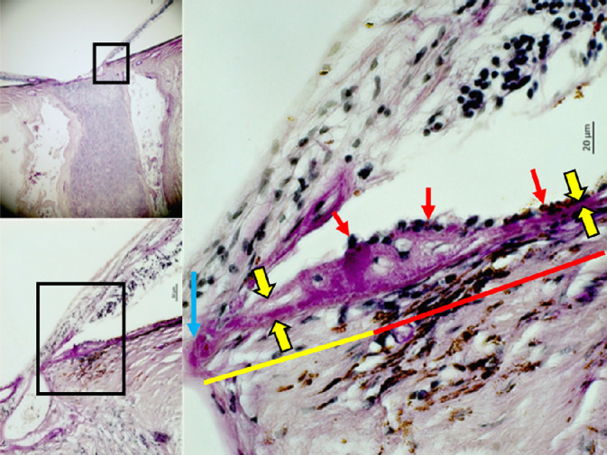
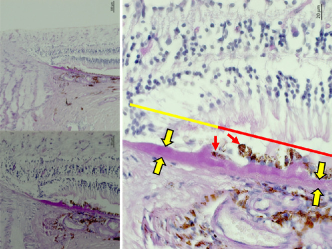
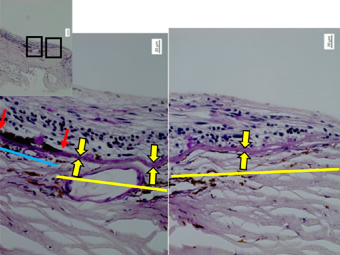
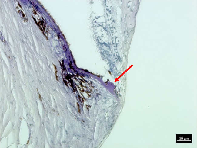
Conclusions: The glaucomatous beta zone in eyes with chronic angle-closure glaucoma (with the alpha zone, parapapillary RPE drusen, thickened BM, and higher RPE cell count in the adjacent alpha zone) differs histologically from the myopic beta zone (characterized by the absence of the alpha zone and parapapillary RPE drusen, unremarkable BM thickness, and unremarkable parapapillary RPE). The differences suggest different etiologies of the glaucomatous versus myopic beta zone.
Conflict of interest statement
Disclosure:
Figures

References
-
- Jonas JB, Nguyen XN, Gusek GC, Naumann GO.. Parapapillary chorioretinal atrophy in normal and glaucoma eyes. I. Morphometric data. Invest Ophthalmol Vis Sci . 1989; 30(5): 908–918. - PubMed
-
- Wang YX, Panda-Jonas S, Jonas JB.. Optic nerve head anatomy in myopia and glaucoma, including parapapillary zones alpha, beta, gamma and delta: histology and clinical features. Prog Retin Eye Res . 2021; 83: 100933. - PubMed
-
- Teng CC, De Moraes CG, Prata TS, Tello C, Ritch R, Liebmann JM.. Beta-zone parapapillary atrophy and the velocity of glaucoma progression. Ophthalmology. 2010; 117(5): 909–915. - PubMed
-
- Park KH, Tomita G, Liou SY, Kitazawa Y.. Correlation between peripapillary atrophy and optic nerve damage in normal-tension glaucoma. Ophthalmology. 1996; 103(11): 1899–1906. - PubMed
-
- Skaat A, De Moraes CG, Bowd C, et al.. Diagnostic Innovations in Glaucoma Study and African Descent and Glaucoma Evaluation Study Groups. African Descent and Glaucoma Evaluation Study (ADAGES): racial differences in optic disc hemorrhage and beta-zone parapapillary atrophy. Ophthalmology. 2016; 123(7): 1476–1483. - PMC - PubMed
MeSH terms
LinkOut - more resources
Full Text Sources
Medical
