. 2023 Jul 10;8(13):e167329.
doi: 10.1172/jci.insight.167329.

The TLR7/IRF-5 axis sensitizes memory CD4+ T cells to Fas-mediated apoptosis during HIV-1 infection

Affiliations

The TLR7/IRF-5 axis sensitizes memory CD4+ T cells to Fas-mediated apoptosis during HIV-1 infection

Liseth Carmona-Pérez et al. JCI Insight. .

Abstract

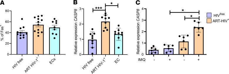
HIV-1 infection is characterized by inflammation and a progressive decline in CD4+ T cell count. Despite treatment with antiretroviral therapy (ART), the majority of people living with HIV (PLWH) maintain residual levels of inflammation, a low degree of immune activation, and higher sensitivity to cell death in their memory CD4+ T cell compartment. To date, the mechanisms responsible for this high sensitivity remain elusive. We have identified the transcription factor IRF-5 to be involved in impairing the maintenance of murine CD4+ T cells during chronic infection. Here, we investigate whether IRF-5 also contributes to memory CD4+ T cell loss during HIV-1 infection. We show that TLR7 and IRF-5 were upregulated in memory CD4+ T cells from PLWH, when compared with naturally protected elite controllers and HIVfree participants. TLR7 was upstream of IRF-5, promoting Caspase 8 expression in CD4+ T cells from ART HIV-1+ but not from HIVfree donors. Interestingly, the TLR7/IRF-5 axis acted synergistically with the Fas/FasL pathway, suggesting that TLR7 and IRF-5 expression in ART HIV-1+ memory CD4+ T cells represents an imprint that predisposes cells to Fas-mediated apoptosis. This predisposition could be blocked using IRF-5 inhibitory peptides, suggesting IRF-5 blockade as a possible therapy to prevent memory CD4+ T cell loss in PLWH.

Keywords: AIDS/HIV; Apoptosis; Immunology; T cells.


Conflict of interest statement

Conflict of interest: The authors have declared that no conflict of interest exists.

Figures

Figure 1
Figure 1. IRF5 is expressed in CD4+ T cells from PHI.
PBMCs from PHI and HIVfree individuals were analyzed by flow cytometry. (A) Representative FACS plots and percentage of IRF-5+ CD4+ T cells. (B) Representative ImageStreamX images of CD4+ T cells expressing IRF5 in the cytoplasm (first 2 lanes) and in the nucleus (third and fourth lane). (CF) Graphs show (C) the frequency of CD4+ T cells whose IRF-5 expression colocalizes with the nucleus; (D) the percentage of IFN-γ+ and IFN-γIRF-5+ CD4+ T cells; (E) the frequency of CD45RA+ and CD45RAIFN-γ+ CD4+ T cells expressing IRF-5 in PHI individuals; and (F) the percentage of annexin V+CD4+ T cells from PHI and HIVfree individuals. Data are presented as the mean ± SD. The Mann-Whitney U test was used for significance. **P < 0.01, ****P < 0.0001, n =10. Graphs represent correlations between (G) annexin V and IRF-5 expression in CD4+ T cells, (H) annexin V expression and CD4+ T cell count, and (I) IRF-5 expression and CD4+ T cell count in PHI donors. The Spearman’s r test was used for significance, *P < 0.05, **P < 0.01, n =10.
Figure 2
Figure 2. IRF5 is expressed in memory and effector CD4+ T cells from individuals on ART.
PBMCs from ART HIV-1+, EC, and HIVfree donors were analyzed by flow cytometry. (A) Representative FACS plots and percentage of IRF-5+ CD4+ T cells. (BD) Graphs show (B) the percentage of CD4+ T cells expressing IRF-5 in the nucleus; (C) the percentage of IRF-5 expression in naive, effector, and memory CD4+ T cell; and (D) the percentage of memory CD4+ T cells positive for annexin V in PBMCs isolated from ART HIV-1+, EC, and HIVfree individuals. Data are presented as the mean ± SD. The Kruskal-Wallis followed by the Dunn’s multiple-comparison tests were used for significance. *P < 0.05, **P < 0.01, ***P < 0.001, ****P < 0.0001, n = 12 (ART HIV-1+ and HIVfree), n = 9 (EC). (E) Graph represents correlation between annexin V and IRF-5 expression in memory CD4+ T cells from patients with HIV-1+ on ART. The Spearman’s r test was used for significance. n =12.
Figure 3
Figure 3. TLR7 is upstream of IRF5.
(A) Graph shows TLR7 mRNA levels in CD4+ T cells from ART HIV-1+, EC, and HIVfree individuals analyzed by qPCR. The Kruskal-Wallis followed by the Dunn’s multiple-comparison tests were used for significance. *P < 0.05, **P < 0.01. n =9. CD4+ T cells purified from ART HIV-1+, EC, and HIVfree donors were treated in vitro with imiquimod (IMQ) or medium alone for 30 hours. (BD) Graphs represent (B) the percentage of IRF-5+, (C) the percentage of apoptotic cells, and (D) the percentage dead memory CD4+ T cells. Data are presented as the mean ± SD. The Friedman’s followed by the Dunn’s multiple-comparison tests were used to determine statistical differences. *P < 0.05, **P < 0.01. n = 6. (E and F) Graphs show correlations between (E) TLR7 and IRF-5 expression and (F) annexin V and TLR7 expression in ART HIV-1+ donors. The Spearman’s r test was used for statistical significance. n =9.
Figure 4
Figure 4. Caspase 8 is downstream of IRF5.
CD4+ T cells from PBMCs of ART HIV-1+, EC, and HIVfree individuals were analyzed ex vivo. (A and B) Graphs show (A) the percentage of Fas+ memory CD4+ T cells determined by flow cytometry and (B) RT-PCR analysis of CASP8 mRNA levels in purified CD4+ T cells. Data are presented as the mean ± SD. The Kruskal-Wallis followed by the Dunn’s multiple-comparison tests were used for significance. *P < 0.05, ***P < 0.001. n =12 (ART HIV-1+ and HIVfree), n =9 (EC). (C) Purified CD4+ T cells from ART HIV-1+ and HIVfree donors were treated in vitro with imiquimod (IMQ) or medium alone for 30 hours. Graph show expression of CASP8 mRNA. Data are presented as the mean ± SD. The Friedman’s followed by the Dunn’s multiple-comparison tests were used for significance. *P < 0.05. n =6.
Figure 5
Figure 5. Activation of the TLR7/IRF-5 axis predisposes memory CD4+ T cells to Fas-mediated apoptosis.
(A) Purified CD4+ T cells from ART HIV-1+ and HIVfree donors were incubated in vitro with rFasL or medium alone for 18 hours. Graph shows IRF-5 expression as measured by flow cytometry. Data are presented as the mean ± SD. The Friedman’s followed by the Dunn’s multiple-comparison tests were used for significance. *P < 0.05. n =6. (B) Scheme of the experimental design. Purified CD4+ T cells were treated with IMQ or medium alone; 12 hours later, rFasL or medium was added to the culture, and the cells were incubated for a further 18 hours for a total of 30 hours of incubation at 37°C. (CE) Graphs show (C) the percentage of apoptotic memory CD4+ T cells, (D) the percentage of dead memory CD4+ T cells, and (E) CASP8 mRNA levels in memory CD4+ T cells after 30-hour stimulation. Data are presented as the mean ± SD. The Wilcoxon test was used for significance. *P < 0.05. n =6.
Figure 6
Figure 6. IRF-5 inhibitory peptides rescue memory CD4+ T cells from Fas-mediated apoptosis.
(AC) Purified CD4+ T cells from ART HIV-1+ and HIVfree individuals were pretreated with 10 μM of IRF-5–CPPs for 30 minutes before stimulation with IMQ and incubated at 37°C for 30 hours. Graphs show (A) the percentage of apoptotic cells, (B) the percentage of dead cells, and (C) CASP8 mRNA expression levels in memory CD4+ T cells from ART HIV-1+ and HIVfree donors. (D) Experimental design. Purified CD4+ T cells from ART HIV-1+ and HIVfree individuals were pretreated with 10 μM of IRF-5–CPPs for 30 minutes, stimulated with or without IMQ for 12 hours, and incubated for a further 18 hours with or without rFasL. (EG) Graphs show (E) the percentage of apoptotic cells, (F) the percentage of dead cells, and (G) CASP8 mRNA expression levels in memory CD4+ T cells from ART HIV-1+ and HIVfree donors. Data are presented as the mean ± SD. The Friedman’s followed by the Dunn’s multiple-comparison tests were used for significance. *P < 0.05. n = 6.
Figure 7
Figure 7. IFN-β and DAMPs promote TLR7 expression on CD4+ T cells.
(A) Purified CD4+ T cells from HIVfree individuals were incubated with IFN-β or with medium alone. Graph shows RT-PCR analysis of TLR7 expression at 0, 6, 12, and 24 hours of treatment. (B) Purified CD4+ T cells from HIVfree individuals were treated with IFN-β in the presence or absence of conditioned medium with 10% v/v apoptotic material (AM, supernatant of staurosporine-treated PBMCs) for 24 hours, before stimulation with αCD3α/CD28 for a further 24 hours. Graphs shows RT-PCR analysis of TLR7 gene expression. (CE) Purified CD4+ T cells from HIVfree individuals were incubated with IFN-β in the presence or absence of conditioned medium with 10% v/v apoptotic material for 24 hours, before adding rFasL for a further 18 hours. Graphs represent (C) the percentage of IRF-5+, (D) the percentage of apoptotic cells, and (E) the percentage of dead CD4+ T cells after incubation with the indicated culture conditions. (FH) Purified CD4+ T cells from HIVfree donors were treated with IFN-β in the presence or absence of conditioned medium with 10% v/v apoptotic material for 24 hours; they were then stimulated with αCD3/αCD28 for a further 24 hours and finally incubated for 18 hours with rFasL. Graphs represent (F) the percentage of IRF5+, (G) the percentage of apoptotic cells, and (H) the percentage of dead CD4+ T cells after incubation as described above. Data are presented as the mean ± SD. The Friedman’s followed by the Dunn’s multiple-comparison tests were used for significance. *P < 0.05. n =6.
Figure 8
Figure 8. Proposed model.
Residual inflammation maintains the upregulation of TLR7 and IRF-5 in memory CD4+ T cells from ART HIV-1+ individuals but not in HIVfree individuals. The activation of the TLR7/IRF-5 axis feeds into the Fas/FasL pathway, predisposing and enhancing Fas-mediated apoptosis only in memory CD4+ T cells from ART HIV-1+ individuals. Finally, we proposed that this mechanism can be blocked by adding IRF-5 inhibitory peptides, which block the predisposition to Fas-mediated cell death. Created with BioRender.com.
Figure 9
Figure 9. Summary of cell death pathways occurring in CD4+ T cells during primary HIV-1 infection or following ART.
(A) A large proportion of CD4+ T cell death during primary HIV-1 infection occurs via apoptosis and comprises extrinsic and intrinsic pathways. Extrinsic apoptosis involves death receptor members of the tumor necrosis factor receptor gene superfamily, such as Fas, TNFR1, and DR5 (TRAILR2). The activation of these receptors by a death ligand results in the activation of Caspase 8. We propose that the TLR7/IRF-5 pathway feeds into these pathways by sensitizing cells to Fas-, TNFR1-, and DR5-mediated apoptosis through the induction of Caspase 8 and IRF-5. Other extrinsic cell death pathways include activation-induced cell death (AICD) and killing by cytotoxic CD8+ T cells (CTL) via secretion of perforin and granzymes. Intrinsic apoptosis includes activation of Caspase 3 by HIV proteins and DNA. Finally, abortively infected CD4+ T cells die by pyroptosis, a highly inflammatory form of programmed cell death that results from the activation of Caspase 1. (B) Although antiretroviral therapy restores CD4+ T cell counts, memory CD4+ T cells from ART HIV-1+ individuals are still prone to apoptosis, despite treatment. These cells mostly die by Fas-mediated apoptosis. The Fas/FasL pathway is strengthened on one hand by the transcription factor FOXO3a, which empowers the “killers,” and, on the other hand, by the TLR7/IRF-5 axis, which weakens the “victims.” Death by pyroptosis in abortively infected cells may also occur. Created with BioRender.com.

Similar articles

Cited by

References

    1. Chun TW, et al. Presence of an inducible HIV-1 latent reservoir during highly active antiretroviral therapy. Proc Natl Acad Sci U S A. 1997;94(24):13193–13197. doi: 10.1073/pnas.94.24.13193. - DOI - PMC - PubMed
    1. Finzi D, et al. Identification of a reservoir for HIV-1 in patients on highly active antiretroviral therapy. Science. 1997;278(5341):1295–1300. doi: 10.1126/science.278.5341.1295. - DOI - PubMed
    1. Wong JK, et al. Recovery of replication-competent HIV despite prolonged suppression of plasma viremia. Science. 1997;278(5341):1291–1295. doi: 10.1126/science.278.5341.1291. - DOI - PubMed
    1. Doitsh G, et al. Cell death by pyroptosis drives CD4 T-cell depletion in HIV-1 infection. Nature. 2014;505(7484):509–514. doi: 10.1038/nature12940. - DOI - PMC - PubMed
    1. Monroe KM, et al. IFI16 DNA sensor is required for death of lymphoid CD4 T cells abortively infected with HIV. Science. 2014;343(6169):428–432. doi: 10.1126/science.1243640. - DOI - PMC - PubMed

Publication types