. 2023 Jun 28;194(1):53-69.
doi: 10.1093/toxsci/kfad049.

Use of the dTAG system in vivo to degrade CDK2 and CDK5 in adult mice and explore potential safety liabilities

Affiliations

Use of the dTAG system in vivo to degrade CDK2 and CDK5 in adult mice and explore potential safety liabilities

Paul Yenerall et al. Toxicol Sci. .

Abstract

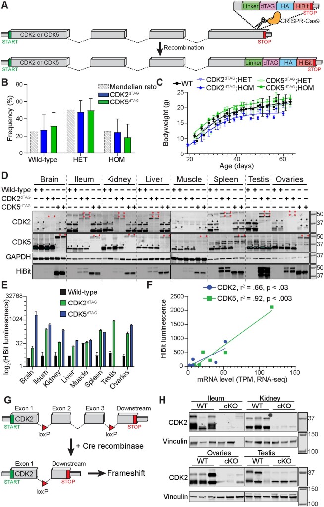
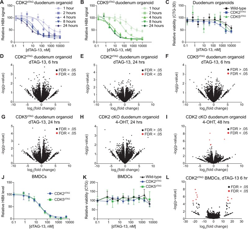
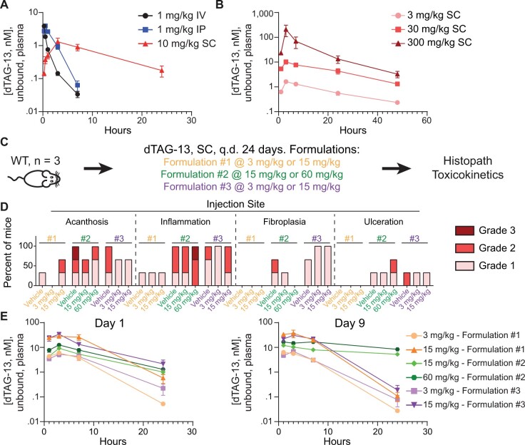
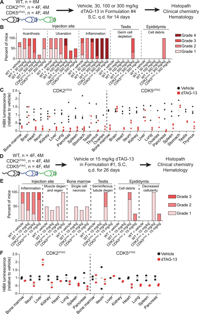
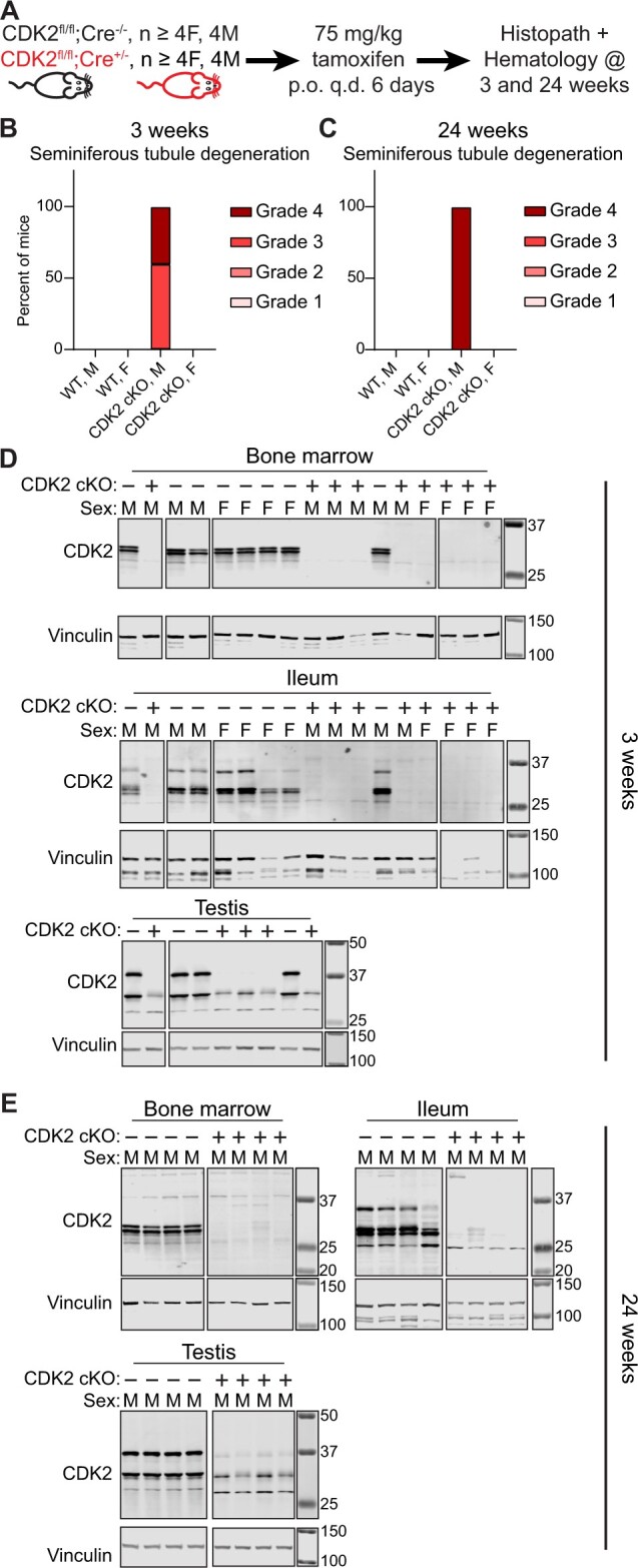
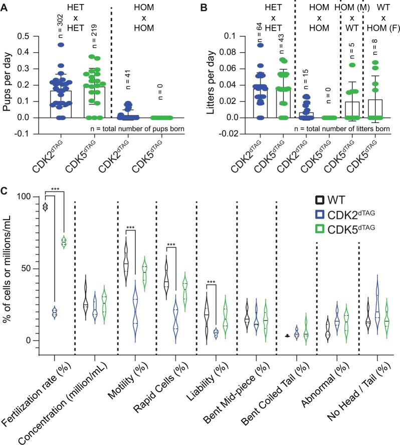
The degradation tag (dTAG) system for target protein degradation can remove proteins from biological systems without the drawbacks of some genetic methods, such as slow kinetics, lack of reversibility, low specificity, and the inability to titrate dosage. These drawbacks can make it difficult to compare toxicity resulting from genetic and pharmacological interventions, especially in vivo. Because the dTAG system has not been studied extensively in vivo, we explored the use of this system to study the physiological sequalae resulting from CDK2 or CDK5 degradation in adult mice. Mice with homozygous knock-in of the dTAG sequence onto CDK2 and CDK5 were born at Mendelian ratios despite decreased CDK2 or CDK5 protein levels in comparison with wild-type mice. In bone marrow cells and duodenum organoids derived from these mice, treatment with the dTAG degrader dTAG-13 resulted in rapid and robust protein degradation but caused no appreciable change in viability or the transcriptome. Repeated delivery of dTAG-13 in vivo for toxicity studies proved challenging; we explored multiple formulations in an effort to maximize degradation while minimizing formulation-related toxicity. Degradation of CDK2 or CDK5 in all organs except the brain, where dTAG-13 likely did not cross the blood brain barrier, only caused microscopic changes in the testis of CDK2dTAG mice. These findings were corroborated with conditional CDK2 knockout in adult mice. Our results suggest that the dTAG system can provide robust protein degradation in vivo and that loss of CDK2 or CDK5 in adult mice causes no previously unknown phenotypes.

Keywords: CDK2; CDK5; PROTAC; degradation; mice; toxicity.


Figures

Figure 1.
Figure 1.
Derivation and validation of CDK2dTAG and CDK5dTAG knock-in mice. A, Knock-in of the dTAG onto the C-terminus of Cdk2 and Cdk5 in the germline of C57BL/6J mice. Cartoon diagram of the strategy to generate dTAG knock-in mice. A DNA sequence encoding a linker, the dTAG, an HA tag and a HiBit sequence were knocked onto the C-terminus of Cdk2 or Cdk5 in the germline of C57BL/6J mice using CRISPR/Cas9 and homology-directed repair. Dashed lines represent introns, boxes represent exons. Not drawn to scale and all exons/introns are not included for illustrative purposes. B, HET × HET crosses of CDK2dTAG or CDK5dTAG mice yield pups with Mendelian ratios. Mendelian ratio is the expected genotype frequency from crossing heterozygous mice. HET, heterozygous; HOM, homozygous. n > 17 litters for CDK2dTAG and CDK5dTAG. Data are displayed as averages ± standard deviations (SDs). C, Developing HOM CDK2dTAG mice tend to weigh less than CDK5dTAG or WT mice. Bodyweight of developing wild-type (WT), CDK2dTAG and CDK5dTAG mice. n > 3 mice for all data points, n > 6 for most data points. Lines fit using a smoothing spline with 4 knots in GraphPad prism. D, CDK2dTAG and CDK5dTAG mice have decreased expression of the dTAG fusion protein. Western blot for CDK2 and CDK5 and far western for the HiBit tag in HOM mice for the indicated genotype. Each lane represents an individual mouse. Highlighted regions are saturated and asterisks at the top right of bands represent the presumed CDK2 or CDK5 proteins (black or red for untagged or dTAG’d proteins, respectively). Mice were approximately 6–10 weeks old. Molecular weight markers are in kDa. E, Expression of CDK2dTAG and CDK5dTAG fusion proteins by the HiBit assay in various tissues. Protein levels of CDK2 and CDK5 as read out via a plate-based HiBit assay. Values are averages ± SD, n= 2. Same protein lysate as from (D). F, Strong correlation between mRNA levels of CDK2 or CDK5 in WT mice and CDK2dTAG or CDK5dTAG protein levels, respectively, in dTAG knock-in mice. Correlation between HiBit signal and mRNA levels (RNA-seq) for CDK2 and CDK5. Tissues shown are brain, ileum, kidney, liver, muscle, testis, and ovaries. For the brain, the HiBit signal is for the entire brain and mRNA level is the average transcripts per million (TPM) from RNA-seq on the cerebellum and cerebral cortex. HiBit data is from (E). G, Knock-in of loxP sites flanking exon 2 and 3 to generate Cdk2 conditional KO mice. Cartoon diagram of the strategy to delete Cdk2. Exons 2 and 3 were flanked by loxP sites, leading to deletions of exon 2 and 3 and a frameshift in CDK2 upon Cre-mediated recombination. Dashed lines represent introns, boxes represent exons. Not drawn to scale and all exons/introns are not included for illustrative purposes. H, Treatment of CDK2 cKO mice with tamoxifen causes loss of CDK2 protein expression. CDK2 cKO mice (genotype Cdk2fl/fl; Rosa26tm(Cre/ERT2)+/−) or “wild-type” mice (genotype Cdk2fl/fl; Rosa26tm(Cre/ERT2−/−)) were administered 75 mg/kg tamoxifen orally for 6 days. Twelve days later tissues were analyzed for CDK2 protein expression by immunoblot. Molecular weight markers are in kDa.
Figure 2.
Figure 2.
Kinetics and consequences of CDK2 or CDK5 destruction using in vitro models of the bone marrow and small intestine. A and B, Kinetics of CDK2 or CDK5 destruction after dTAG-13 administration in duodenum organoids derived from HOM CDK2dTAG or CDK5dTAG mice. HOM CDK2dTAG (A) and HOM CDK5dTAG (B) duodenum organoids were treated as indicated and then the HiBit assay was performed to determine the amount of CDK2 (A) or CDK5 (B) protein. Values shown are normalized to DMSO treatment and are averages ± standard error of the mean (SEM) for 3 biological replicates. C, Degradation of CDK2 or CDK5 in CDK2dTAG or CDK5dTAG duodenum organoids does not affect viability. Duodenum organoids derived from WT, CDK2dTAG or CDK5dTAG HOM mice were treated with dTAG-13 for 4 days, then viability was measured by CellTiter-Glo 3D. Values shown are normalized to DMSO treatment and are averages ± SEM for 3 biological replicates. D and E, No significant changes in gene transcription after degrading CDK2 in CDK2dTAG duodenum organoids. Duodenum organoids derived from HOM CDK2dTAG mice were treated for 6 h (D) or 24 h (E) with 250 nM dTAG-13 or DMSO, QuantSeq was performed, and differential gene expression analysis was performed between the 2 groups. n = 3 biological replicates. FDR, false discovery rate. F and G, No significant changes in gene transcription after degrading CDK5 in CDK5dTAG duodenum organoids. Duodenum organoids derived from HOM CDK5dTAG mice were treated for 6 h (F) or 24 h (G) with 250 nM dTAG-13 or DMSO, QuantSeq was performed, and differential gene expression analysis was performed between the 2 groups. n = 3 biological replicates. (H and I) No significant changes in gene transcription after CDK2 KO in CDK2 cKO duodenum organoids. Duodenum organoids derived from HOM CDK2 cKO mice were treated for 24 h (H) or 48 h (I) with 1 µM 4-hydroxytamoxifen (4-OHT) or DMSO, QuantSeq was performed, and differential gene expression analysis was performed between the 2 groups. n = 3 biological replicates. J, Potent degradation of CDK2 or CDK5 in BMDCs from CDK2dTAG and CDK5dTAG mice. Bone marrow cells were derived from multiple CDK2dTAG or CDK5dTAG HOM mice, pooled, treated with dTAG-13 in vitro for 6 h at the indicated concentrations, and the HiBit assay was performed to determine levels of CDK2 or CDK5 protein. Values are normalized to DMSO treatment and are averages ± SEM for 3 technical replicates. K, No change in viability after degrading CDK2 or CDK5 in BMDCs from CDK2dTAG or CDK5dTAG mice. Bone marrow cells were derived from 3–4 WT, CDK2dTAG or CDK5dTAG HOM mice, pooled, treated with dTAG-13 in vitro for 4 days at the indicated concentrations, and cell viability measured via CellTiter-Glo. Values are normalized to DMSO treatment and are averages ± SEM for 3 technical replicates. L, No significant changes in gene transcription after degrading CDK2 in BMDCs from CDK2dTAG mice. BMDCs from HOM CDK2dTAG were treated with 250 nM dTAG-13 or DMSO for 24 h, QuantSeq was performed, and differential gene expression analysis was performed between the 2 groups. n = 3 technical replicates. BMDCs, bone marrow derived cells.
Figure 3.
Figure 3.
Delivering dTAG-13 in vivo for toxicity studies. A, Determining the optimal route to deliver dTAG-13 in vivo. A single dose of dTAG-13 was given intravenously (IV) dissolved in an IV formulation, intraperitoneally (IP) dissolved in Formulation no. 1, or subcutaneously (SC) dissolved in Formulation no. 2, and levels of unbound dTAG-13 in plasma were quantified over time. n = 4 mice per group (2 males, 2 females); values shown are averages of the estimated unbound plasma levels of dTAG-13 ± SEM. B, Linear dose-exposure relationship for dTAG-13 delivered SC using Formulation no. 2. Pharmacokinetics of various doses of dTAG-13 dissolved in Formulation no. 2 and delivered SC. n = 6 male mice per group. Values shown are averages of the estimated unbound plasma levels of dTAG-13 ± SEM. C–E, Testing multiple dTAG-13 formulations in WT mice to identify a formulation suitable for repeat dose toxicity studies. (C) study outline for treating 3 male WT mice with dTAG-13 delivered in 3 different formulations. (D) histopath results of the injection site after 24 days of dosing. H&E slides from all mice in all groups were evaluated by a board-certified veterinary anatomic pathologist. Findings deemed secondary to injection site toxicity are displayed in Supplementary Figure 3. E, Estimated unbound plasma levels of dTAG-13 after 1 day or 9 days of dosing. Values are displayed as averages ± SEM and n = 3. q.d., once daily. Formulation composition can be found in Materials and Methods.
Figure 4.
Figure 4.
Degradation of CDK2 or CDK5 outside the brain in adult mice only causes a morphological change in the testis of CDK2dTAG mice. A–C, Potent degradation of CDK2 or CDK5 for 14 days using dTAG-13 delivered in Formulation no. 4 only causes a morphological change in the testis. A, Study outline for treating HOM WT, CDK2dTAG or CDK5dTAG mice with dTAG-13 delivered in Formulation no. 4 for 14 days. B, CDK2-specific and injection site histopathology results following 14 days of dTAG-13 treatment using Formulation no. 4. For most findings, H&E slides from 5 to 8 mice were evaluated by a board-certified veterinary anatomic pathologist. Results from male and female mice were combined due to a lack of gender specific differences. Findings deemed secondary to injection site toxicity are displayed in Supplementary Figure 5. C, Levels of CDK2 or CDK5 via a plate-based HiBit assay in HOM CDK2dTAG or CDK5dTAG mice after 2 days of dosing dTAG-13 or vehicle in vivo using Formulation no. 4. Data points are from individual mice and are relative to the average HiBit signal from vehicle-treated mice. A dashed line separates results from CDK2dTAG and CDK5dTAG mice. For most tissues, results from 2 male and 2 female mice are shown. M, male; F, female. ND, not determined. D–F, Modest degradation of CDK2 or CDK5 for 26 days using dTAG-13 delivered in Formulation no. 1 only causes a morphological change in the testis. D, Study outline for treating HOM CDK2dTAG or CDK5dTAG mice with dTAG-13 in Formulation no. 1 for 26 days. E, Summary of all histopathologic findings. For most findings, H&E slides from at least 4 mice per gender were evaluated by a board-certified veterinary anatomic pathologist. Results from male and female mice were combined due to lack of gender differences. Degen, degeneration, regen, regeneration. F, Levels of CDK2 or CDK5 via a plate-based HiBit assay in HOM CDK2dTAG or CDK5dTAG mice after 2 days of dosing dTAG-13 or vehicle in vivo using Formulation no. 1. Values are from individual mice and are relative to the average HiBit signal from vehicle treated mice. A dashed line separates results from CDK2dTAG and CDK5dTAG mice. For most tissues, results from 1 male and 1 female mouse are shown.
Figure 5.
Figure 5.
Conditional knockout of CDK2 in adult mice confirms lack of a novel morphological change after CDK2 loss. A–C, Conditional knockout of CDK2 in adult mice using Cre-mediated recombination only causes a morphological change in the testis. A, Study outline. CDK2 cKO (genotype Cdk2fl/fl; Rosa26tm(Cre/ERT2)+/−) or WT (genotype Cdk2fl/fl; Rosa26tm(Cre/ERT2−/−) mice were administered 75 mg/kg tamoxifen by oral gavage once per day for 6 days and mice were analyzed 3 weeks or 24 weeks after tamoxifen administration. B, Summary of histopathology results 3 weeks after deletion of CDK2. H&E slides from at least 4 mice per gender were evaluated by a board-certified veterinary anatomic pathologist. C, Summary of histopathology results 24 weeks after deletion of CDK2. H&E slides from at least 8 mice per gender were evaluated by a board-certified veterinary anatomic pathologist. D and E, Treatment of CDK2 cKO mice with tamoxifen yields potent and long-lasting loss of CDK2 protein. D, Western blot for CDK2 in the bone marrow, ileum or testis 3 weeks after WT (“CDK2 cKO –”)or CDK2 cKO (“CDK2 cKO +”) mice have been administered 75 mg/kg tamoxifen for 6 days. E, Western blot for CDK2 in the bone marrow, ileum, or testis 24 weeks after WT or CDK2 cKO mice have been administered 75 mg/kg tamoxifen for 6 days. For each tissue, all samples were ran and transferred on the same gel/membrane but images were cropped to remove failed or unrelated samples. Molecular weight markers are in kDa.
Figure 6.
Figure 6.
Homozygous CDK2dTAG and CDK5dTAG mice have reduced fecundity. A, Breeding of HOM CDK2dTAG and CDK5dTAG mice produces less pups than HET mice. Number of pups born per day from crossing heterozygotes (HET × HET, left) or homozygous (HOM × HOM, right) CDK2dTAG or CDK5dTAG mice. n = number of pups born. B, Breeding of HOM CDK2dTAG or CDK5dTAG mice produces less litters than HET or WT mice. Number of litters born per day from crossing heterozygotes (HET × HET), homozygous (HOM × HOM), or homozygous and wild-type (HOM × WT) CDK2dTAG or CDK5dTAG mice. n is number of litters born. C, Sperm from HOM CDK2dTAG and CDK5dTAG mice are less fertile than WT mice. For fertilization rates, oocytes from WT mice were inseminated with sperm from HOM WT, CDK2dTAG or CDK5dTAG mice and the next day the total number of 2 cell embryos was counted to determine the fertilization rate. For sperm morphological analyses, mice were euthanized and sperm was collected from the caudal epididymis and vas deferens, chilled, and analyzed on an IVOS machine. Liability is the percent of normal sperm in the sample. Data shown are 2 independent runs on the IVOS machine from 4 mice (2 mice per run). *** = p < .01, 2-way ANOVA with Dunnett’s multiple comparison test.

References

    1. Abuhashem A., Lee A. S., Joyner A. L., Hadjantonakis A. K. (2022). Rapid and efficient degradation of endogenous proteins in vivo identifies stage-specific roles of RNA pol II pausing in mammalian development. Dev. Cell. 57, 1068–1080.e6. - PMC - PubMed
    1. Aulbach A., Vitsky A., Arndt T., Ramaiah L., Logan M., Siska W., Cregar L., Tripathi N., Adedeji A., Provencher A., et al. (2019). Interpretative considerations for clinical pathology findings in nonclinical toxicology studies. Vet. Clin. Pathol. 48, 383–388. - PubMed
    1. Banaszynski L. A., Sellmyer M. A., Contag C. H., Wandless T. J., Thorne S. H. (2008). Chemical control of protein stability and function in living mice. Nat. Med. 14, 1123–1127. - PMC - PubMed
    1. Berthet C., Aleem E., Coppola V., Tessarollo L., Kaldis P. (2003). CDK2 knockout mice are viable. Curr. Biol. 13, 1775–1785. - PubMed
    1. Bondeson D. P., Mullin-Bernstein Z., Oliver S., Skipper T. A., Atack T. C., Bick N., Ching M., Guirguis A. A., Kwon J., Langan C., et al. (2022). Systematic profiling of conditional degron tag technologies for target validation studies. Nat. Commun. 13, 5495. - PMC - PubMed