Review
. 2023 May 26;132(11):1521-1545.
doi: 10.1161/CIRCRESAHA.123.321563. Epub 2023 May 25.

HDL Function and Atherosclerosis: Reactive Dicarbonyls as Promising Targets of Therapy

Affiliations
Review

HDL Function and Atherosclerosis: Reactive Dicarbonyls as Promising Targets of Therapy

MacRae F Linton et al. Circ Res. .

Abstract

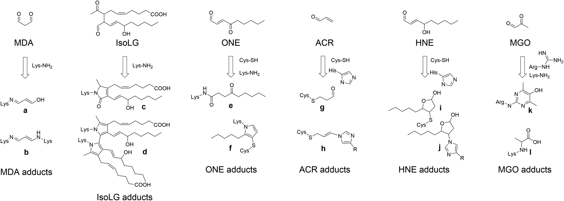
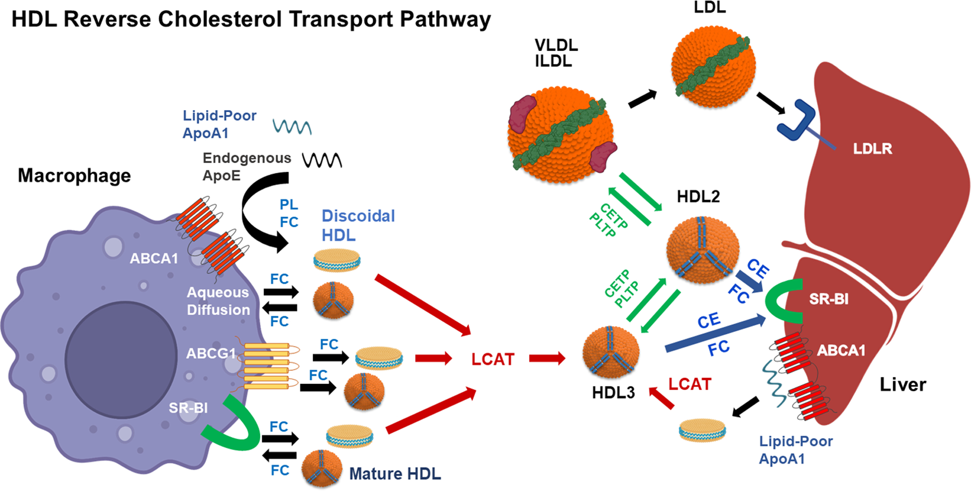
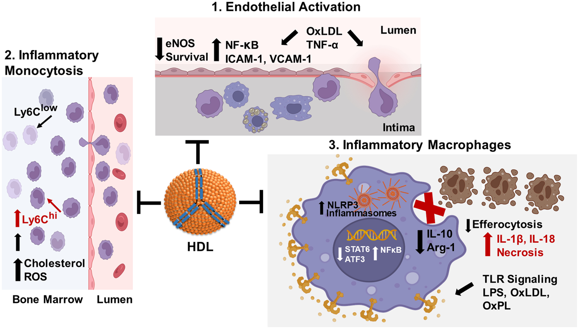
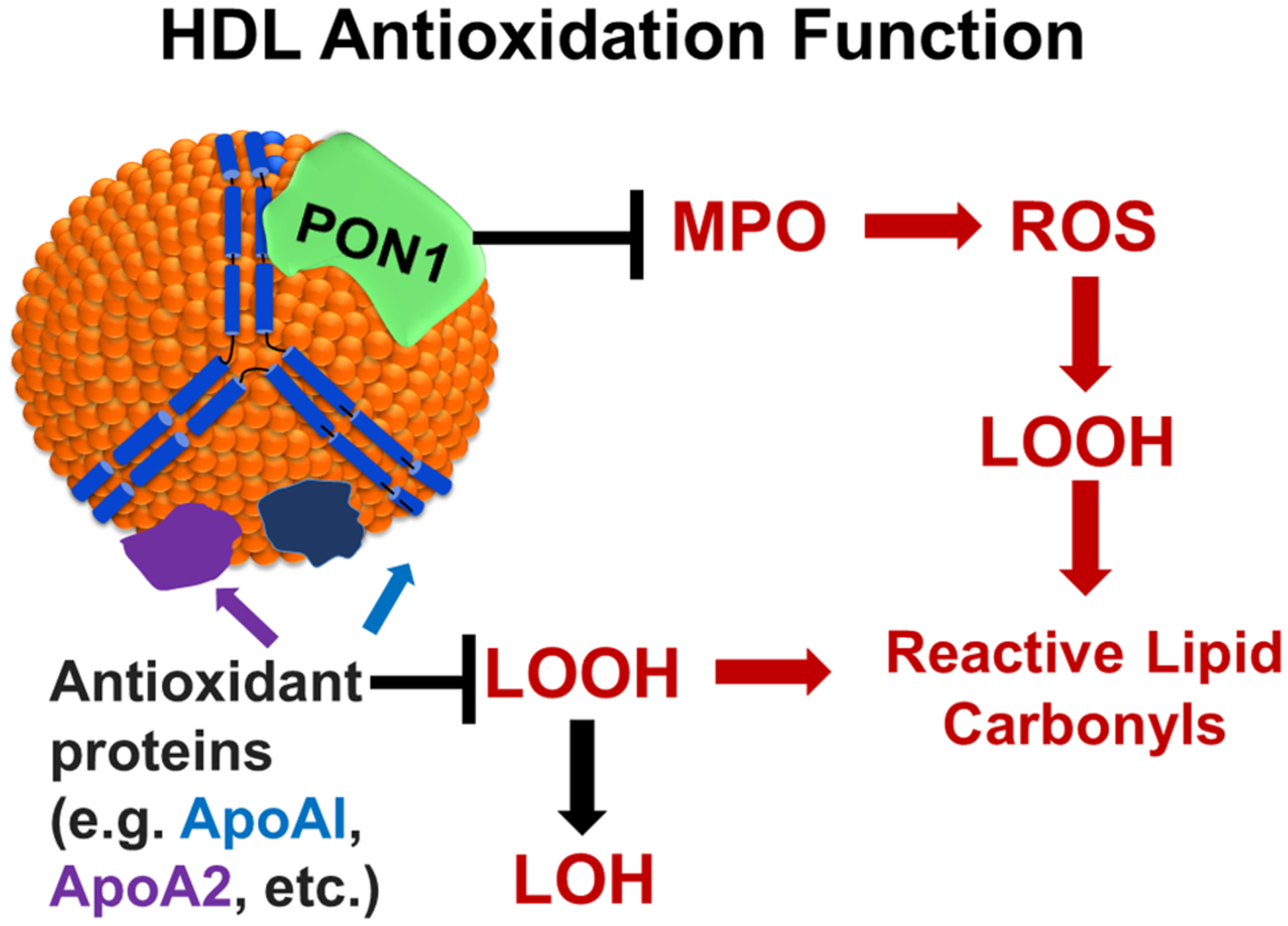
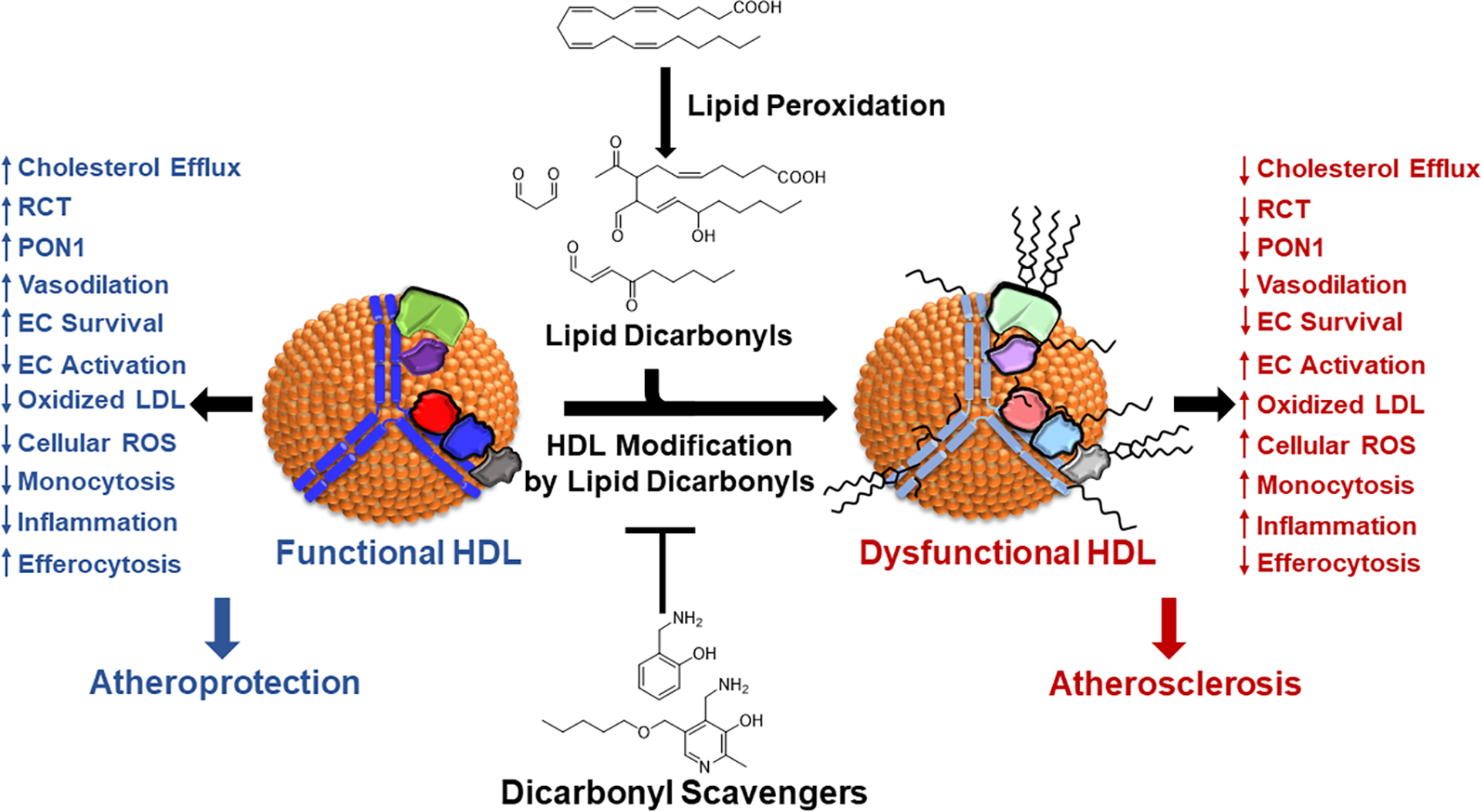
Epidemiologic studies detected an inverse relationship between HDL (high-density lipoprotein) cholesterol (HDL-C) levels and atherosclerotic cardiovascular disease (ASCVD), identifying HDL-C as a major risk factor for ASCVD and suggesting atheroprotective functions of HDL. However, the role of HDL-C as a mediator of risk for ASCVD has been called into question by the failure of HDL-C-raising drugs to reduce cardiovascular events in clinical trials. Progress in understanding the heterogeneous nature of HDL particles in terms of their protein, lipid, and small RNA composition has contributed to the realization that HDL-C levels do not necessarily reflect HDL function. The most examined atheroprotective function of HDL is reverse cholesterol transport, whereby HDL removes cholesterol from plaque macrophage foam cells and delivers it to the liver for processing and excretion into bile. Indeed, in several studies, HDL has shown inverse associations between HDL cholesterol efflux capacity and ASCVD in humans. Inflammation plays a key role in the pathogenesis of atherosclerosis and vulnerable plaque formation, and a fundamental function of HDL is suppression of inflammatory signaling in macrophages and other cells. Oxidation is also a critical process to ASCVD in promoting atherogenic oxidative modifications of LDL (low-density lipoprotein) and cellular inflammation. HDL and its proteins including apoAI (apolipoprotein AI) and PON1 (paraoxonase 1) prevent cellular oxidative stress and LDL modifications. Importantly, HDL in humans with ASCVD is oxidatively modified rendering HDL dysfunctional and proinflammatory. Modification of HDL with reactive carbonyl species, such as malondialdehyde and isolevuglandins, dramatically impairs the antiatherogenic functions of HDL. Importantly, treatment of murine models of atherosclerosis with scavengers of reactive dicarbonyls improves HDL function and reduces systemic inflammation, atherosclerosis development, and features of plaque instability. Here, we discuss the HDL antiatherogenic functions in relation to oxidative modifications and the potential of reactive dicarbonyl scavengers as a therapeutic approach for ASCVD.

Keywords: atherosclerosis; lipoproteins, HDL; macrophages; malondialdehyde; peroxidase.


Conflict of interest statement

Disclosures M.F. Linton and S.S. Davies are inventors on a patent for the use of 2-hydroxybenzylamine and related dicarbonyl scavengers for the treatment of cardiovascular disease (US 11 389 414). The other authors report no conflicts.

Figures

Figure 1.
Figure 1.. Lipid carbonyls react with amino acid residues to form adducts and crosslinks.
MDA (Malondialdehyde), IsoLG (isolevuglandins), ONE (4-oxo-nonenal), ACR (acrolein), HNE (4-hydroxy-nonenal), and MGO (methylglyoxal) react with their preferred amino acid targets including Lys-NH2 (lysine), Cys-SH (cysteine), His-imidazole (histidine), or Arg-guanidine (arginine) to form various adducts and crosslinks. Crosslinks form when the same lipid carbonyl reacts with two closely adjacent amino acids. In general, multiple species of adducts can form for each lipid carbonyls, two of the most important for each reactive lipid carbonyl are shown here. Adducts shown are: a. MDA N-propenal-Lys monoadduct, b. MDA Lys-1-amino-3-iminopropene-Lys crosslink, c. IsoLG-lactam-Lys monoadduct, d. IsoLG-dipyrrole-Lys crosslink, e. ONE N-4-ketoamide-Lys monoadduct, f. ONE-Lys-pyrrole-Cys crosslink, g. ACR-S-propanol-Cys monoadduct, h. ACR-His-propenamine-Cys crosslink, i. HNE-S-hemiacetal-Cys monoadduct, j. HNE-N-hemiacetal-His monoadduct, k. MGO Argpyrimidine monoadduct, and l. MGO-carboxy-ethyl-Lys.
Figure 2.
Figure 2.. Critical steps of the HDL reverse cholesterol transport (RCT) pathway.
The first step, which is rate limiting, is the removal of free cholesterol (FC) from macrophage foam cells. FC is released from macrophages by four mechanisms. ABCA1 (ATP-binding cassette transporter A1) releases PL (phospholipid) and FC to lipid-poor apoAI or endogenous apoE that is secreted by macrophage foam cells. FC is released to the discoidal HDL formed by ABCA1 and to mature HDL by ABCG1, SR-BI (scavenger receptor-class BI), and aqueous diffusion. The flux of FC between cells and HDL is bidirectional. FC influx occurs by SR-BI and aqueous diffusion. The FC in discoidal HDL particles is esterified by LCAT (lecithin:cholesterol acyltransferase) to form mature HDL3. Plasma HDL is remodeled by CETP (cholesteryl ester transfer protein) and PLTP (phospholipid transfer protein). PLTP transfers PL between VLDL and HDL and among HDL particles. CETP transfers triglyceride from VLDL/IDL to HDL3 to form larger HDL2 particles. CETP also transfers HDL CE to VLDL and IDL to form LDL, which is then internalized by the hepatic LDLR (LDL receptor) for cholesterol routing into bile. An alternative pathway for hepatic delivery of cholesterol and routing to bile is the selective uptake of HDL CE and the influx of HDL FC via SR-BI. CETP also transfers oxidized lipids from LDL to HDL for delivery to the liver by SR-BI. Nascent HDL is synthesized by interaction of hepatocyte or enterocyte (not shown) derived apoAI with hepatic or intestinal ABCA1, and then nascent HDL is routed to peripheral tissue to act as a FC acceptor. Created with BioRender.com.
Figure 3.
Figure 3.. The anti-inflammatory functions of HDL.
1. HDL suppresses endothelial activation in response to proinflammatory stimuli such as TNF-α and oxLDL. Proinflammatory stimuli activate NF‐κB (nuclear factor kappa B), which increases expression of monocyte adhesion proteins and chemotactic/inflammatory cytokines, thereby increasing monocyte recruitment and endothelial cell death. HDL signaling pathways stimulate nitric oxide production to decrease NF‐κB activation and maintain endothelial barrier integrity. 2. HDL prevents inflammatory monocytosis. Increased LDL and oxLDL, resulting from hypercholesterolemia, induce inflammatory signaling and ROS (reactive oxygen species) leading to expansion of bone marrow granulocyte monocyte progenitors that give rise to inflammatory Ly6Chigh monocytes. HDL signaling pathways suppress ROS production and inflammatory signaling and expansion of Ly6Chigh monocytes. 3. HDL prevents the macrophage proinflammatory phenotype. LPS, oxLDL, and other TLR (toll-like receptor) ligands activate NF‐κB leading to increased expression of inflammatory cytokines and NLRP3 (nucleotide oligomerization domain-like receptor protein with pyrin domain containing 3) inflammasome components resulting in decreased efferocytosis of apoptotic cells and necrotic death. HDL suppresses TLR signaling and NF‐κB activation via cholesterol efflux dependent and independent pathways to promote activation of Atf3 (activating transcription factor 3) and STAT6 (signal transducer and activator of transcription 6), thereby reducing activation of inflammasomes and enhancing efferocytosis of apoptotic cells by promoting expression of Arg-1 (arginase-1) and IL-10. Created with BioRender.com.
Figure 4.
Figure 4.. The antioxidant functions of HDL.
A number of proteins associated with HDL including apoAI and apoA2 prevent the formation of reactive lipid carbonyls by reducing LOOH (lipid hydroperoxides) to LOH (lipid hydroxides). MPO (myeloperoxidase) generates ROS (reactive oxygen species) leading to sequential formation of LOOH and reactive lipid carbonyls. The association of PON1 (paraoxonase 1) with apoAI both inhibits MPO activity and also directly inhibits formation of lipid hydroperoxides and reactive lipid carbonyls.
Figure 5.
Figure 5.. Current classes of small molecule scavengers available to target reactive lipid carbonyls.
Thiol-based scavengers capture ACR (acrolein), ONE (4-oxo-nonenal), and HNE (4-hydroxy-nonenal). Imidazole-based scavengers capture ACR, ONE, HNE, and MGO (methylglyoxal). 2-aminomethylphenol-based dicarbonyl scavengers capture IsoLG (isolevuglandins), ONE, MDA (malondialdehyde), and MGO.
Figure 6.
Figure 6.. Dicarbonyl scavengers protect HDL functionality.
Under oxidative conditions, lipid peroxidation generates lipid dicarbonyls that react with HDL proteins to render HDL dysfunctional. Dicarbonyl scavengers can intercept these lipid dicarbonyls because the primary amines of these scavengers is even more reactive with dicarbonyls than the lysyl residues of proteins are reactive with the dicarbonyls. Thus, these scavengers protect HDL proteins from modification and thereby prevent HDL dysfunction. Shown are the atheroprotective functions of HDL that dicarbonyl scavengers can protect against dysfunction including EC (endothelial cell) activation and survival.

References

    1. Gordon T, Castelli WP, Hjortland MC, Kannel WB and Dawber TR. High density lipoprotein as a protective factor against coronary heart disease. The Framingham Study. Am J Med. 1977;62:707–14. - PubMed
    1. Gordon DJ, Probstfield JL, Garrison RJ, Neaton JD, Castelli WP, Knoke JD, Jacobs DR Jr., Bangdiwala S and Tyroler HA. High-density lipoprotein cholesterol and cardiovascular disease. Four prospective American studies. Circulation. 1989;79:8–15. - PubMed
    1. Rohatgi A, Westerterp M, von Eckardstein A, Remaley A and Rye KA. HDL in the 21st Century: A Multifunctional Roadmap for Future HDL Research. Circulation. 2021;143:2293–2309. - PMC - PubMed
    1. Voight BF, Peloso GM, Orho-Melander M, Frikke-Schmidt R, Barbalic M, Jensen MK, Hindy G, Holm H, Ding EL, Johnson T, Schunkert H, Samani NJ, Clarke R, Hopewell JC, Thompson JF, Li M, Thorleifsson G, Newton-Cheh C, Musunuru K, Pirruccello JP, Saleheen D, Chen L, Stewart A, Schillert A, Thorsteinsdottir U, Thorgeirsson G, Anand S, Engert JC, Morgan T, Spertus J, Stoll M, Berger K, Martinelli N, Girelli D, McKeown PP, Patterson CC, Epstein SE, Devaney J, Burnett MS, Mooser V, Ripatti S, Surakka I, Nieminen MS, Sinisalo J, Lokki ML, Perola M, Havulinna A, de Faire U, Gigante B, Ingelsson E, Zeller T, Wild P, de Bakker PI, Klungel OH, Maitland-van der Zee AH, Peters BJ, de Boer A, Grobbee DE, Kamphuisen PW, Deneer VH, Elbers CC, Onland-Moret NC, Hofker MH, Wijmenga C, Verschuren WM, Boer JM, van der Schouw YT, Rasheed A, Frossard P, Demissie S, Willer C, Do R, Ordovas JM, Abecasis GR, Boehnke M, Mohlke KL, Daly MJ, Guiducci C, Burtt NP, Surti A, Gonzalez E, Purcell S, Gabriel S, Marrugat J, Peden J, Erdmann J, Diemert P, Willenborg C, Konig IR, Fischer M, Hengstenberg C, Ziegler A, Buysschaert I, Lambrechts D, Van de Werf F, Fox KA, El Mokhtari NE, Rubin D, Schrezenmeir J, Schreiber S, Schafer A, Danesh J, Blankenberg S, Roberts R, McPherson R, Watkins H, Hall AS, Overvad K, Rimm E, Boerwinkle E, Tybjaerg-Hansen A, Cupples LA, Reilly MP, Melander O, Mannucci PM, Ardissino D, Siscovick D, Elosua R, Stefansson K, O’Donnell CJ, Salomaa V, Rader DJ, Peltonen L, Schwartz SM, Altshuler D and Kathiresan S. Plasma HDL cholesterol and risk of myocardial infarction: a mendelian randomisation study. Lancet. 2012;380:572–80. - PMC - PubMed
    1. Karjalainen MK, Holmes MV, Wang Q, Anufrieva O, Kahonen M, Lehtimaki T, Havulinna AS, Kristiansson K, Salomaa V, Perola M, Viikari JS, Raitakari OT, Jarvelin MR, Ala-Korpela M and Kettunen J. Apolipoprotein A-I concentrations and risk of coronary artery disease: A Mendelian randomization study. Atherosclerosis. 2020;299:56–63. - PubMed

Publication types