. 2023 Jun;31(6):861-873.
doi: 10.1016/j.jsps.2023.04.005. Epub 2023 Apr 13.

Design, physicochemical characterisation, and in vitro cytotoxicity of cisplatin-loaded PEGylated chitosan injectable nano / sub-micron crystals

Affiliations

Design, physicochemical characterisation, and in vitro cytotoxicity of cisplatin-loaded PEGylated chitosan injectable nano / sub-micron crystals

Muhammad H Sultan et al. Saudi Pharm J. 2023 Jun.

Abstract

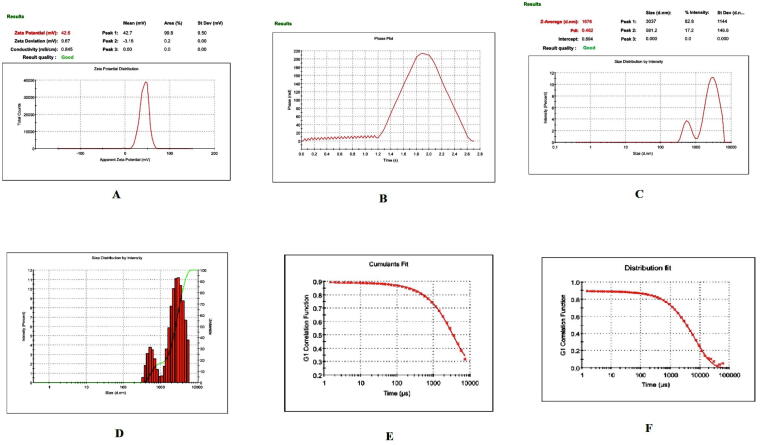
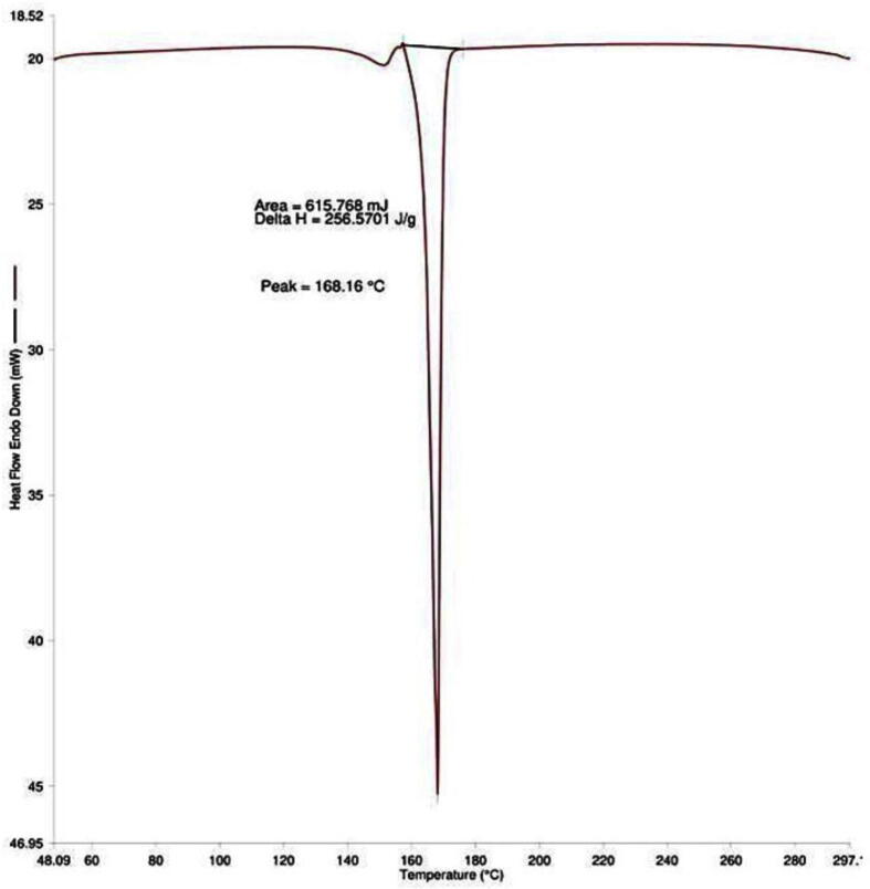
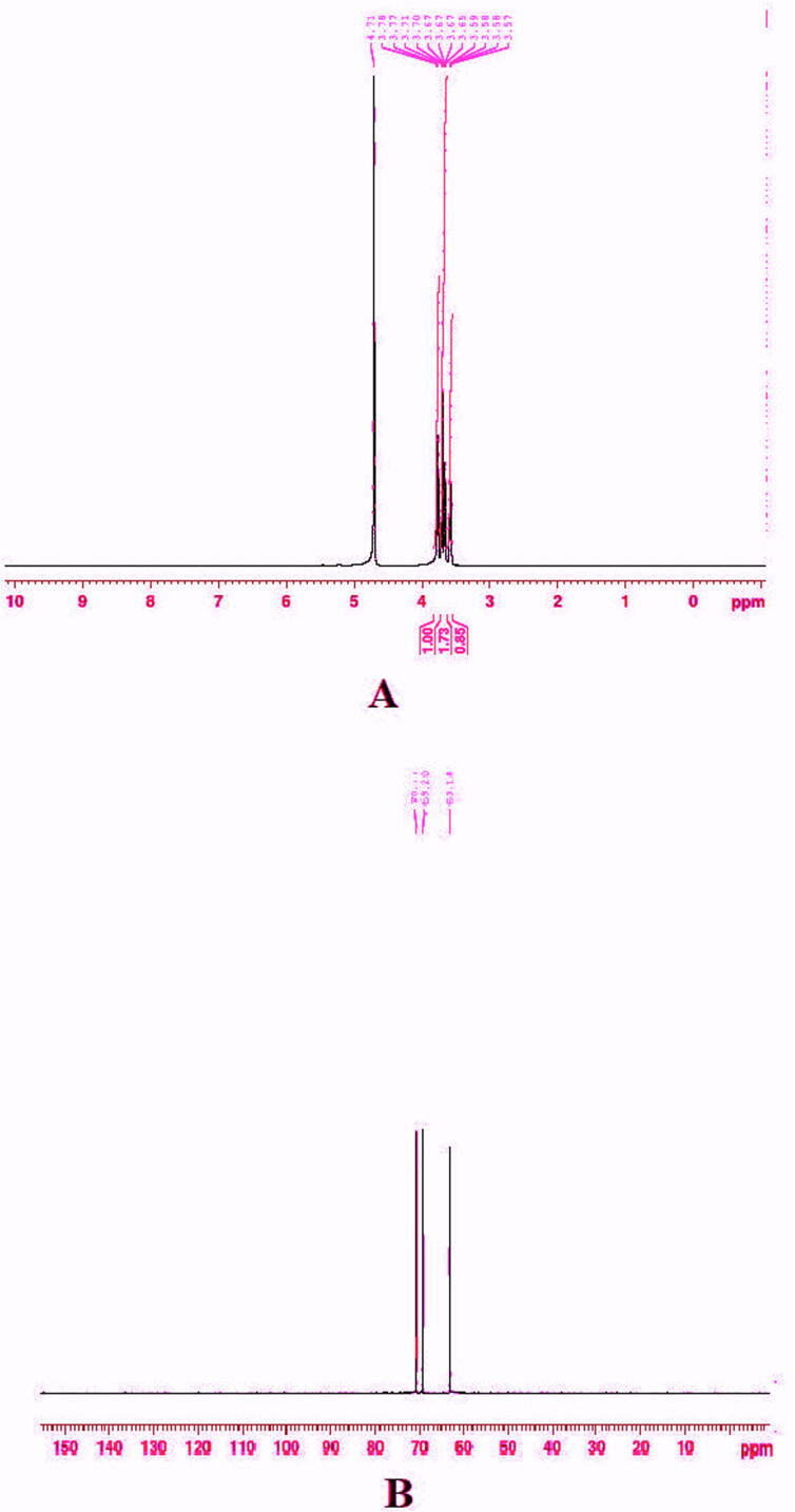
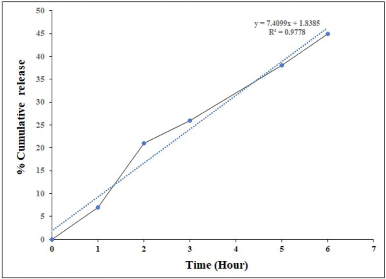
The study aimed to develop cisplatin-loaded PEGylated chitosan nanoparticles. The optimal batch of cisplatin-loaded PEGylated chitosan nanoparticles had a + 49.9 mV zeta potential, PDI of 0.347, and % PDI of 58.9. Nanoparticle zeta size was 741.4 z. d.nm, the size in diameter was 866.7 ± 470.5 nm, and nanoparticle conductivity in colloidal solution was 0.739 mS/cm. Differential scanning calorimetry (DSC) revealed that cisplatin-loaded PEGylated chitosan nanoparticles had sharp endothermic peaks at temperatures at 168.6 °C. The thermogravimetric analysis (TGA) showed the weight loss of cisplatin-loaded PEGylated chitosan nanoparticles, which was observed as 95% at 262.76 °C. XRD investigation on cisplatin-loaded PEGylated chitosan nanoparticles exhibited distinct peaks at 2θ as 9.7°, 20.4°, 22.1°, 25.3°, 36.1°, 38.1°, 39.5°, 44.3°, and 64.5°, confirming crystalline structure. The 1H NMR analysis showed the fingerprint region of cisplatin-loaded PEGylated chitosan nanoparticles as 0.85, 1.73, and 1.00 ppm in the proton dimension and de-shielded proton peaks appeared at 3.57, 3.58, 3.58, 3.59, 3.65, 3.67, 3,67, 3,67, 3.70, 3.71, 3.77, 3.78 and 4.71 ppm. The 13C NMR spectrum showed specified peaks at 63.18, 69.20, and 70.77 ppm. The FT-IR spectra of cisplatin loaded PEGylated nanoparticles show the existence of many fingerprint regions at 3186.52, 2931.68, 1453.19, 1333.98, 1253.71, 1085.19, 1019.60, 969.98, 929.53, 888.80, 706.13, and 623.67 cm-1. The drug release kinetics of cisplatin loaded PEGylated chitosan nanoparticles showed zero order kinetics with 48% of drug release linearity fashion which has R2 value of 0.9778. Studies on the MCF-7 ATCC human breast cancer cell line in vitro revealed that the IC50 value 82.08 µg /mL. Injectable nanoparticles had good physicochemical and cytotoxic properties. This method is novel since the application of the PEGylation processes leads to an increased solubility of chitosan nanoparticles at near neutral pH.

Keywords: Cancer; Cisplatin; Cytotoxicity; Nanoparticles; Pegylated Chitosan; Polydispersity Index; Size; Zetapotential.


Conflict of interest statement

The authors declare that they have no known competing financial interests or personal relationships that could have appeared to influence the work reported in this paper.

Figures

Fig. 1
Fig. 1
Physical characterization of cisplatin-loaded PEGylated chitosan nanoparticles of batch 1 (A) Zetapotential analysis (B) Phase plot of particulate colloidal system (C) Particle Size distribution analysis through intensity (D) Particle Size distribution analysis (E) Cumulative fit of particulate colloidal system (F) Particle distribution fit of particulate colloidal system.
Fig. 2
Fig. 2
Scanning electron micrograph study of batch 1 (A) Cisplatin-loaded PEGylated chitosan nanoparticles of batch 1 under 8,000 × magnification (B) Cisplatin-loaded PEGylated chitosan nanoparticles of batch 1 under 20,000 × magnification.
Fig. 3
Fig. 3
Physical characterization of cisplatin-loaded PEGylated chitosan nanoparticles of batch 2 (A) Zetapotential analysis (B) Phase plot of particulate colloidal system (C) Particle Size distribution analysis through intensity (D) Particle Size distribution analysis (E) Cumulative fit of particulate colloidal system (F) Particle distribution fit of particulate colloidal system.
Fig. 4
Fig. 4
Scanning electron micrograph study of batch 2 (A) The scanning electron micrograph of cisplatin-loaded PEGylated chitosan nanoparticles of batch 2 under 3000 × magnification (B) The scanning electron micrograph of cisplatin-loaded PEGylated chitosan nanoparticles of batch 1 under 3,000 × magnification.
Fig. 5
Fig. 5
Physical characterization of cisplatin-loaded PEGylated chitosan nanoparticles of batch 3 (A) Zetapotential analysis (B) Phase plot of particulate colloidal system (C) Particle Size distribution analysis through intensity (D) Particle Size distribution analysis (E) Cumulative fit of particulate colloidal system (F) Particle distribution fit of particulate colloidal system.
Fig. 6
Fig. 6
Scanning electron micrograph study of batch 3 (A) The scanning electron micrograph of cisplatin-loaded PEGylated chitosan nanoparticles of batch 1 under 6, 000 × magnification (B) The scanning electron micro-graph of cisplatin-loaded PEGylated chitosan nanoparticles of batch 3 under 15,000 × magnification.
Fig. 7
Fig. 7
Differential scanning calorimetry analysis of cisplatin-loaded PEGylated chitosan nanoparticles of ideal batch.
Fig. 8
Fig. 8
Thermo gravity analysis of cisplatin-loaded PEGylated chitosan nanoparticles of ideal batch.
Fig. 9
Fig. 9
XRD analysis of cisplatin-loaded PEGylated chitosan nanoparticles of ideal batch.
Fig. 10
Fig. 10
NMR analysis. (A) 1H NMR analysis of lyophilized cisplatin-loaded PEGylated chitosan nanoparticles of ideal batch (B) 13CNMR analysis of lyophilized cisplatin-loaded PEGylated chitosan nanoparticles of ideal batch.
Fig. 11
Fig. 11
FT-IR spectroscopy analyisis. (A) FT-IR spectrum of Chitosan polymer (B) FT-IR cisplatin-loaded PEGylated chitosan nanoparticles.
Fig. 12
Fig. 12
In vitro release profile. Release kinetics of lyophilized cisplatin-loaded PEGylated chitosan nanoparticles of ideal batch.
Fig. 13
Fig. 13
Cytotoxicity study. Dose-response curve of lyophilized cisplatin-loaded PEGylated chitosan nanoparticles of ideal batch against MCF-7 breast cancer cells.

References

    1. Abouelhag H.A., Sivakumar S.M., Bagul U.S., Mohamed Eltyep E., Safhi M.M. Preparation and physical characterization of cisplatin-chitosan nanoparticles by zeta nanosizer “prime step for formulation and development”. Int. J. Pharm. Sci. Res. 2017;10:4245–4249. doi: 10.13040/IJPSR.0975-8232.8(10).4245-49. - DOI
    1. Ahmed A.M., Ali M.R., Noor A.K. Use of biocomposite adsorbents for the removal of methylene blue dye from aqueous solution. Am. J. Mater. Sci. 2016;6(5):135–146. doi: 10.5923/j.materials.20160605.03. - DOI
    1. Alexandri E., Ahmed R., Siddiqui H., Choudhary M.L., Tsiafoulis C.G., Gerothanassis I.P. High resolution NMR spectroscopy as a structural and analytical tool for unsaturated lipids in solution. Molecules. 2017;22:1663. doi: 10.3390/molecules22101663. - DOI - PMC - PubMed
    1. Amidi M., Mastrobattista E., Wim J., Wim E.H. Chitosan-based delivery systems for protein therapeutics and antigens. Adv. Drug Deliv. Rev. 2010;62(1):59–82. doi: 10.1016/j.addr.2009.11.009. - DOI - PubMed
    1. Anand M., Sathyapriya P., Maruthupandy M., Beevi A.H. Synthesis of chitosan nanoparticles by TPP and their potential mosquito larvicidal application. Front. Lab. Med. 2018;2:72–78. doi: 10.1016/j.flm.2018.07.003. - DOI

LinkOut - more resources