. 2023 May 9:10:1128921.
doi: 10.3389/fmed.2023.1128921. eCollection 2023.

GADD34 activates p53 and may have utility as a marker of atherosclerosis

Affiliations

GADD34 activates p53 and may have utility as a marker of atherosclerosis

Go Tomiyoshi et al. Front Med (Lausanne). .

Abstract

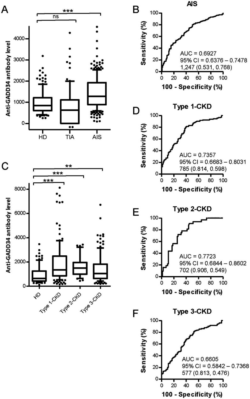
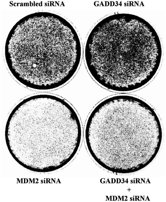
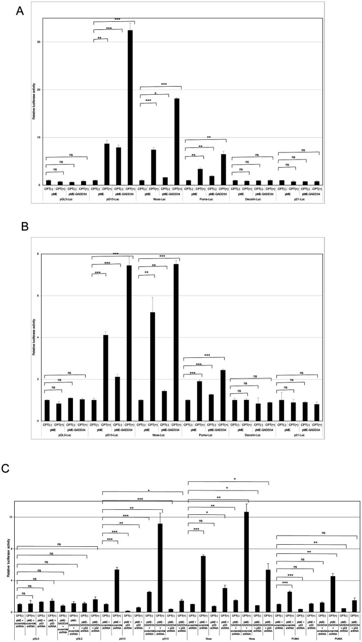
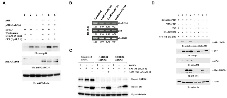
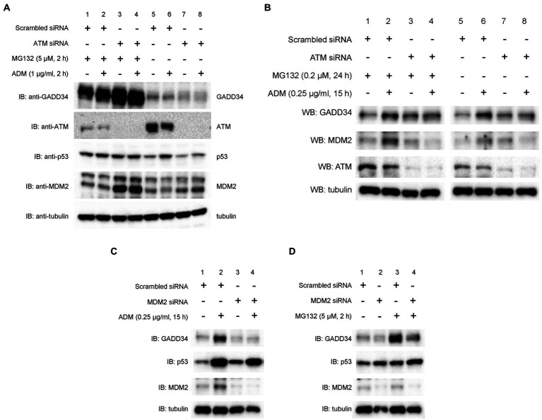
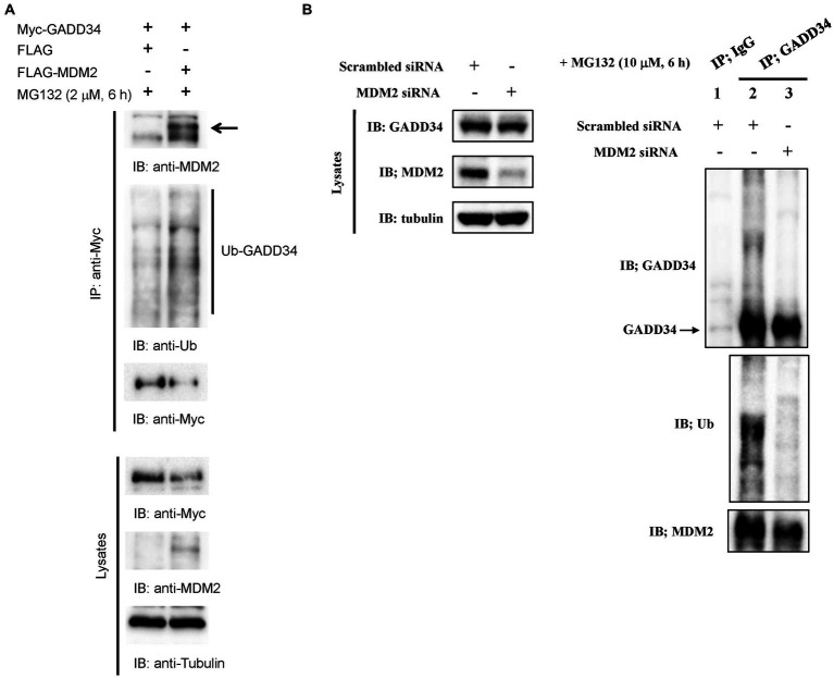
We previously identified growth arrest and DNA-damage-inducible gene 34 (GADD34) as a marker of ischemic stroke. In the present study, serum levels of anti-GADD34 antibodies were found to be significantly higher in patients with acute ischemic stroke or chronic kidney disease compared to healthy donors. We then examined the biological function of GADD34 by transfection into U2OS human osteosarcoma and U87 human glioblastoma cells. Knockdown of GADD34 by siRNA resulted in enhanced cell proliferation, which was reversed by co-knockdown of MDM2. Luciferase reporter assays revealed that the transactivation ability of p53 enhanced by genotoxic anticancer drugs such as camptothecin and etoposide was further potentiated by enforced expression of GADD34 but attenuated by co-transfection with p53 shRNA expression plasmids. Western blotting demonstrated increased p53 protein levels after treatment with camptothecin, which was also potentiated by GADD34 but suppressed by GADD34 siRNA, ATM siRNA, and ATM inhibitor wortmannin. GADD34 levels also increased in response to treatment with camptothecin or adriamycin, and this increase was attenuated by MDM2 siRNA. Immunoprecipitation with anti-GADD34 antibody followed by Western blotting with anti-MDM2 antibodies indicated ubiquitination of GADD34 is mediated by MDM2. Accordingly, GADD34 may function as a ubiquitination decoy to reduce p53 ubiquitination and increase p53 protein levels. Increased neuronal cell death due to activation of p53 by GADD34 may account for the elevated serum levels of anti-GADD34 antibodies observed in patients with acute ischemic stroke.

Keywords: GADD34; atherosclerosis; ischemic stroke; p53; ubiquitination.


Conflict of interest statement

This work was performed in collaboration with Fujikura Kasei Co., Ltd. GT, RN, NS, and HK are employee of Fujikura Kasei Co., Ltd. The remaining authors declare that the research was conducted in the absence of any commercial or financial relationships that could be construed as a potential conflict of interest.

Figures

Figure 1
Figure 1
Comparison of serum GADD34 antibody levels between healthy donors (HDs) and patients with stroke or chronic kidney disease (CKD) by AlphaLISA. Serum samples from HDs and patients with AIS were obtained from Chiba Prefectural Sawara Hospital. Samples from patients with CKD were obtained from the Kumamoto cohort. Serum antibody levels in HDs and patients with transient ischemic attack (TIA) and acute ischemic stroke (AIS) (A) and patients with CKD (C) measured by AlphalLISA are shown by box-whisker plot. CKD types 1, 2, and 3 represent diabetic CKD, nephrosclerosis, and glomerulonephritis, respectively. Box plots display the 10th, 20th, 50th, 80th, and 90th percentiles. Total numbers of males and females, mean age ± standard deviation (SD), mean antibody levels ± SD, cutoff values, total numbers of individuals with anti-GADD34 antibodies, proportions (%) of individuals with anti-GADD34 antibodies, and p-values from HDs versus patients with TIA, AIS, and CKD are summarized and shown in Supplementary Tables S2, S3, respectively. p-values were calculated using the Kruskal-Wallis test. **, p < 0.01; ***, p < 0.001; ns, not significant. Receiver operating characteristic curve (ROC) analyses were performed to evaluate the utility of GADD34 antibodies in detecting AIS (B), type-1 CKD (D), type-2 CKD (E), and type-3 CKD (F). Values for area under the curve (AUC), 95% confidence intervals (CI), and cutoff values for serum anti-GADD34 antibody levels are shown. Sensitivity (left) and specificity (right) values are shown in parentheses.
Figure 2
Figure 2
Knockdown of GADD34 increases cell proliferation. Human osteosarcoma U2OS cells were transfected with GADD34 siRNA and/or MDM2 siRNA. Scrambled siRNA was used as a control. Cells were cultured for 2 weeks, fixed, and stained with Giemsa.
Figure 3
Figure 3
Activation of camptothecin (CPT)-activated p53 reporters by GADD34. (A) U2OS human osteosarcoma cells and (B) U87 human glioblastoma cells (5 × 104 cells) were co-transfected with p53-responsive reporter plasmids (pG13-Luc, Noxa-Luc, PUMA-Luc, Decorin-Luc, and p21-Luc; 100 ng), transfection standard SV40-Rluc (10 ng), and the expression plasmid (500 ng) of pME-GADD34 or control empty vector pME-18S (pME). The control reporter plasmid, pGL3-Luc, was also used. The effects of knockdown of p53 were also examined by co-transfection with p53 shRNA or scrambled shRNA (C). Cells were cultured for 24 h and then treated with CPT (1 μM) for 24 h. Relative firefly luciferase activities versus Renilla luciferase activities were measured in cell extracts. Error bars represent SD (n = 3). *, p < 0.05; **, p < 0.01; ***, p < 0.001; ns, not significant.
Figure 4
Figure 4
Increased p53 protein levels in the presence of GADD34. (A) Synergistic up-regulation of p53 protein by GADD34 and CPT. U2OS cells were transfected with pME-GADD34 or pME control vector and cultured for 48 h. Cells were pretreated with wortmannin (25 μM) or dimethyl sulfoxide (DMSO) for 30 min and then treated with CPT (2 μM) for 2 h. Total cell extracts were analyzed by Western blotting using anti-p53, anti-GADD34, and anti-α-tubulin antibodies. (B) Decreased GADD34 mRNA levels following treatment with siRNA. U2OS cells were transfected with three siRNAs including GADD34 siRNA1, siRNA2, and siRNA3, and cultured for 2 days. Then, total RNA was isolated and used as a template for reverse transcription followed by polymerase chain reaction (RT-PCR). Amplified DNA fragments of GADD34, p53, and control GAPDH are shown. Numbers indicate the relative amounts of DNA fragments versus cells transfected with scrambled siRNA. (C) Decreased GADD34 protein levels following treatment with siRNA. U2OS cells were transfected with GADD34 siRNA2, GADD34 siRNA3, and control scrambled siRNA and cultured for 33 h. Cells were then treated with CPT (0.5 μM), adriamycin (ADM; 0.25 μg/mL), or solvent DMSO (0.1%) for 15 h. Total cell extracts were analyzed by Western blotting using anti-GADD34, anti-p53, and anti-α-tubulin. (D) Increased p53 phosphorylation in the presence of GADD34. U2OS cells were transfected with ATM siRNA or control scrambled siRNA, cultured for 24 h, transfected with Myc-tagged GADD34 or control Myc-tagged expression vector (pcDNA-My), cultured for 24 h, then treated with CPT for further 24 h. Cells were harvested and subjected to Western blotting using anti-p53, anti-ATM, anti-Myc, and anti-β-actin antibodies.
Figure 5
Figure 5
Suppression of ADM-induced increases in GADD34 protein levels by ATM siRNA and MDM2 siRNA. U2OS cells were transfected with (A,B) ATM siRNA, (C,D) MDM2 siRNA, or control scrambled siRNA and cultured for 24 h. (A) Cells were then treated with MG132 (5 μM) and/or ADM (1 μg/mL) for 2 h. (B) Cells were pretreated with MG132 (0.2 μM) for 9 h and then treated with ADM (0.25 μg/mL) for 15 h. Cells were treated with (C) ADM (0.25 μg/mL) for 15 h or (D) MG132 (5 μM) for 2 h. Total cell lysates were analyzed by Western blotting using anti-GADD34, anti-MDM2, anti-ATM, anti-p53, and anti-α-tubulin antibodies.
Figure 6
Figure 6
Ubiquitination of GADD34 via MDM2. (A) Increased ubiquitination of GADD34 following treatment with genotoxic anticancer drugs. U2OS cells were co-transfected with pcDNA-Myc-GADD34 and FLAG-MDM2 or the control FLAG vector and cultured for 48 h. Cells were then treated with MG132 (2 μM) for 6 h and then harvested. Cell extracts were immunoprecipitated using anti-Myc antibodies followed by Western blotting (IB) using anti-MDM2, anti-ubiquitin (Ub), and anti-Myc antibodies. Lysates without immunoprecipitation were analyzed using anti-Myc, anti-MDM2, and anti-α-tubulin antibodies. (B) Attenuation of GADD34 ubiquitination by MDM2 siRNA. A549 human lung carcinoma cells were transfected with MDM2 siRNA or control scrambled siRNA, cultured for 48 h, and then treated with MG132 (10 μM) for 6 h. Left panel, Western blotting of cell lysates using anti-GADD34, anti MDM2, and anti-α-tubulin antibodies. Right panel, cell lysates were immunoprecipitated with anti-GADD34 antibodies or control IgG and analyzed by Western blotting using anti-GADD34, anti-ubiquitin (Ub), anti-MDM2 antibodies.

Similar articles

Cited by

References

    1. Greenblatt MS, Bennett WP, Hollstein M, Harris CC. Mutations in the p53 tumor suppressor gene: clues to cancer etiology and molecular pathogenesis. Cancer Res. (1994) 54:4855–78. PMID: - PubMed
    1. Varley JM, Evans DG, Birch JM. Li-Fraumeni syndrome–a molecular and clinical review. Br J Cancer. (1997) 76:1–14. doi: 10.1038/bjc.1997.328, PMID: - DOI - PMC - PubMed
    1. Donehower LA, Harvey M, Slagle BL, McArthur MJ, Montgomery CA, Jr, Butel JS, et al. . Mice deficient for p53 are developmentally normal but susceptible to spontaneous tumours. Nature. (1992) 356:215–21. doi: 10.1038/356215a0, PMID: - DOI - PubMed
    1. El-Deiry WS, Tokino T, Velculescu VE, Levy DB, Parsons R, Trent JM, et al. . WAF1, a potential mediator of p53 tumor suppression. Cell. (1993) 75:817–25. doi: 10.1016/0092-8674(93)90500-p, PMID: - DOI - PubMed
    1. Miyashita T, Reed JC. Tumor suppressor p53 is a direct transcriptional activator of the human bax gene. Cell. (1995) 80:293–9. doi: 10.1016/0092-8674(95)90412-3, PMID: - DOI - PubMed