

Stroke recurrence is associated with unfavorable intracranial venous outflow in patients with symptomatic intracranial atherosclerotic large vessel severe stenosis or occlusion
- PMID: 37228413
- PMCID: PMC10203233
- DOI: 10.3389/fneur.2023.1156315
Stroke recurrence is associated with unfavorable intracranial venous outflow in patients with symptomatic intracranial atherosclerotic large vessel severe stenosis or occlusion
Abstract
Objective: The purpose of this study was to investigate the predictive value of intracranial venous outflow for recurrent cerebral ischemic events (RCIE) in patients with symptomatic intracranial atherosclerotic large-vessel severe stenosis or occlusion (sICAS-S/O).
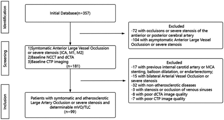
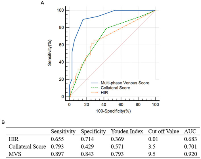
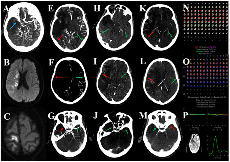
Methods: This retrospective study included sICAS-S/O patients with anterior circulation who underwent dynamic computed tomography angiography (dCTA) and computed tomography perfusion (CTP). Arterial collaterals were evaluated using the pial arterial filling score for dCTA data, tissue-level collaterals (TLC) were assessed using the high-perfusion intensity ratio (HIR, Tmax >10 s/Tmax >6 s), and cortical veins were evaluated using the multi-phase venous score (MVS) for the vein of Labbé (VOL), sphenoparietal sinus (SPS), and superficial cerebral middle vein (SCMV). The relationships between multi-phase venous outflow (mVO), TLC, and 1-year RCIE were analyzed.
Results: Ninety-nine patients were included, 37 of whom had unfavorable mVO (mVO-) and 62 of whom had favorable mVO (mVO+). Compared with the mVO+ patients, mVO- patients had a higher admission National Institutes of Health Stroke Scale (NIHSS) score (median, 4 [interquartile range (IQR), 0-9] vs. 1 [IQR, 0-4]; p = 0.048), larger ischemic volume (median, 74.3 [IQR, 10.1-177.9] vs. 20.9 [IQR, 5-86.4] mL; p = 0.042), and worse tissue perfusion (median, 0.04 [IQR, 0-0.17] vs. 0 [IQR, 0-0.03]; p = 0.007). Multivariate regression analysis showed that mVO- was an independent predictor of 1-year RCIE.
Conclusion: For patients with sICAS-S/O of the anterior circulation, unfavorable intracranial venous outflow is a potential imaging indicator for predicting higher 1-year RCIE risk.
Keywords: collaterals; cortical vein; intracranial atherosclerotic disease; perfusion; stenosis.
Copyright © 2023 Gao, Zhang, Lin, Yang, Yao, Cheng, Cai and Huang.
Conflict of interest statement
The authors declare that the research was conducted in the absence of any commercial or financial relationships that could be construed as a potential conflict of interest.
Figures
Similar articles
-
Favorable Venous Outflow Profiles Correlate With Favorable Tissue-Level Collaterals and Clinical Outcome.Stroke. 2021 May;52(5):1761-1767. doi: 10.1161/STROKEAHA.120.032242. Epub 2021 Mar 8. Stroke. 2021. PMID: 33682452
-
Unfavorable venous outflow correlates with poor prognosis in acute ischemic stroke due to large vessel occlusion (AIS-LVO) patients assessed dynamically and quantitatively based on four-dimensional computed tomography angiography/perfusion (4D-CTA/CTP).Quant Imaging Med Surg. 2025 Apr 1;15(4):2865-2880. doi: 10.21037/qims-24-669. Epub 2025 Mar 28. Quant Imaging Med Surg. 2025. PMID: 40235806 Free PMC article.
-
Venous Outflow Profiles Are Linked to Cerebral Edema Formation at Noncontrast Head CT after Treatment in Acute Ischemic Stroke Regardless of Collateral Vessel Status at CT Angiography.Radiology. 2021 Jun;299(3):682-690. doi: 10.1148/radiol.2021203651. Epub 2021 Apr 6. Radiology. 2021. PMID: 33825511
-
Intravenous tPA (Tissue-Type Plasminogen Activator) Correlates With Favorable Venous Outflow Profiles in Acute Ischemic Stroke.Stroke. 2022 Oct;53(10):3145-3152. doi: 10.1161/STROKEAHA.122.038560. Epub 2022 Jun 23. Stroke. 2022. PMID: 35735008
-
Stent-Retriever Thrombectomy and Rescue Treatment of M1 Occlusions Due to Underlying Intracranial Atherosclerotic Stenosis: Cohort Analysis and Review of the Literature.Cardiovasc Intervent Radiol. 2019 Jun;42(6):863-872. doi: 10.1007/s00270-019-02187-9. Epub 2019 Mar 11. Cardiovasc Intervent Radiol. 2019. PMID: 30859286 Review.
References
-
- Zaidat OO, Fitzsimmons BF, Woodward BK, Wang Z, Killer-Oberpfalzer M, Wakhloo A, et al. . Effect of a balloon-expandable intracranial stent vs medical therapy on risk of stroke in patients with symptomatic intracranial stenosis: the VISSIT randomized clinical trial. JAMA. (2015) 313:1240–8. doi: 10.1001/jama.2015.1693, PMID: - DOI - PubMed
-
- Derdeyn CP, Chimowitz MI, Lynn MJ, Fiorella D, Turan TN, Janis LS, et al. . Aggressive medical treatment with or without stenting in high-risk patients with intracranial artery stenosis (SAMMPRIS): the final results of a randomised trial. Lancet. (2014) 383:333–41. doi: 10.1016/S0140-6736(13)62038-3, PMID: - DOI - PMC - PubMed
LinkOut - more resources
Full Text Sources